Your cart
There are no more items in your cart


Cranial arterial circulation - Erler Zimmer 3D anatomy Series MP1650
erler zimmer
EZ-MP1650
€2,344.47
Tax included
Made in ultra-high resolution 3D printing in full color.
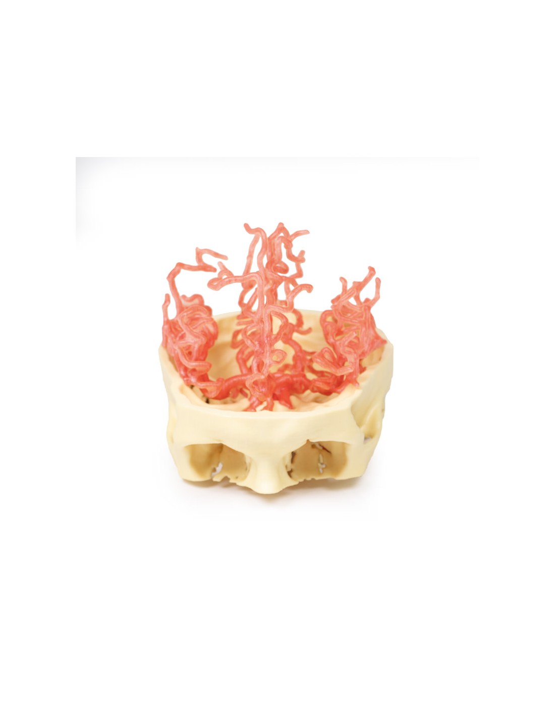
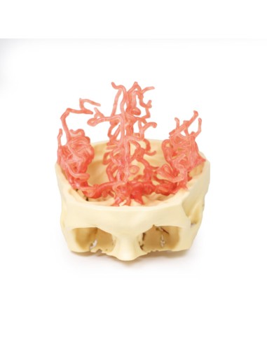
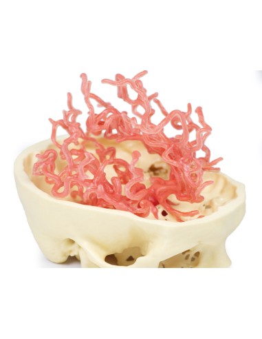
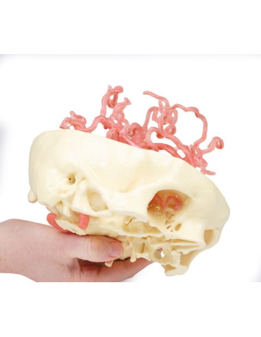
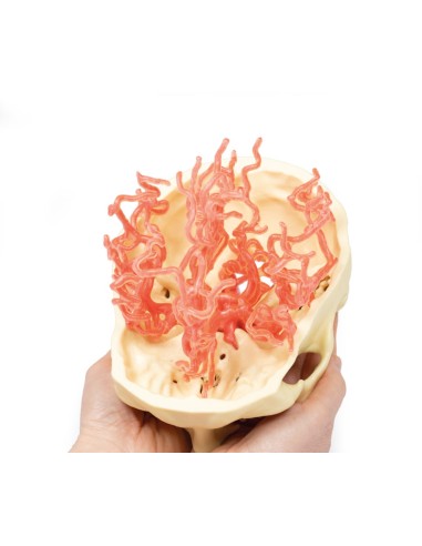
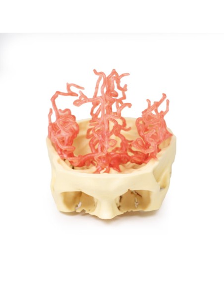
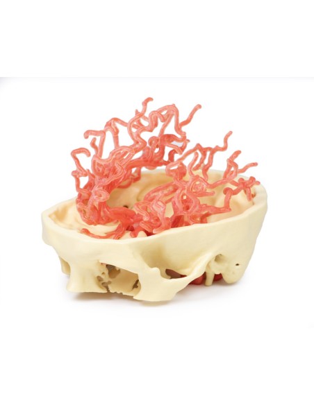
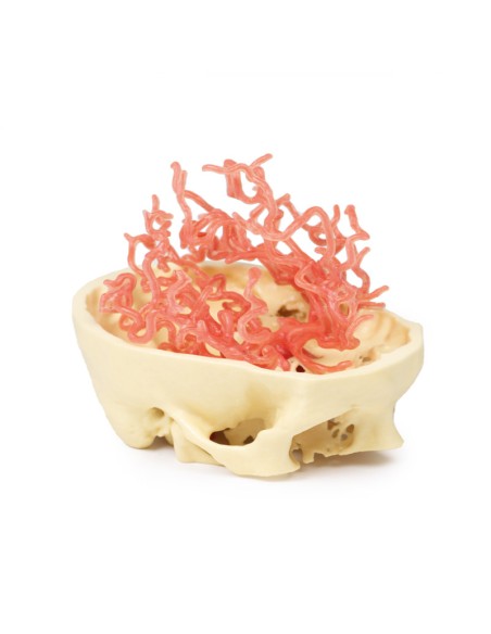
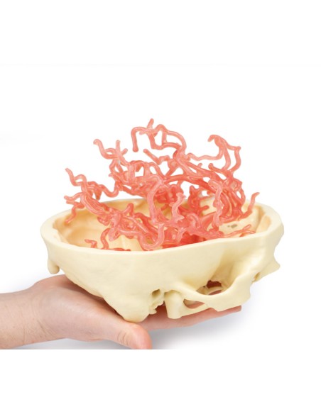
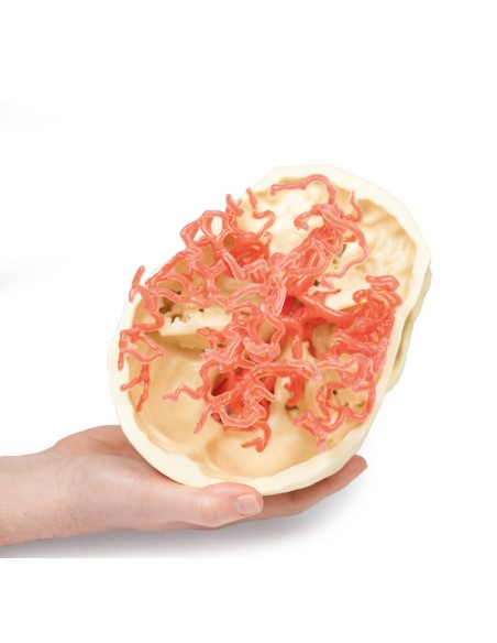
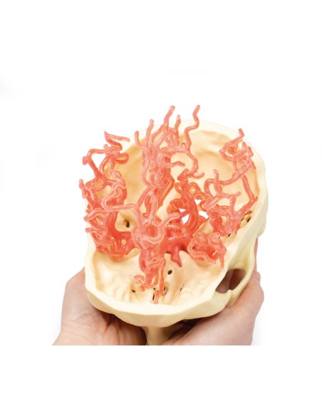
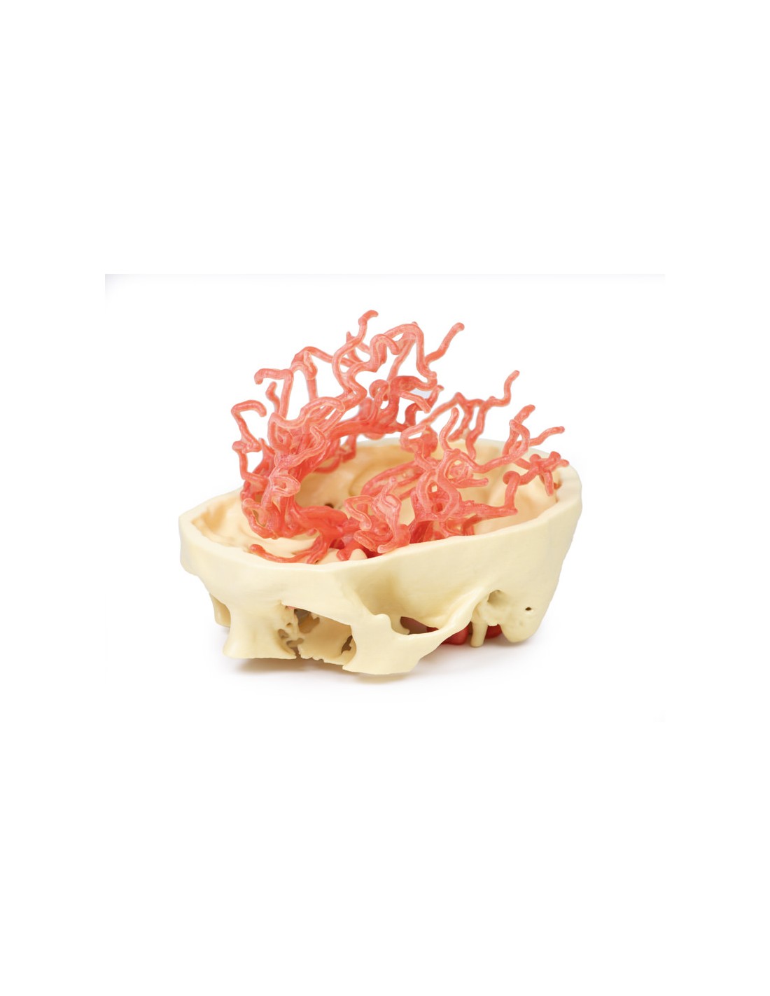
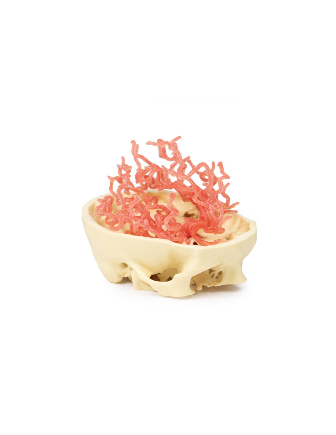
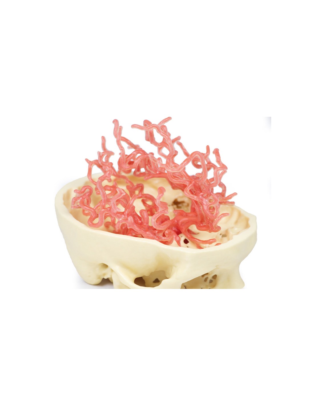
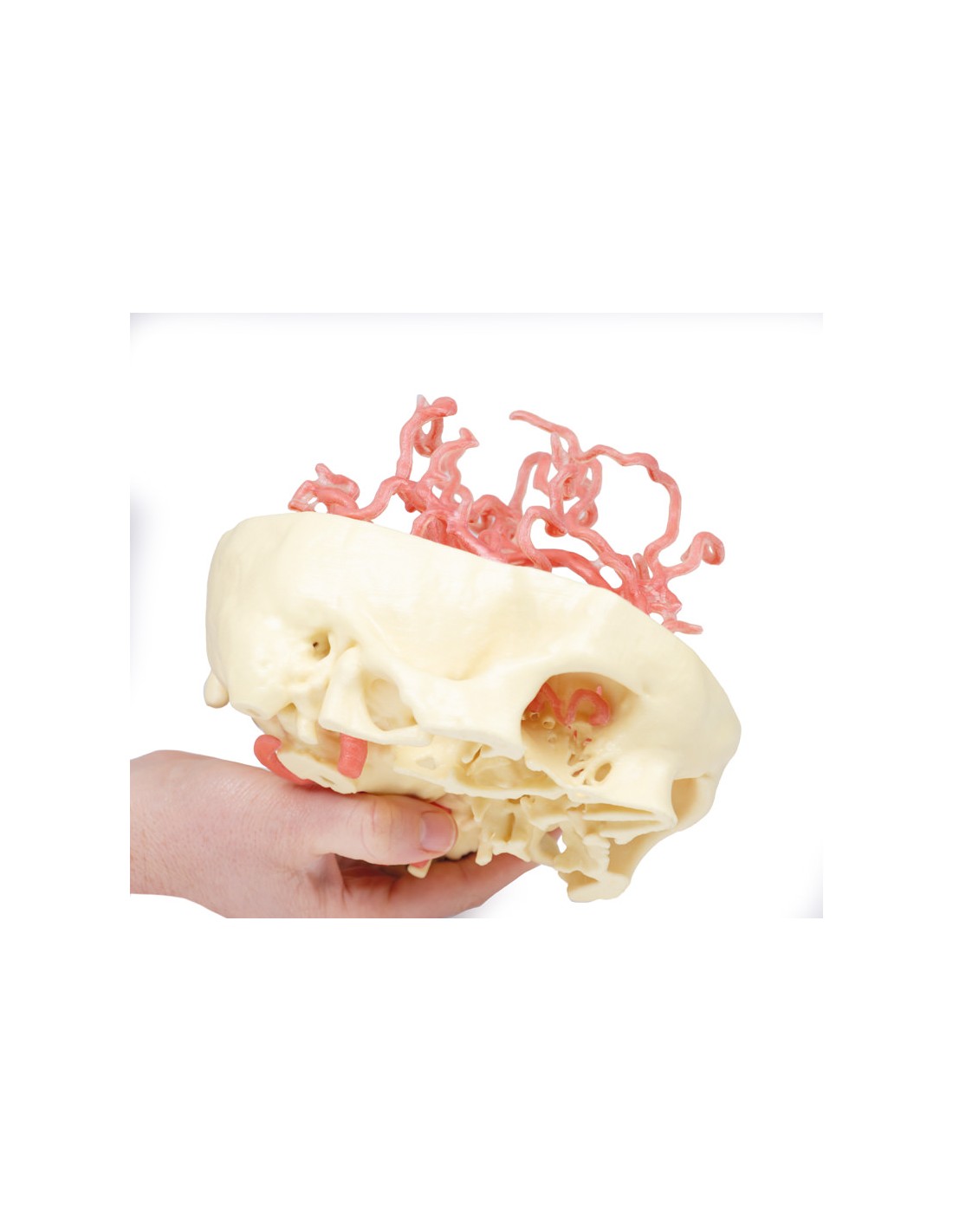
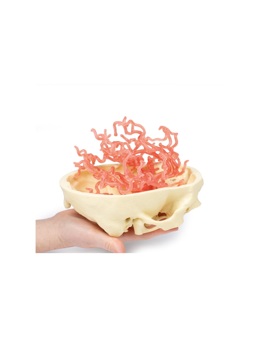
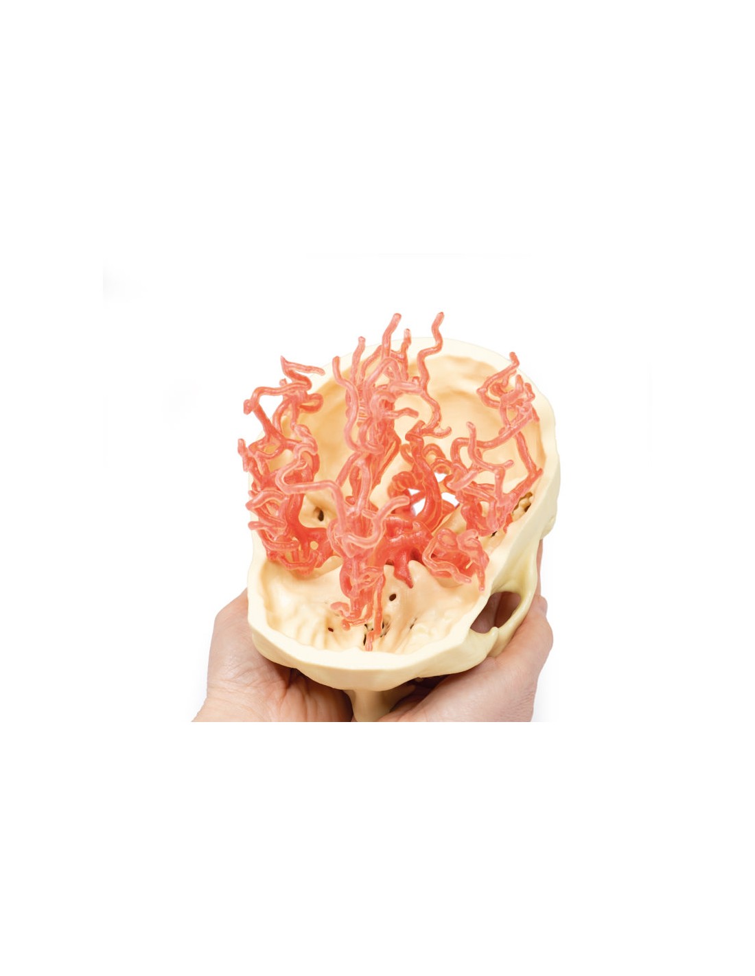
Cranial Arterial Circulation - Erler Zimmer 3D anatomy Series MP1650
This model of Cranial Arterial Circulation is part of the exclusive Monash 3D anatomy series, a comprehensive series of human dissections reproduced with ultra-high resolution color 3D printing.
This 3D print presents an expanded version of the same dataset that underlies our Circle of Willis 3D print derived from careful segmentation of angiographic data. Like our Circle of Willis print, this model shows the internal carotid and vertebral arteries entering the skull, branching into the intracranial arteries that supply the brain. This more expanded 3D print of the internal carotid and vertebral artery and their branches, including the Circle of Willis, shows the complete branching model of the cerebral and cerebellar arteries. This includes the pericallosal arteries (from the anterior brain) with its named branches, the superior and inferior divisions of the middle brain (including the sulcal, temporal and parietal arteries) and the branches of the posterior cerebral artery.
What advantages does the Monash University anatomical dissection collection offer over plastic models or plastinated human specimens?
- Each body replica has been carefully created from selected patient X-ray data or human cadaver specimens selected by a highly trained team of anatomists at the Monash University Center for Human Anatomy Education to illustrate a range of clinically important areas of anatomy with a quality and fidelity that cannot be achieved with conventional anatomical models-this is real anatomy, not stylized anatomy.
- Each body replica has been rigorously checked by a team of highly trained anatomists at the Center for Human Anatomy Education, Monash University, to ensure the anatomical accuracy of the final product.
- The body replicas are not real human tissue and therefore not subject to any barriers of transportation, import, or use in educational facilities that do not hold an anatomy license. The Monash 3D Anatomy dissection series avoids these and other ethical issues that are raised when dealing with plastinated human remains.
No reviews
Tap to zoom