Your cart
There are no more items in your cart


Cranial venous circulation - Erler Zimmer 3D anatomy Series MP1645
erler zimmer
EZ-MP1645
€2,647.77
Tax included
Made in ultra-high resolution 3D printing in full color.
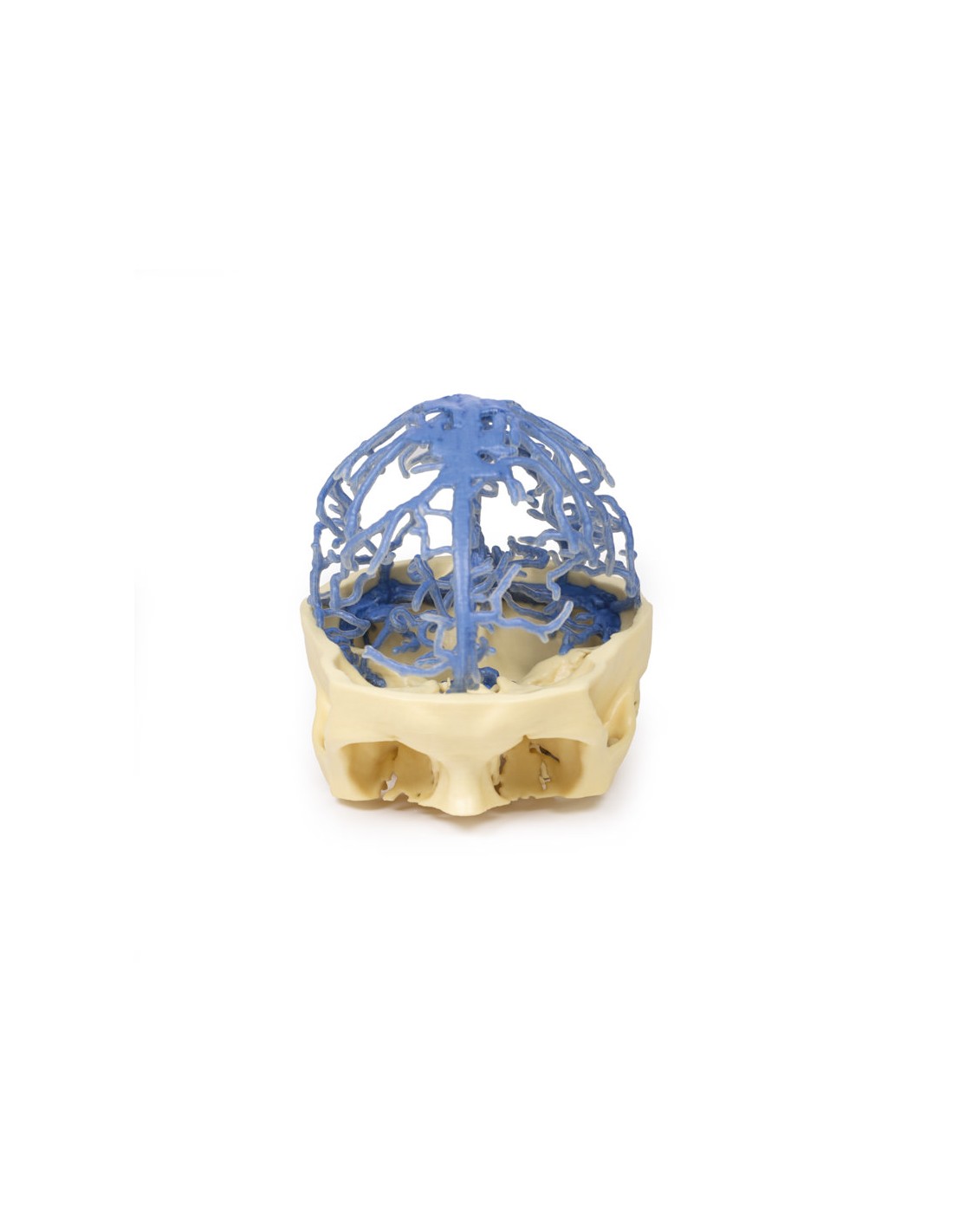
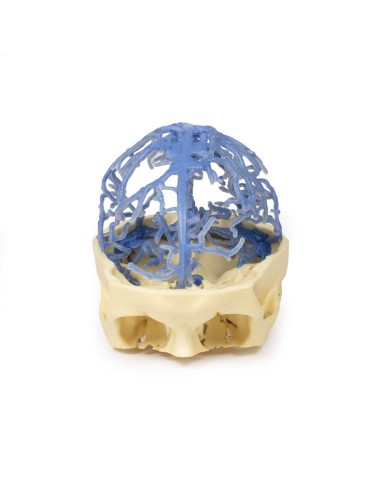
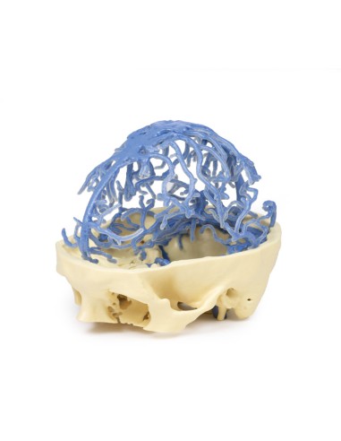
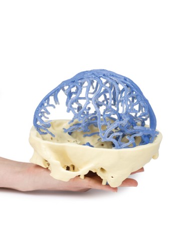
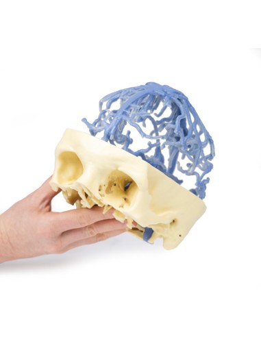
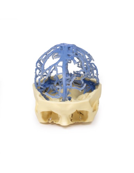
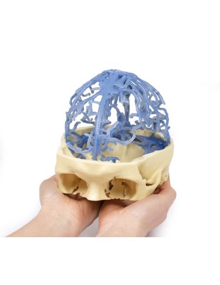
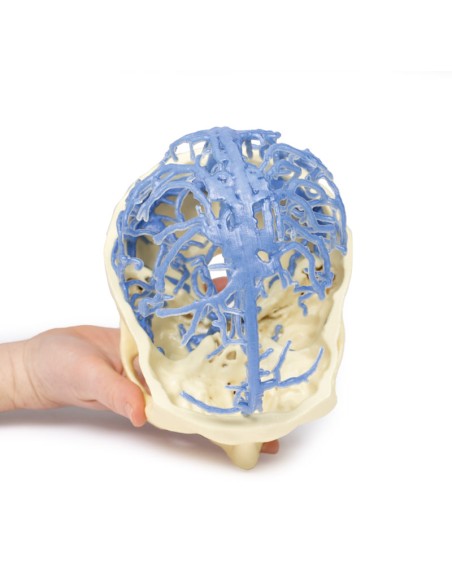
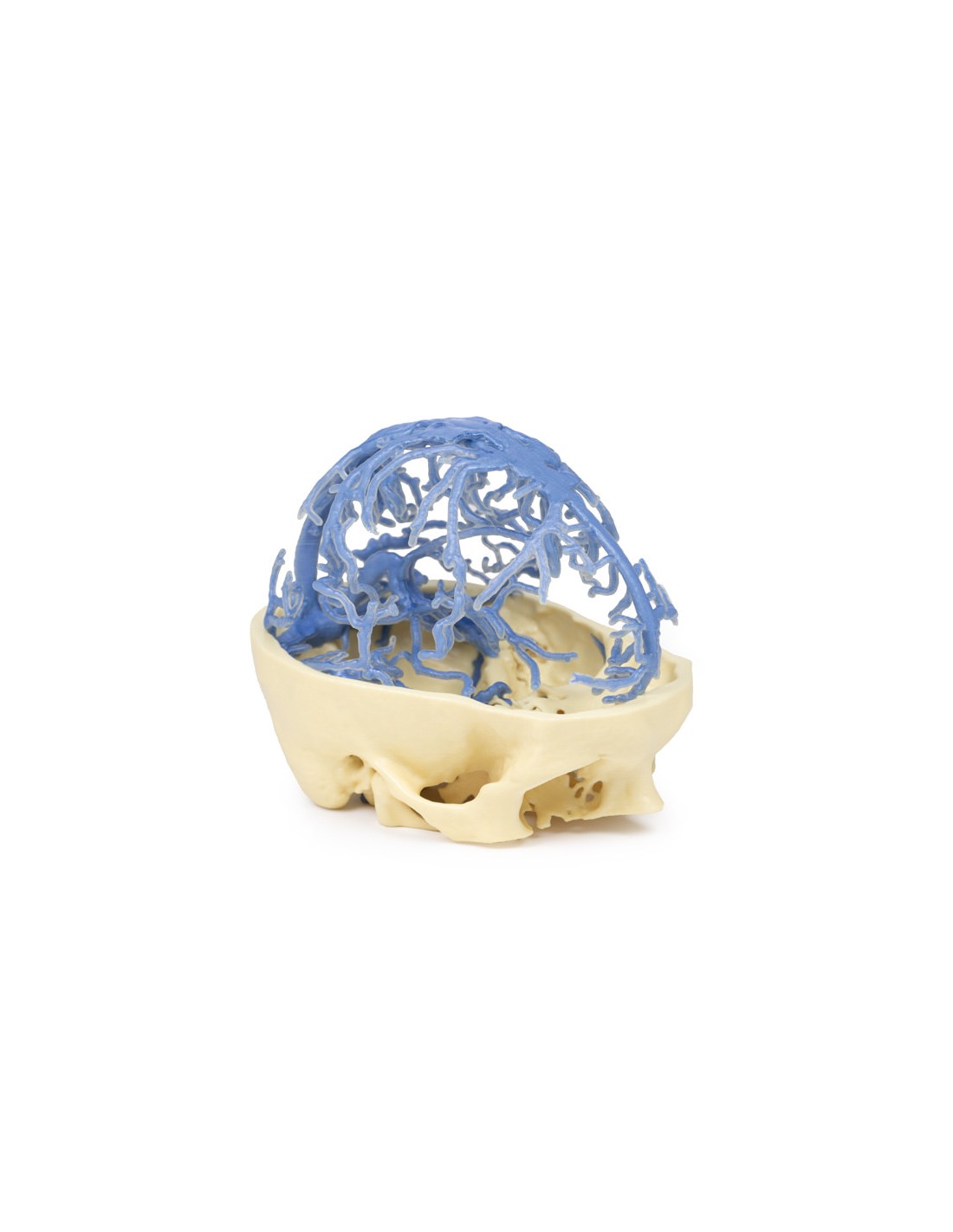
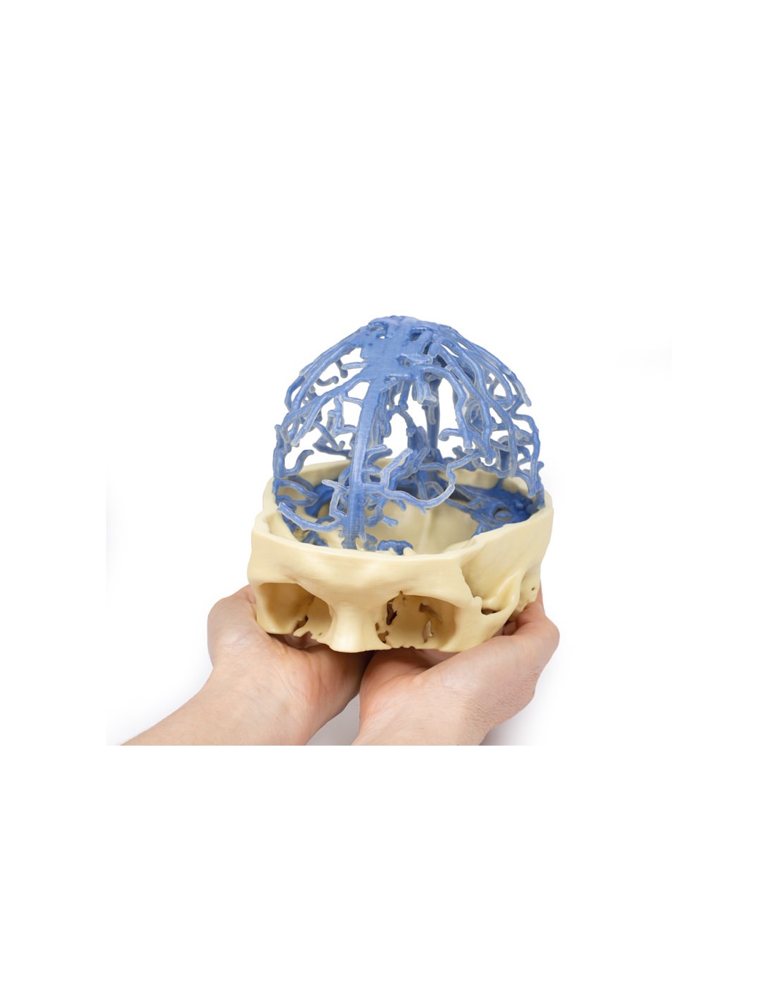
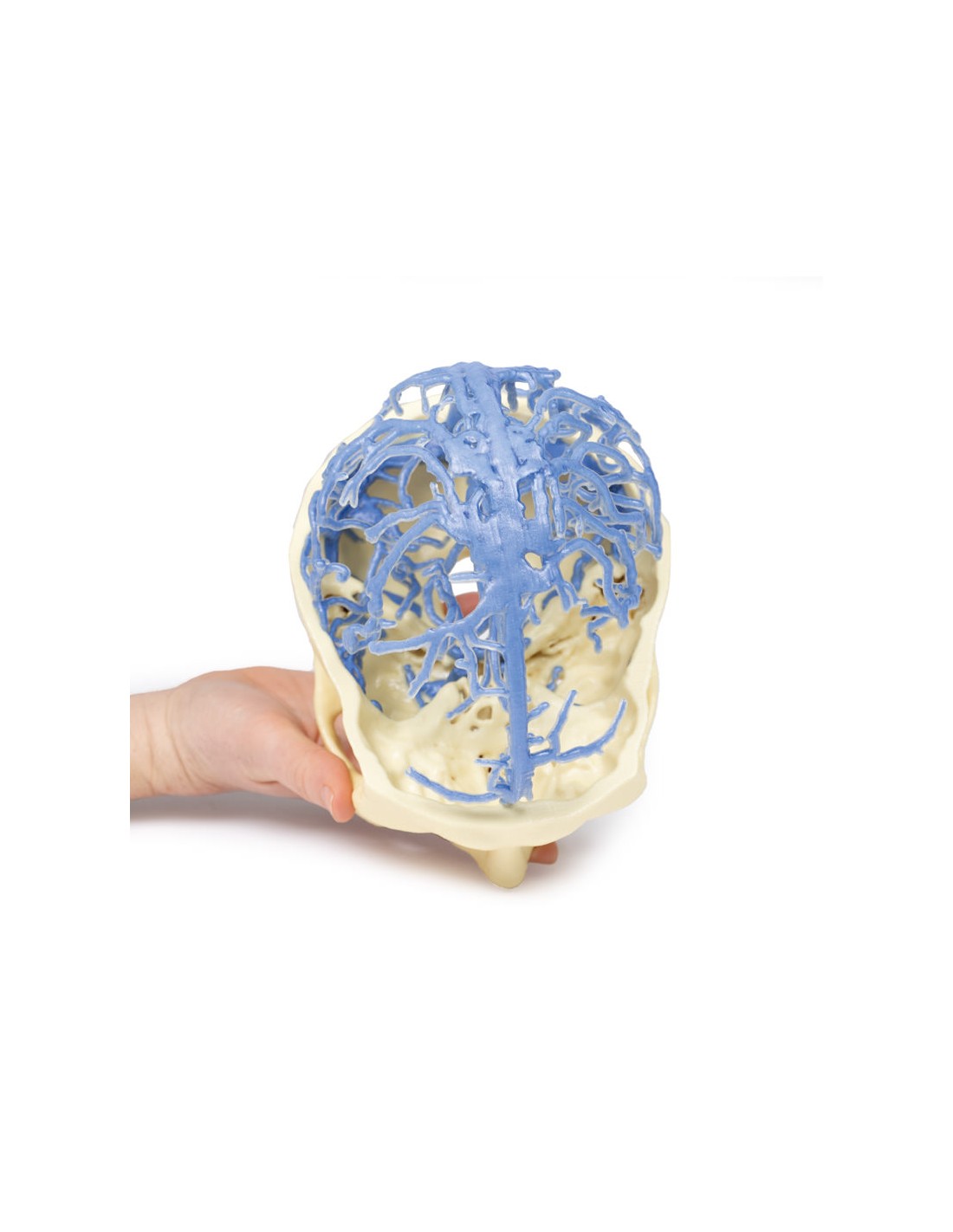
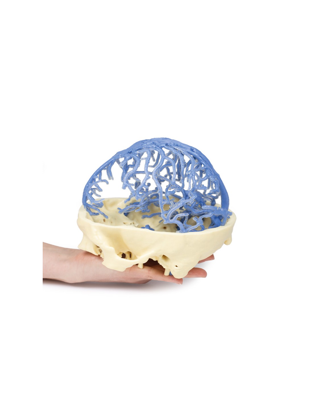
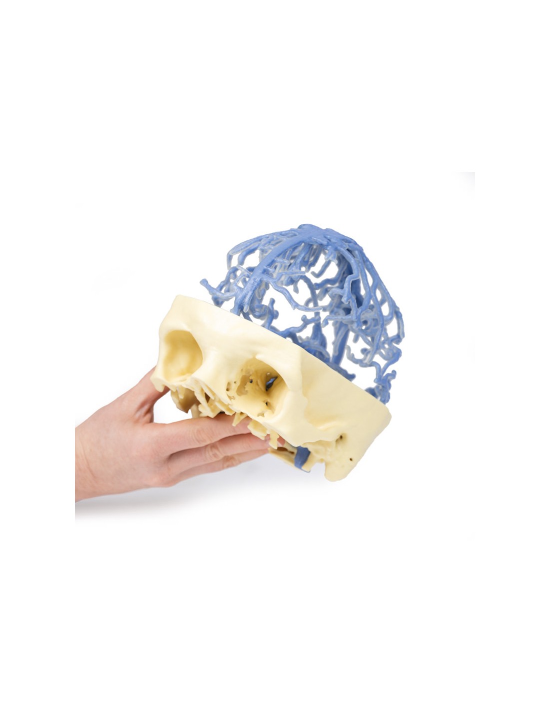
Venous Circulation of the Skull - Erler Zimmer 3D anatomy Series MP1645
This model of Arterial and Venous Circulation is part of the exclusive Monash 3D anatomy series, a comprehensive series of human dissections reproduced with ultra-high resolution color 3D printing.
This 3D print features the same dataset that underlies our Circle of Willis and cranial arterial circulation 3D prints and is derived from careful segmentation of angiographic data. Here, the dural venous sinus network was segmented based on structures visible from the contrast medium circulation in the late phase of filling. Consequently, while most of the sinuses are present, the lack of contrast in the anterior portions of the venous system means that some structures are not as clear in the pattern as might be expected, for example, the cavernous sinus and inferior petrous sinus.
Visible are the extensive network of dural veins and venous lacunae, which drain toward the midline into the superior sagittal sinus. Deep to this network of sinus veins is the great cerebral vein that drains with the inferior sagittal sinus into the straight sinus, which then converges with the superior sagittal sinus at the confluence of the sinuses. Several dural veins drain into the left and right transverse sinuses as they pass anteriorly to the petrous portion of the temporal bone. The sigmoid sinuses can be seen in the posterior cranial fossa before exiting the skull at the jugular foramen and forming the internal jugular vein (visible on the lower surface of the skull).
What advantages does the Monash University anatomical dissection collection offer over plastic models or plastinated human specimens?
- Each body replica has been carefully created from selected patient X-ray data or human cadaver specimens selected by a highly trained team of anatomists at the Monash University Center for Human Anatomy Education to illustrate a range of clinically important areas of anatomy with a quality and fidelity that cannot be achieved with conventional anatomical models-this is real anatomy, not stylized anatomy.
- Each body replica has been rigorously checked by a team of highly trained anatomists at the Center for Human Anatomy Education, Monash University, to ensure the anatomical accuracy of the final product.
- The body replicas are not real human tissue and therefore not subject to any barriers of transportation, import, or use in educational facilities that do not hold an anatomy license. The Monash 3D Anatomy dissection series avoids these and other ethical issues that are raised when dealing with plastinated human remains.
No reviews
Tap to zoom