Your cart
There are no more items in your cart


Structures of the plantar surface of the foot - Erler Zimmer 3D anatomy Series MP1900
erler zimmer
EZ-MP1900
Out-of-Stock
€1,317.84
Tax included
Made in ultra-high resolution 3D printing in full color.
Structures of the plantar surface of the foot - Erler Zimmer 3D anatomy Series - MP1900
This model is part of the exclusive Monash 3D anatomy series, a comprehensive series of human dissections reproduced with ultra-high resolution color 3D printing.
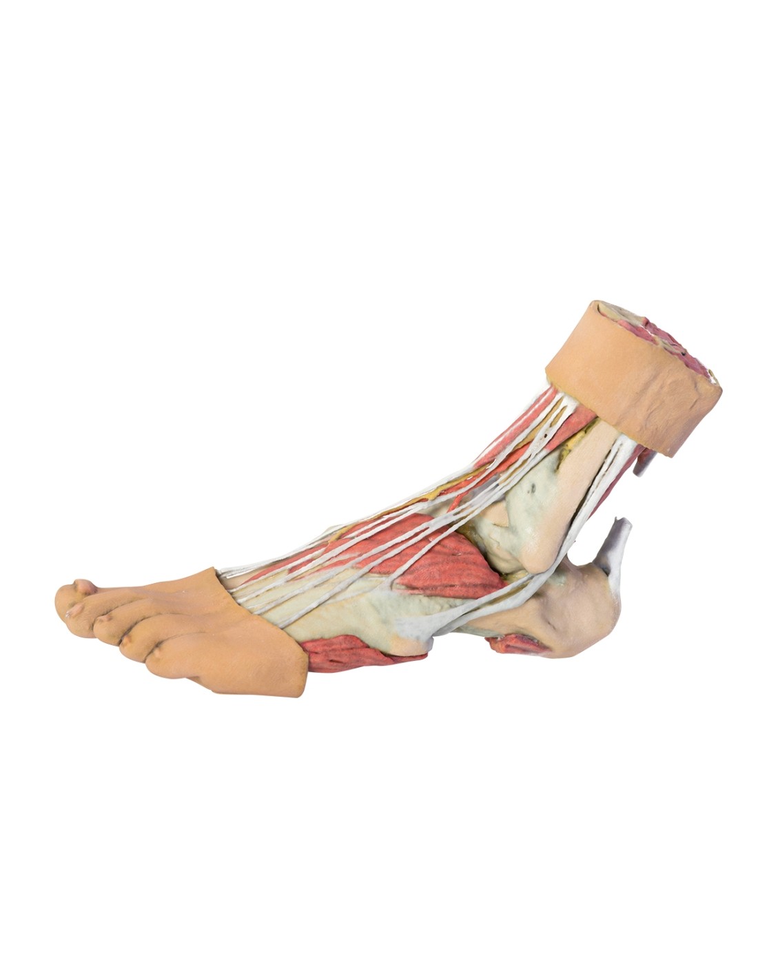
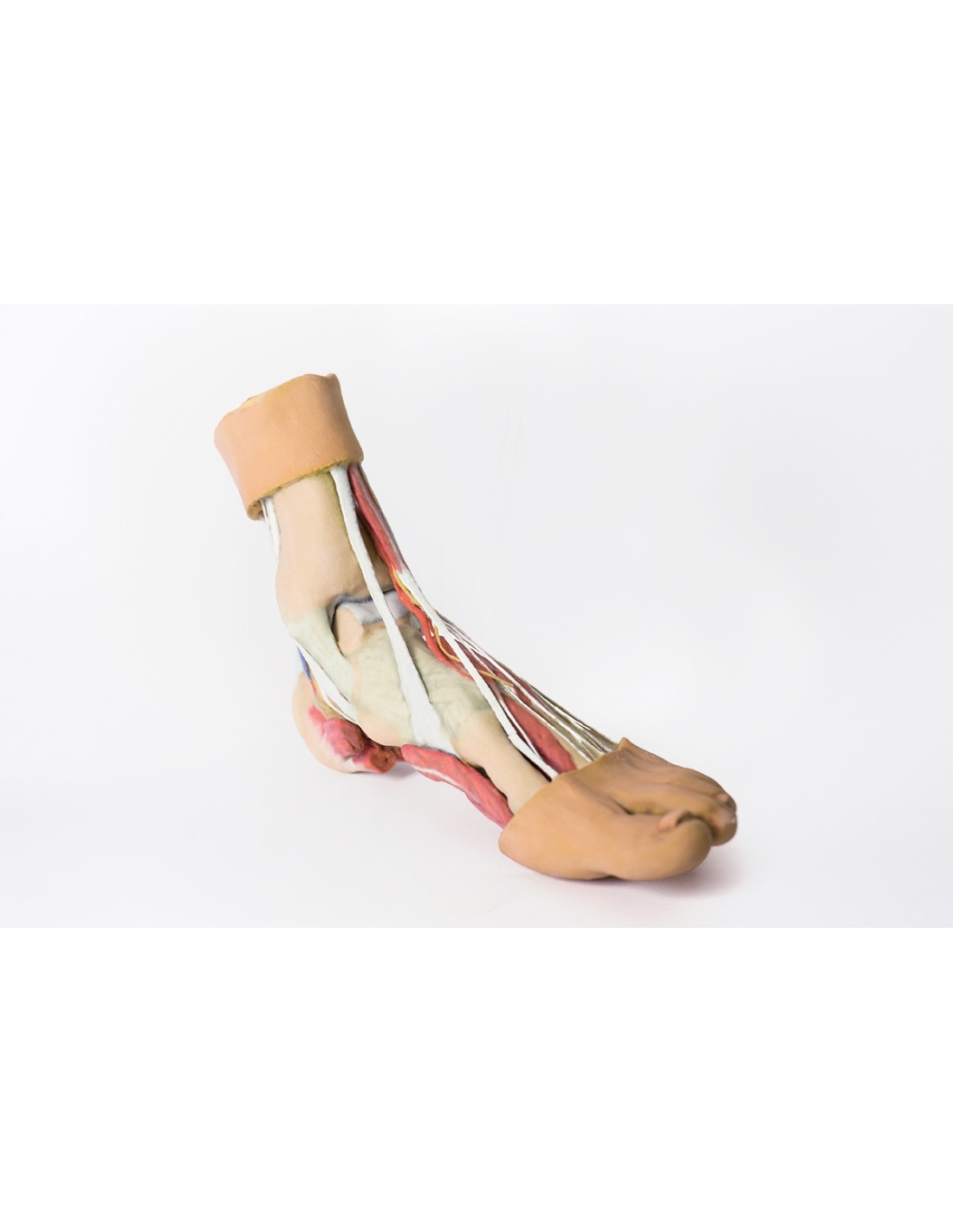
This 3D printed dissection model shows the anatomy of a right distal leg and the deep structures of the plantar surface of the foot.
In the proximal part, the tibia, fibula, interosseous membrane and leg muscles are distinguishable in cross section.
Medially, at the level of the ankle joint, the long tendons of the dorsal and plantar flexors are visible superficially to the capsular and extra capsular ligaments.
Detailed anatomical description upon request.
No reviews
Tap to zoom