
Your cart
There are no more items in your cart


Liver with vessels and gallbladder - Erler Zimmer 3D anatomy Series MP1136
erler zimmer
EZ-MP1136
€2,321.66
Tax included
Made in ultra-high resolution 3D printing in full color.
Liver with Vessels and Gallbladder - Erler Zimmer 3D anatomy Series MP1136
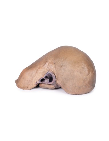
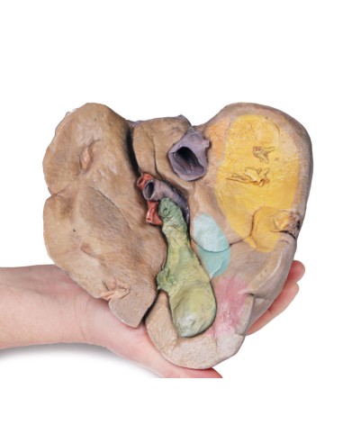
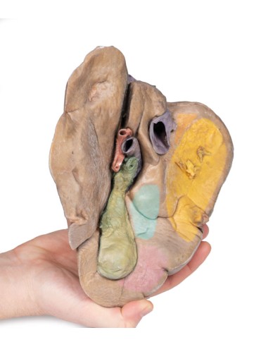
This Liver Dissection with Vessels and Gallbladder model is part of the exclusive Monash 3D anatomy series, a comprehensive series of human dissections reproduced with ultra-high resolution color 3D printing.
The size and shape of this specimen varies slightly from a typical liver. It is less wedge-shaped and longer in the supero-inferior dimension (in the posterior view this would result in greater vertical height). Normally, a liver measures less than 16 cm at the midclavicular line.
This specimen measures about 18 cm at the midclavicular line, suggesting some degree of hepatomegaly. However, it is worth mentioning that some measurement bias may have occurred depending on the fixation and care of the specimen-and it should be noted that the accuracy of estimating liver size using a single parameter is limited. Diagnostic liver measurements of hepatomegaly vary depending on normal anatomic variation in liver size and morphology, the method of measurement, and patient characteristics such as sex and BMI.
An alternative explanation could be normal anatomical variation. However, this specimen does not fit the description of the most common anatomical variations confused with hepatomegaly: Riedel's lobe (a downward projection of the right lobe), "beavertail" liver (an elongated left lobe), or a papillary process protruding from the caudate lode.
What advantages does the Monash University anatomical dissection collection offer over plastic models or plastinated human specimens?
- Each body replica has been carefully created from selected patient X-ray data or human cadaver specimens selected by a highly trained team of anatomists at the Monash University Center for Human Anatomy Education to illustrate a range of clinically important areas of anatomy with a quality and fidelity that cannot be achieved with conventional anatomical models-this is real anatomy, not stylized anatomy.
- Each body replica has been rigorously checked by a team of highly trained anatomists at the Center for Human Anatomy Education, Monash University, to ensure the anatomical accuracy of the final product.
- The body replicas are not real human tissue and therefore not subject to any barriers of transportation, import, or use in educational facilities that do not hold an anatomy license. The Monash 3D Anatomy dissection series avoids these and other ethical issues that are raised when dealing with plastinated human remains.
No reviews
Tap to zoom
