Your cart
There are no more items in your cart


Dissection of Spleen and Pancreas - Erler Zimmer 3D anatomy Series MP1135
erler zimmer
EZ-MP1135
€1,013.21
Tax included
Made in ultra-high resolution 3D printing in full color.
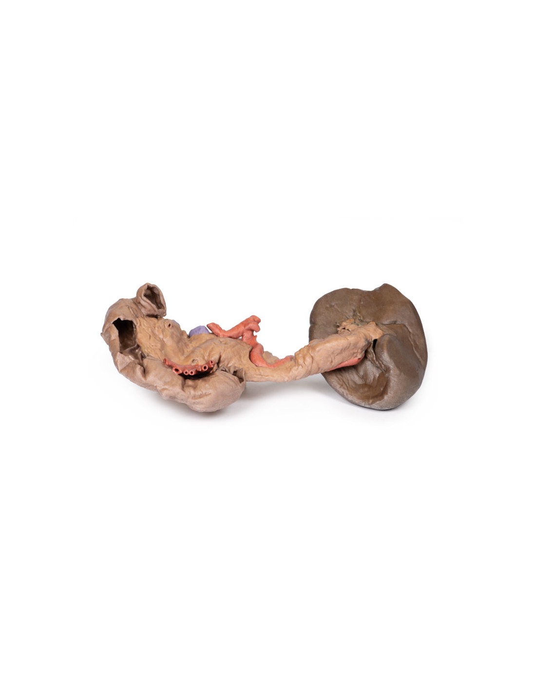
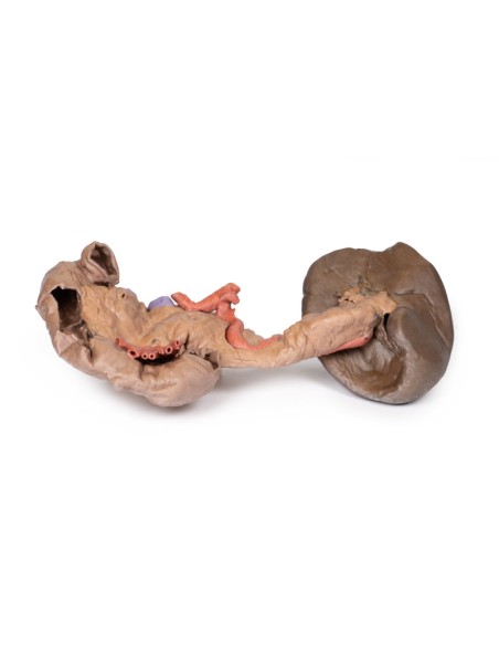
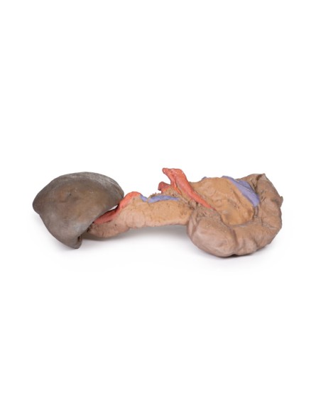
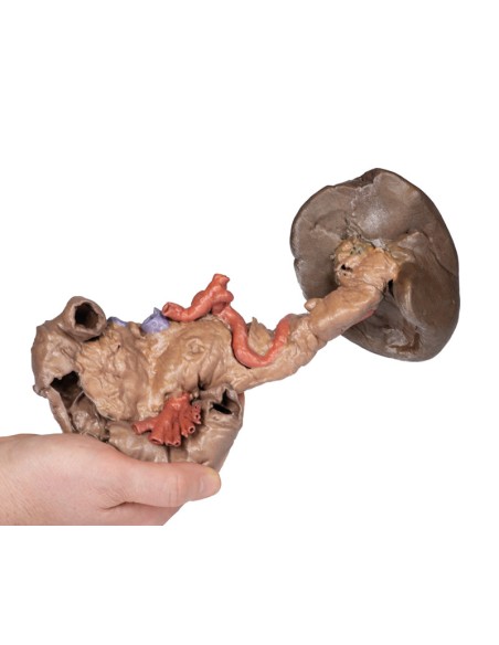
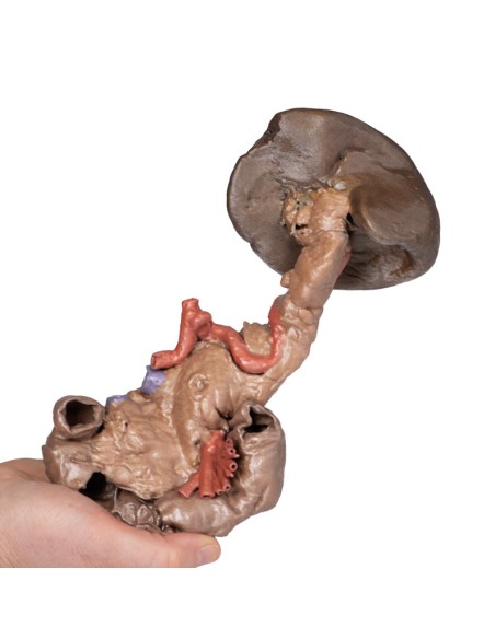
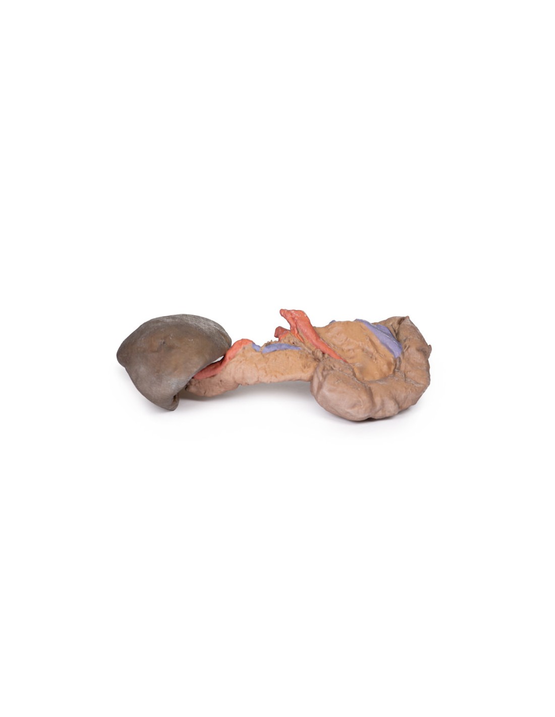
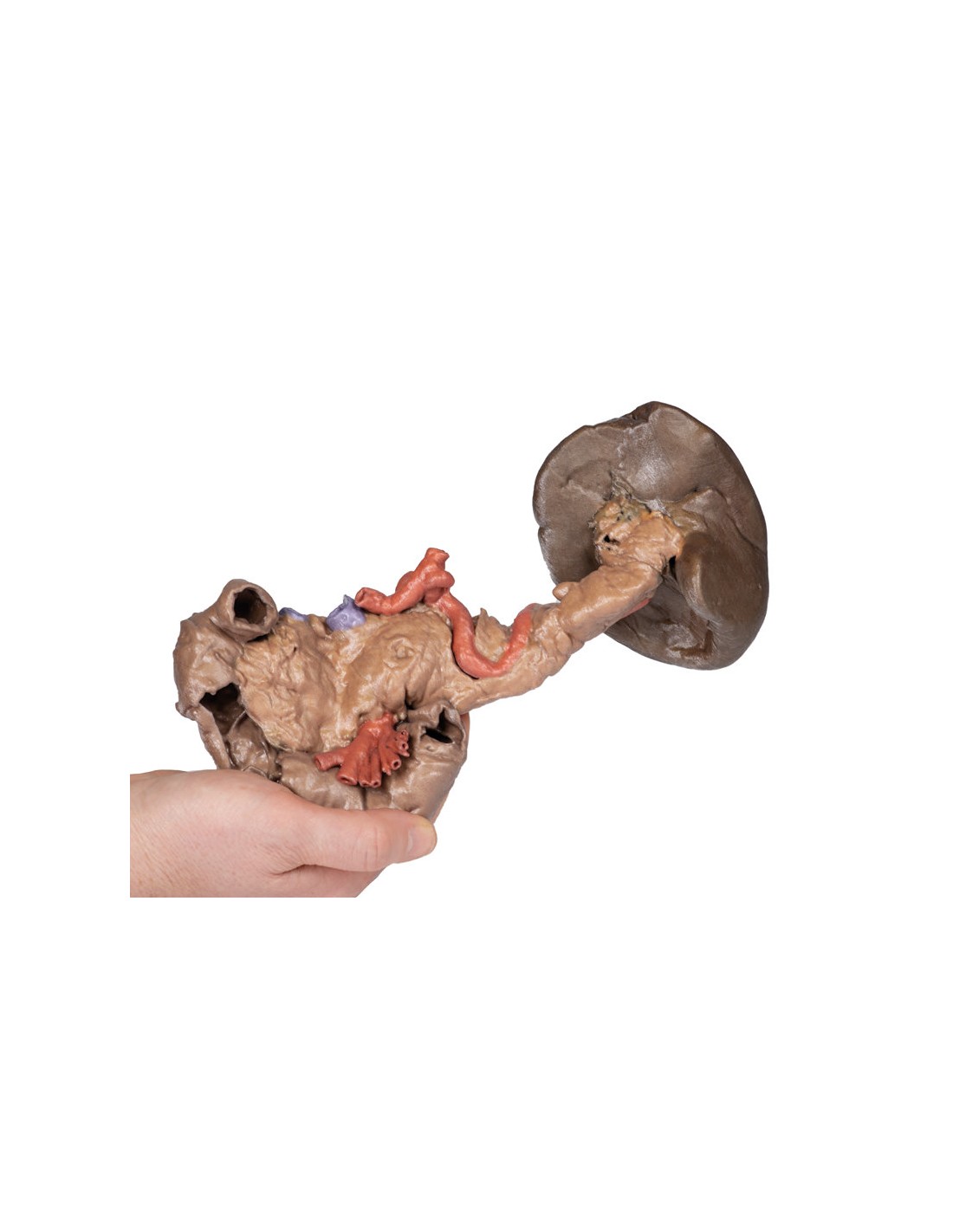
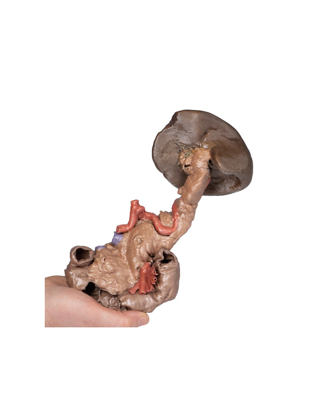
Spleen and Pancreas Dissection - Erler Zimmer 3D anatomy Series MP1135
This Spleen and Pancreas Dissection model is part of the exclusive Monash 3D anatomy series, a comprehensive series of human dissections reproduced with ultra-high resolution color 3D printing.
This 3D model preserves the deep organs of the anterior intestine: the descending, horizontal and ascending duodenum, pancreas and spleen. A small window in the duodenum has been opened to allow a view of the circular folds within this proximal part of the small intestine (and contrasts the strong ruge development seen in the stomach; see AW 42). The head of the pancreas is preserved in its normal position within the curvature of the duodenum, and a distinct uncus can be seen at the distal margin and adjacent to the exit of the superior mesenteric artery (already divided into its many named branches). Along the superior margin of the body of the pancreas, the celiac trunk has been dissected by the descending abdominal aorta. The complete splenic artery can be observed in its tortuous path from its origin to the spleen, as well as the bases of the left gastric artery and the common hepatic artery. Immediately adjacent to the celiac trunk is a small portion of the splenic vein, which emerges from within the capsule of the pancreas, and is partly visible along its course near the splenic artery. A small portion of the superior mesenteric vein adheres to the posterior face of the pancreas, representing the path of the vessel before it normally joins the splenic and forms the hepatic portal vein. The tail of the pancreas is invested in the capsule of the spleen and obscures any branching of the splenic artery before entering the organ (as seen in our other spleen models in A8 and AW 34, which provides further information on the anatomy and associations of this organ). Immediately adjacent to the celiac trunk is a small portion of the splenic vein, which emerges from within the pancreatic capsule and is partly visible along its course near the splenic artery. A small portion of the superior mesenteric vein adheres to the posterior face of the pancreas, representing the path of the vessel before it normally joins the splenic and forms the hepatic portal vein. The tail of the pancreas is invested in the capsule of the spleen and obscures any branching of the splenic artery before entering the organ (as seen in our other spleen models in A8 and AW 34, which provides further information on the anatomy and associations of this organ). Immediately adjacent to the celiac trunk is a small portion of the splenic vein, which emerges from within the pancreatic capsule and is partly visible along its course near the splenic artery. A small portion of the superior mesenteric vein adheres to the posterior face of the pancreas, representing the path of the vessel before it normally joins the splenic and forms the hepatic portal vein. The tail of the pancreas is invested in the capsule of the spleen and obscures any branching of the splenic artery before it enters the organ (as seen in our other spleen models in A8 and AW 34, which provides further information on the anatomy and associations of this organ). and is partly visible along its path near the splenic artery. A small portion of the superior mesenteric vein adheres to the posterior aspect of the pancreas, representing the path of the vessel before it normally joins the splenic and forms the hepatic portal vein. The tail of the pancreas is invested in the capsule of the spleen and obscures any branching of the splenic artery before it enters the organ (as seen in our other spleen models in A8 and AW 34, which provides further information on the anatomy and associations of this organ). and is partly visible along its path near the splenic artery. A small portion of the superior mesenteric vein adheres to the posterior aspect of the pancreas, representing the path of the vessel before it normally joins the splenic and forms the hepatic portal vein. The tail of the pancreas is invested in the capsule of the spleen and obscures any branching of the splenic artery before entering the organ (as seen in our other spleen models in A8 and AW 34, which provides further information on the anatomy and associations of this organ).
What advantages does the Monash University anatomical dissection collection offer over plastic models or plastinated human specimens?
- Each body replica has been carefully created from selected patient X-ray data or human cadaver specimens selected by a highly trained team of anatomists at the Monash University Center for Human Anatomy Education to illustrate a range of clinically important areas of anatomy with a quality and fidelity that cannot be achieved with conventional anatomical models-this is real anatomy, not stylized anatomy.
- Each body replica has been rigorously checked by a team of highly trained anatomists at the Center for Human Anatomy Education, Monash University, to ensure the anatomical accuracy of the final product.
- The body replicas are not real human tissue and therefore not subject to any barriers of transportation, import, or use in educational facilities that do not hold an anatomy license. The Monash 3D Anatomy dissection series avoids these and other ethical issues that are raised when dealing with plastinated human remains.
No reviews
Tap to zoom