Your cart
There are no more items in your cart


Dissection of the abdomen with inguinal hernia - Erler Zimmer 3D anatomy Series MP1133
erler zimmer
EZ-MP1133
€8,309.66
Tax included
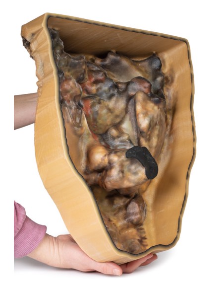
Made in ultra-high resolution 3D printing in full color.
Dissection of the abdomen with inguinal hernia - Erler Zimmer 3D anatomy Series MP1133
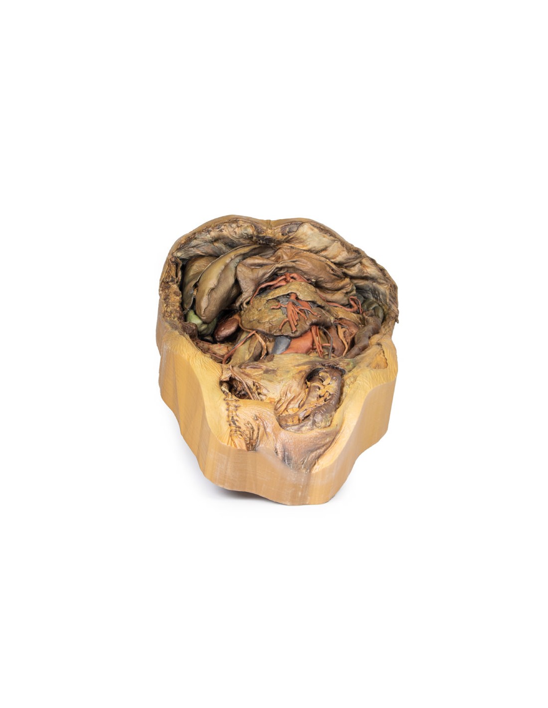
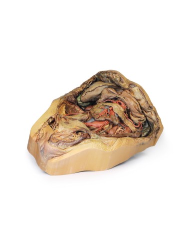
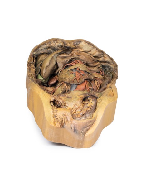
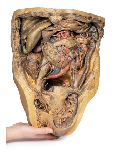
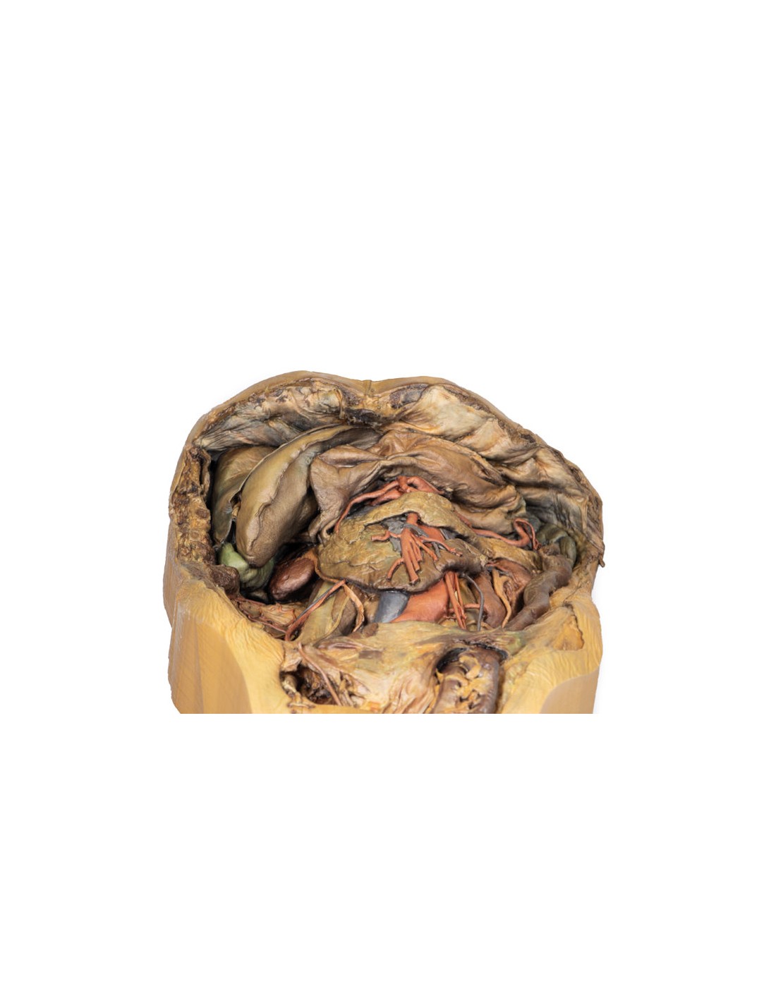
This model of Dissection of the Abdomen with Inguinal Hernia is part of the exclusive Monash 3D anatomy series, a comprehensive series of human dissections reproduced with ultra-high resolution color 3D printing.
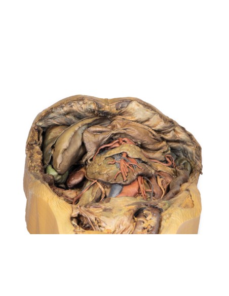
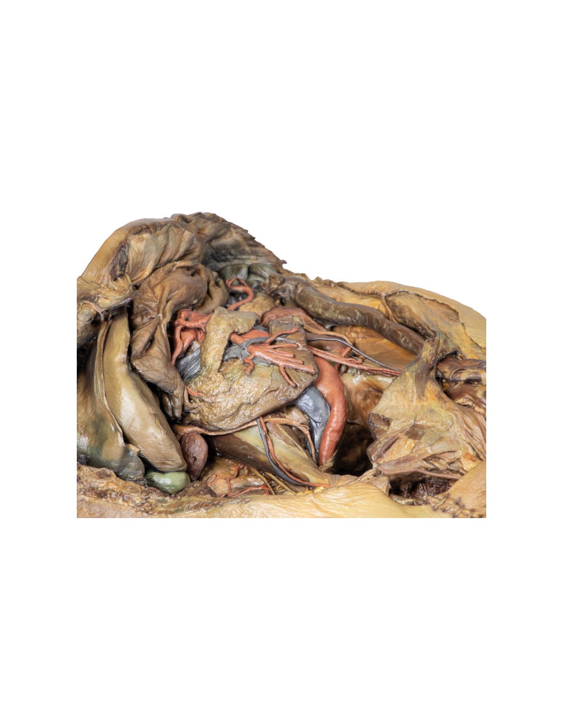
Diaphragm and xiphoid process
The diaphragm was attached to the upper edge of the dissected specimen with sutures to provide an unobstructed view of the abdomen. The xiphoid process is located in the middle of this sutured border.
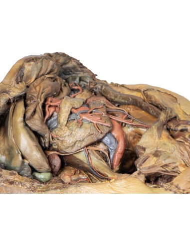
Liver and gallbladder
The liver in the right hypochondrium has been pushed laterally to reveal the kidney posterior to it.
The falciform ligament divides the right and left anatomic lobes of the liver and the round ligament wraps around, which is a remnant of the umbilical vein that is present during fetal development.
Below the round ligament at the lower edge of the liver in this model, the gallbladder is sandwiched between the anatomic lobes of the liver.
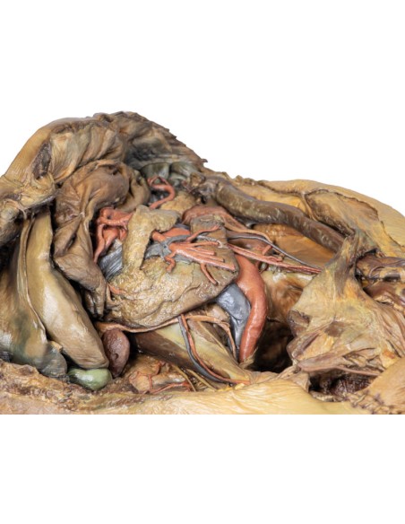
Stomach and splenic vasculature
The deflated stomach has been deviated superiorly to reveal the splenic artery and vein.
The tortuous course of the splenic artery and vein can be seen as it approaches the spleen, issuing numerous branches that enter the hilum of the spleen.
Spleen and pancreas
The spleen is located in the left hypochondrium of the specimen. Its gastric imprint indicates where the greater curvature of the stomach would normally be.
Toward the lower pole of the spleen, the tail of the pancreas is fused with the hilum of the spleen. Unlike the rest of the organ, the tail of the pancreas is intraperitoneal.
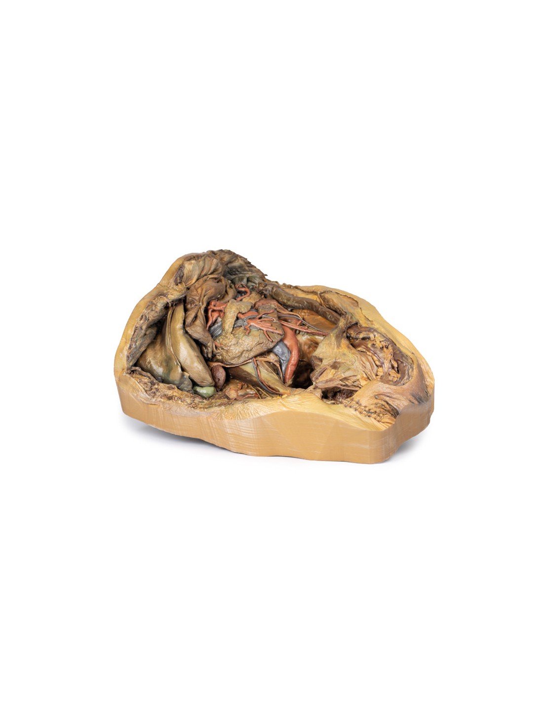
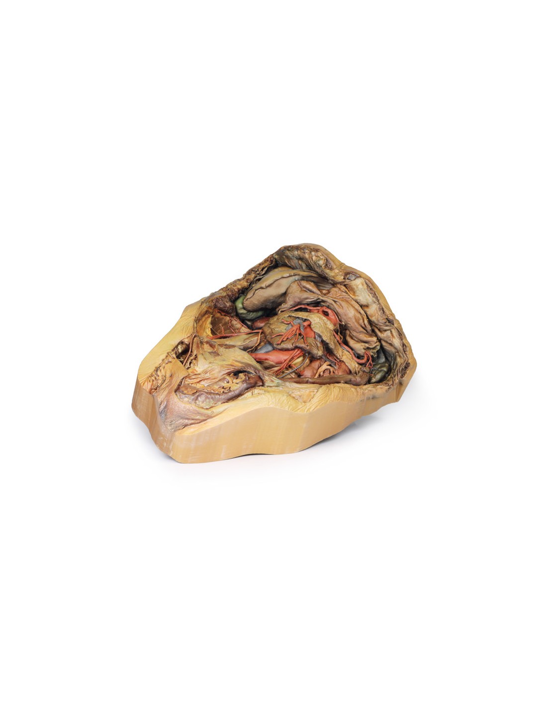
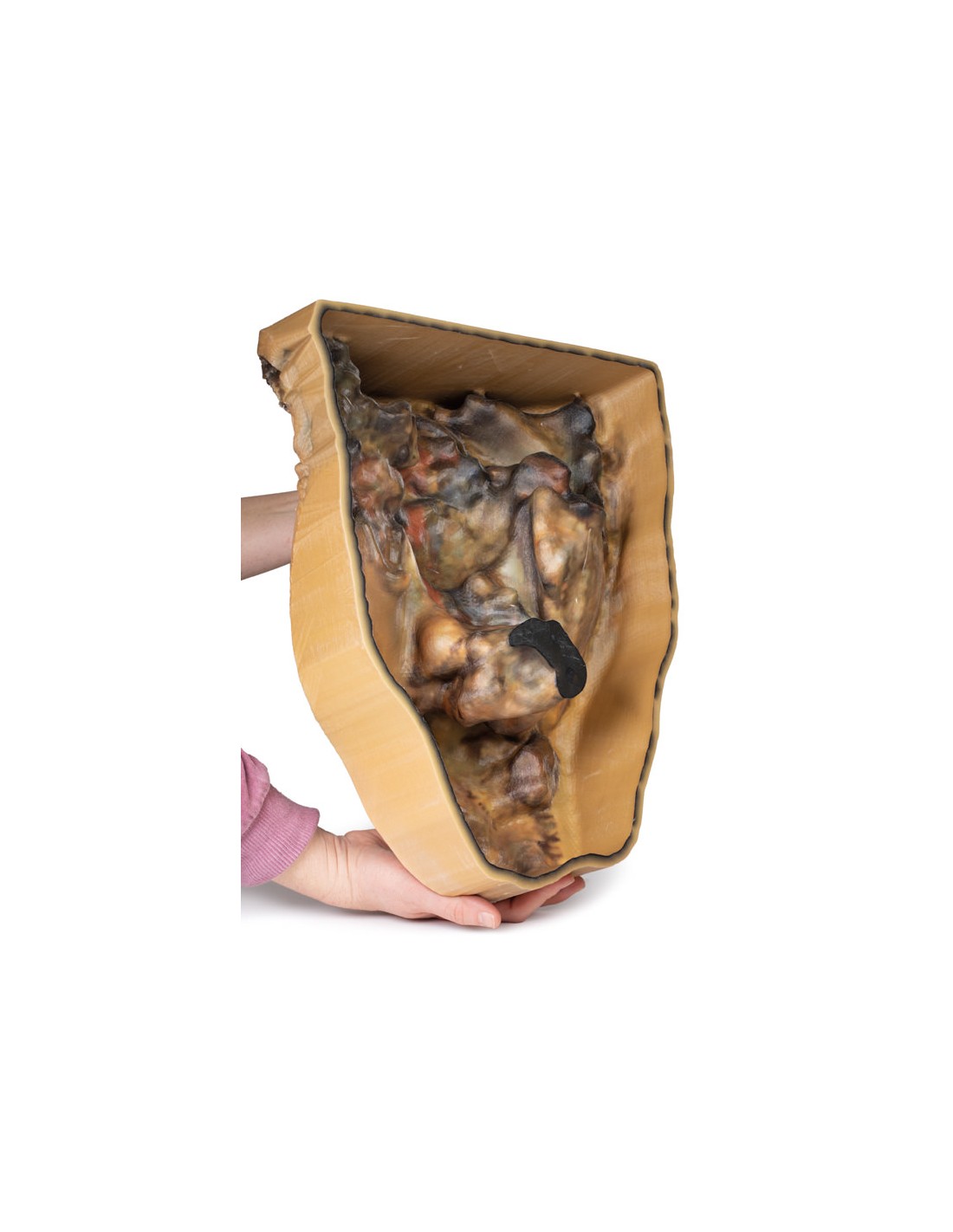
Kidneys
The kidneys are mainly retroperitoneal, however, in this specimen the peritoneum that usually covers these organs has been removed.
Normally, the right kidney is displaced inferiorly from the liver relative to the left kidney. However, in this specimen the right kidney is both higher and smaller than the left.
The left kidney is abnormally large and is supplied by two accessory renal arteries arising directly from the abdominal aorta. These connect just above the hilum and also to the lower pole of the kidney.
Adrenal glands
The left adrenal gland is detached from its usual position on the upper pole of the kidney. The middle adrenal artery originates directly from the aorta, to the left of the celiac trunk, while the inferior adrenal artery is derived from the left renal artery: both supply the adrenal gland. The superior adrenal artery has been obscured by connective tissue.
Rectum and bladder
Although most of the peritoneum in the abdomen has been removed below the level of the sacral prominence (S1), a layer of peritoneum remains intact, overlapping the rectum and bladder. In particular, this is the first part of the rectum, which is intraperitoneal.
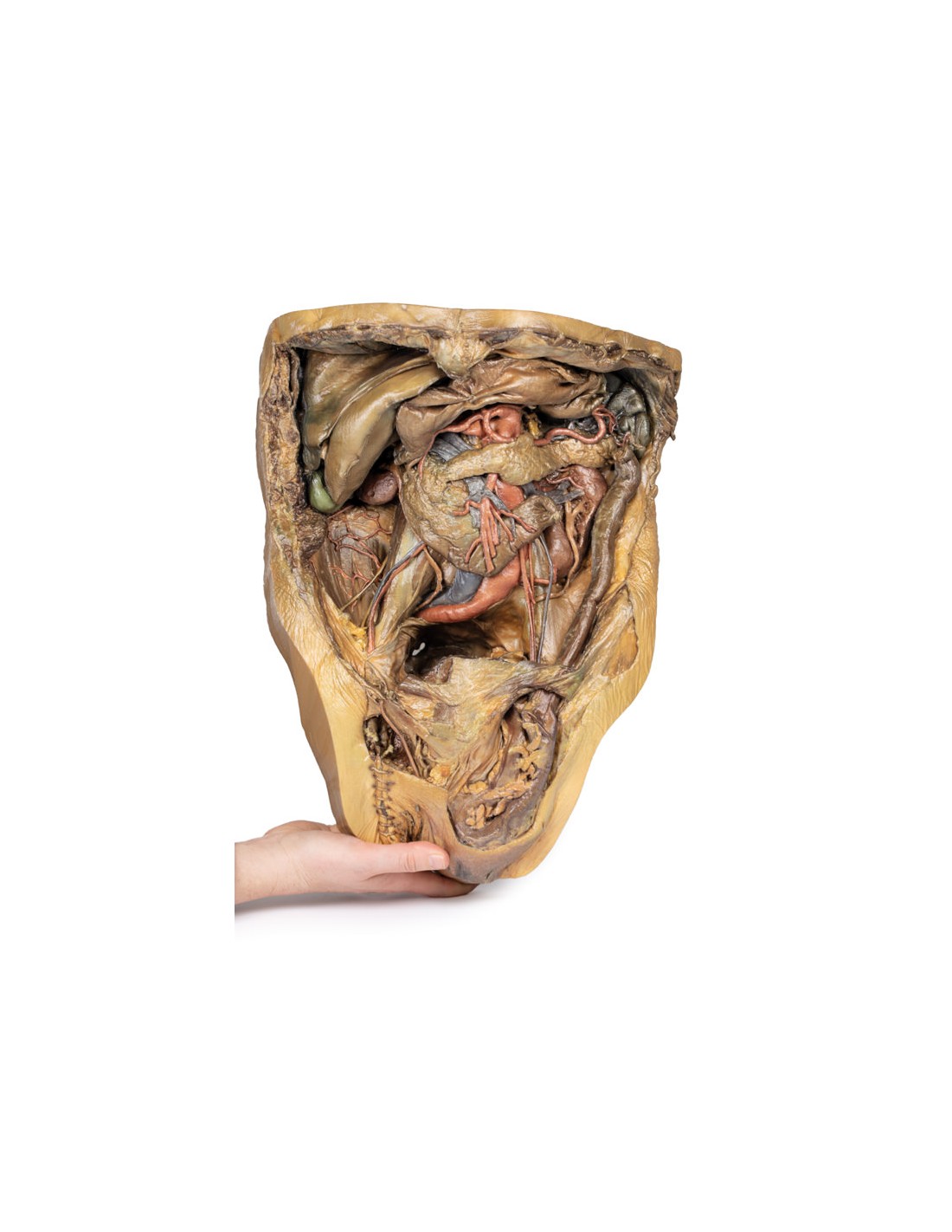
Gastrointestinal tract
The end of the ascending duodenum and descending colon at the left colic flexure were tied with twine, with the intestines in between removed to provide a better view of the abdomen.
Pelvic region
In this specimen, the sigmoid colon herniated indirectly through the inguinal canal.
On the right, the ductus deferens emerges from the superficial inguinal ring and flows to the right scrotum and then attaches to the right testis. The rest of the contents of the right spermatic cord were removed from this specimen.
The sutures observed below the ductus deferens are the remnants of the embalming process. These indicate that the right femoral artery was used as an entry point.
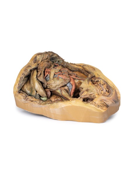
Abdominal vasculature
The celiac trunk can be seen just below the reflex stomach.
Typically, the celiac trunk has three main branches; left gastric, splenic and common hepatic, to supply the anterior intestine.
However, in this 3D model, the celiac trunk gives off both right and left gastric branches, the splenic artery and a gastroduodenal branch that divides to become two superior pancreaticoduodenal arteries. The hepatic artery proper emerges directly from the abdominal aorta, independent of the above branches, and gives rise to the right inferior phrenic artery.
The ilio-lumbar artery can be seen emerging deep at the right psoas, anastomizing with branches of the right deep circumflex iliac artery that runs along the iliac crest.
Do you have questions about this product?
What advantages does the Monash University anatomical dissection collection offer over plastic models or plastinated human specimens?
- Each body replica has been carefully created from selected patient X-ray data or human cadaver specimens selected by a highly trained team of anatomists at the Monash University Center for Human Anatomy Education to illustrate a range of clinically important areas of anatomy with a quality and fidelity that cannot be achieved with conventional anatomical models-this is real anatomy, not stylized anatomy.
- Each body replica has been rigorously checked by a team of highly trained anatomists at the Center for Human Anatomy Education, Monash University, to ensure the anatomical accuracy of the final product.
- The body replicas are not real human tissue and therefore not subject to any barriers of transportation, import, or use in educational facilities that do not hold an anatomy license. The Monash 3D Anatomy dissection series avoids these and other ethical issues that are raised when dealing with plastinated human remains.
No reviews
Tap to zoom