Your cart
There are no more items in your cart


Abdominal vasculature - Erler Zimmer 3D anatomy Series MP1131
erler zimmer
EZ-MP1131
€7,226.67
Tax included
Made in ultra-high resolution 3D printing in full color.
Abdominal Vasculature - Erler Zimmer 3D anatomy Series MP1131
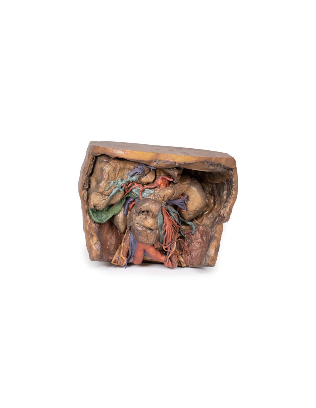
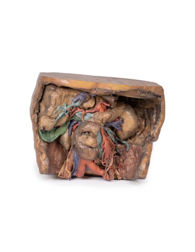
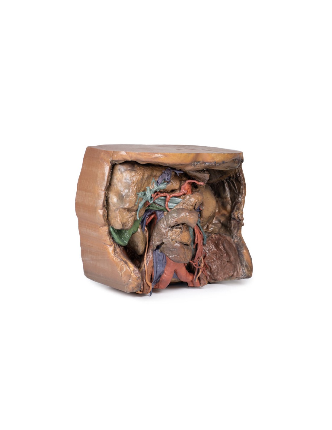
This model of the abdominal vasculature is part of the exclusive Monash 3D anatomy series, a comprehensive series of human dissections reproduced with very high resolution color 3D printing.
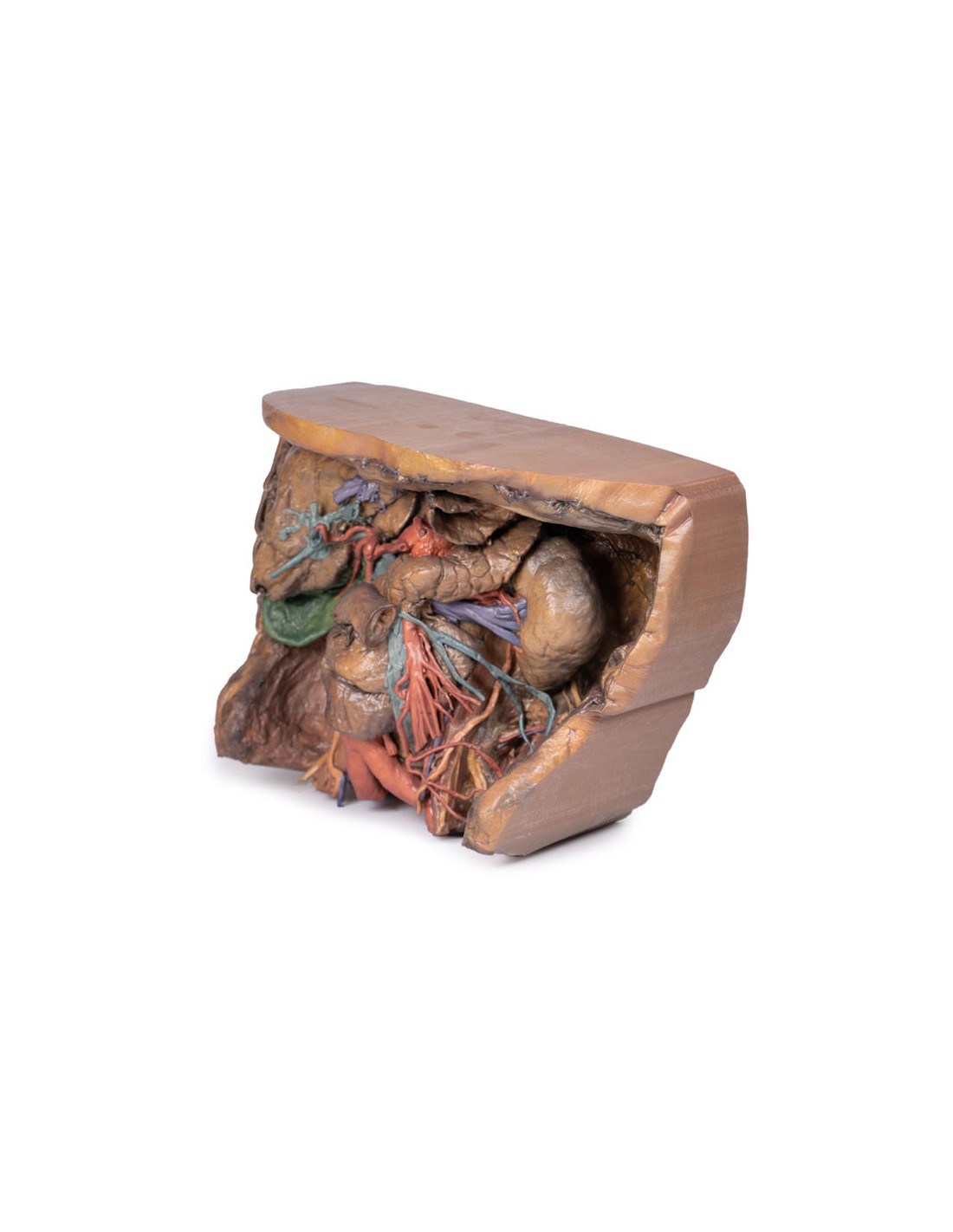
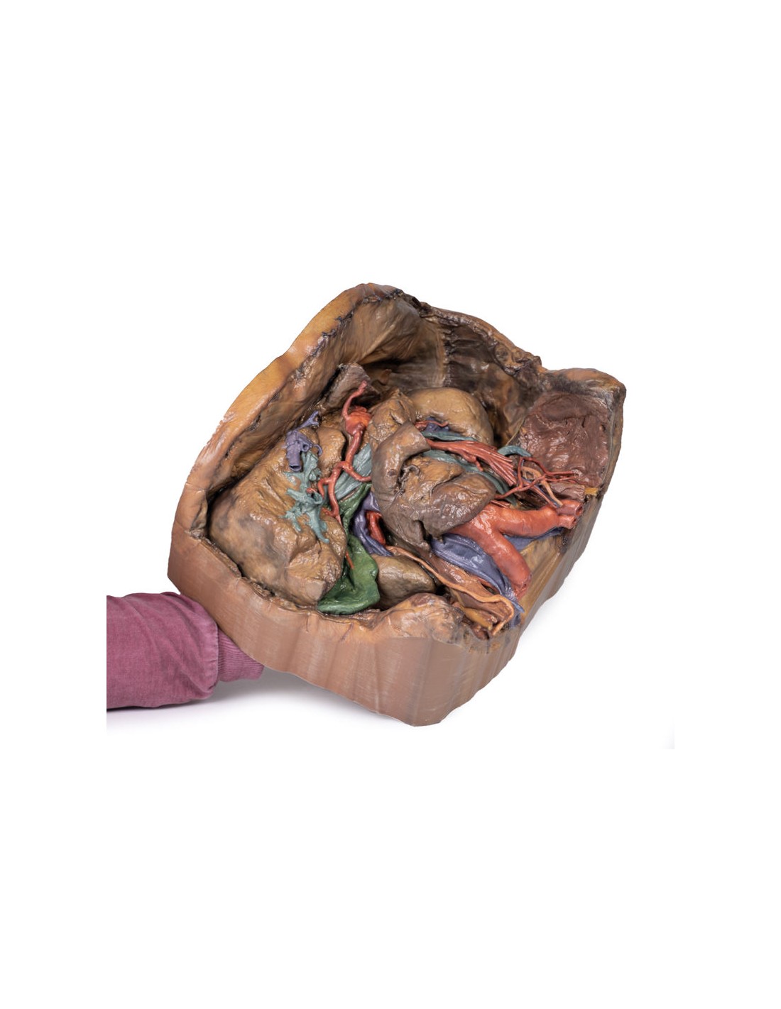
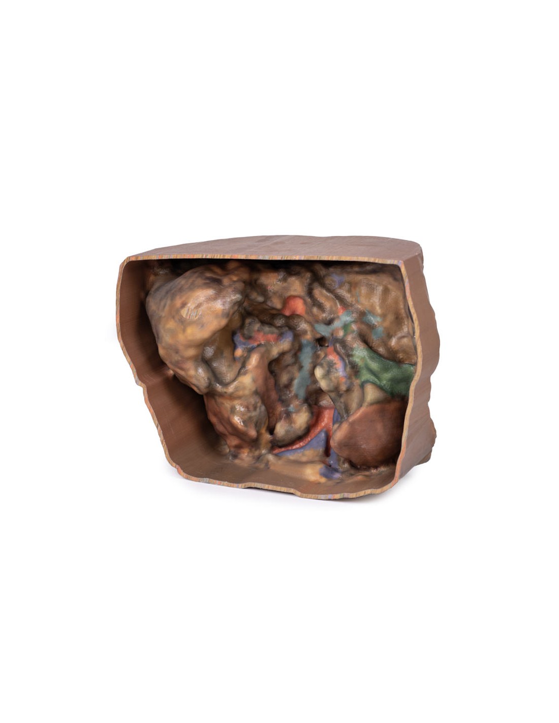
This 3D model represents one of the largest and most complex in the series, consisting of a partial torso from the diaphragm to the proximal thigh with a complete abdominal cavity preserving several levels of dissection. This 3-D model also records the rare simultaneous occurrence of indirect and direct inguinal hernias, allowing consideration of the anatomical basis for both conditions. Given the scale of the dissection, this 3D model description is divided into discrete parts based on views and regions.
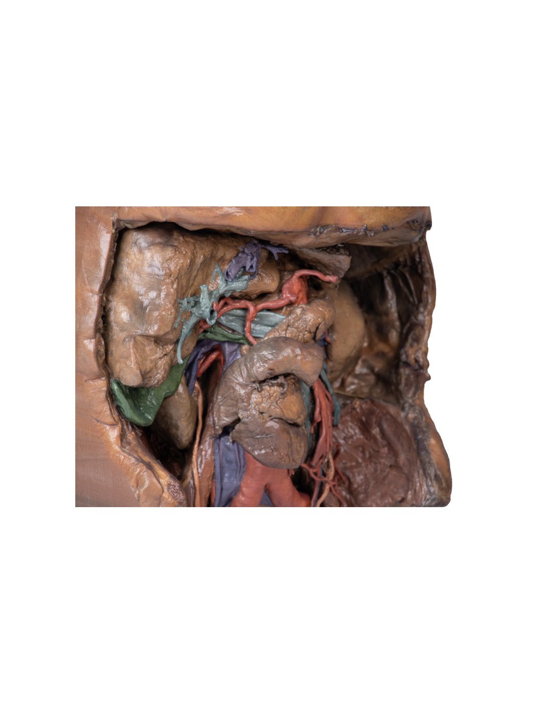
Celiac trunk
Providing the embryological anterior intestine, the celiac trunk arises from the T12 spinal level. Branches that can be seen in this specimen include the left gastric artery arising from the left portion of the celiac trunk; remnants of the splenic artery arising from the celiac trunk and visible by passing to the left hypochondrium; the common hepatic artery, located to the right of the celiac trunk and emanating key branches; the gastroduodenal artery, which branches inferiorly into the right gastric artery and provides an anastomosis to the superior mesenteric artery via the superior pancreaticoduodenal artery; and the right hepatic artery, which begins after the gastroduodenal artery and branches to form the left hepatic artery, the first branch of the right hepatic artery; the right hepatic artery, located inferiorly, which eventually gives rise to the cystic artery,
mesenteric artery
superior and inferior mesenteric artery Providing the middle and posterior intestines, respectively, the superior mesenteric artery and inferior mesenteric artery arise at the L1 and L3 vertebral levels, respectively.
Although both have key branches, this specimen does not preserve them in their entirety. In the specimen, the superior mesenteric artery can be seen exiting below the pancreas, dividing into many branches, and the descending inferior mesenteric artery can be seen to the left of the abdominal aorta. The left colic artery, moving laterally, can be seen leaving the IMA to give rise to the marginal arteries that supply the colon.
Venous system of the abdomen
The superior mesenteric vein can be seen posterior to the superior mesenteric artery, which is considerably less tubular than its arterial counterpart.
In the specimen, the left anatomic lobe of the liver has been removed, exposing the branches of the portal vein. These will supply nutrients from the gastrointestinal system to the hepatocytes, which will then connect to the venous system via the hepatic veins. This will then meet the Inferior Vena Cava.
Ilo of the kidney
The right kidney shows typical anatomy, as opposed to the left kidney which shows anatomical variations. At the right kidney we see the right renal vein, the uppermost, merging directly with the IVC, the right renal artery, the lowest, passing deep to the IVC from its origin from the abdominal aorta, and the right ureter, which runs superficial to the right renal artery to eventually travel inferiorly. The left kidney has unique variations at the hilum with key structures as follows. The left renal vein, very inferior (as opposed to the usual superior) and is highly divided. The left renal artery, more superior (as opposed to the usual inferior) and the left ureter, can be seen descending from the hilum and medial to the kidney.
Muscles, nerves and other vessels
The psoas major and iliacus muscle can be seen on both sides of the specimen, and surrounding them, key branches of the lumbar plexus can be seen, particularly on the left side. The iliohypogastric nerve, continuing laterally as the most superior of the nerves present, and the ilioinguinal nerve, inferior to the iliohypogastric, is directed toward the inguinal canal. The femoral nerve, which originates deep and enters in lateral view at the psoas major, and the genitofemoral nerve, runs surface to the psoas major, dividing into the genital and femoral branches of innervation.
Medial to the psoas major, the left testicular artery and left testicular vein can be seen (as this is a male specimen). While the artery will receive blood directly from the aorta, the left testicular vein drains to the left renal vein.
Right testicular vasculature can also be observed, however, the right testicular vein drains directly into the IVC.
The branch of the ilio-lumbar artery that anastomizes with the iliac circumflex artery can be observed passing under the testicular artery and vein and under the ureter.
Gallbladder
Just below the liver, the gallbladder can be observed with the cystic artery moving inferiorly to meet it. The cystic duct can also be seen moving from the gallbladder, meeting the common hepatic duct that moves from the liver to form the common bile duct.
What advantages does the Monash University anatomical dissection collection offer over plastic models or plastinated human specimens?
- Each body replica has been carefully created from selected patient X-ray data or human cadaver specimens selected by a highly trained team of anatomists at the Monash University Center for Human Anatomy Education to illustrate a range of clinically important areas of anatomy with a quality and fidelity that cannot be achieved with conventional anatomical models-this is real anatomy, not stylized anatomy.
- Each body replica has been rigorously checked by a team of highly trained anatomists at the Center for Human Anatomy Education, Monash University, to ensure the anatomical accuracy of the final product.
- The body replicas are not real human tissue and therefore not subject to any barriers of transportation, import, or use in educational facilities that do not hold an anatomy license. The Monash 3D Anatomy dissection series avoids these and other ethical issues that are raised when dealing with plastinated human remains.
No reviews
Tap to zoom