Your cart
There are no more items in your cart

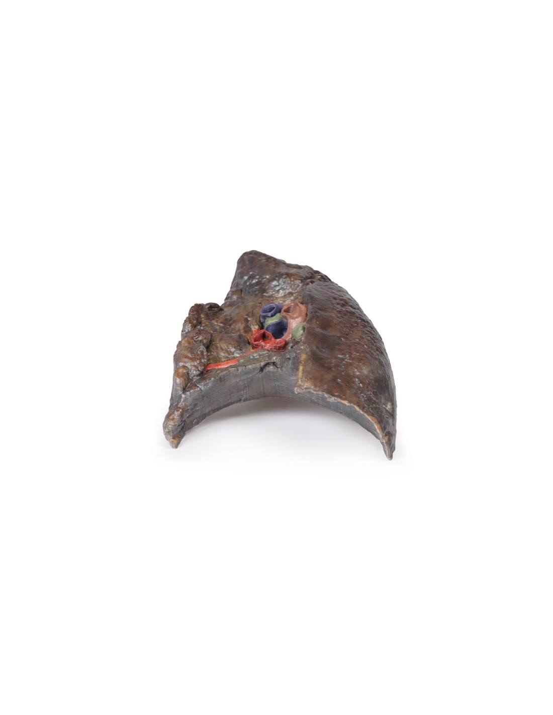
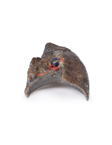
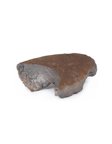
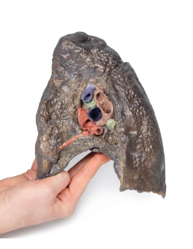
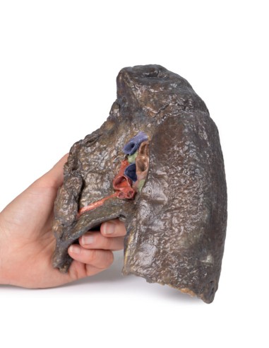
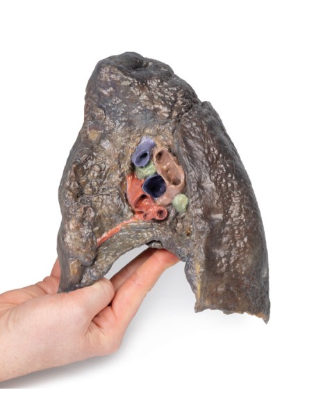
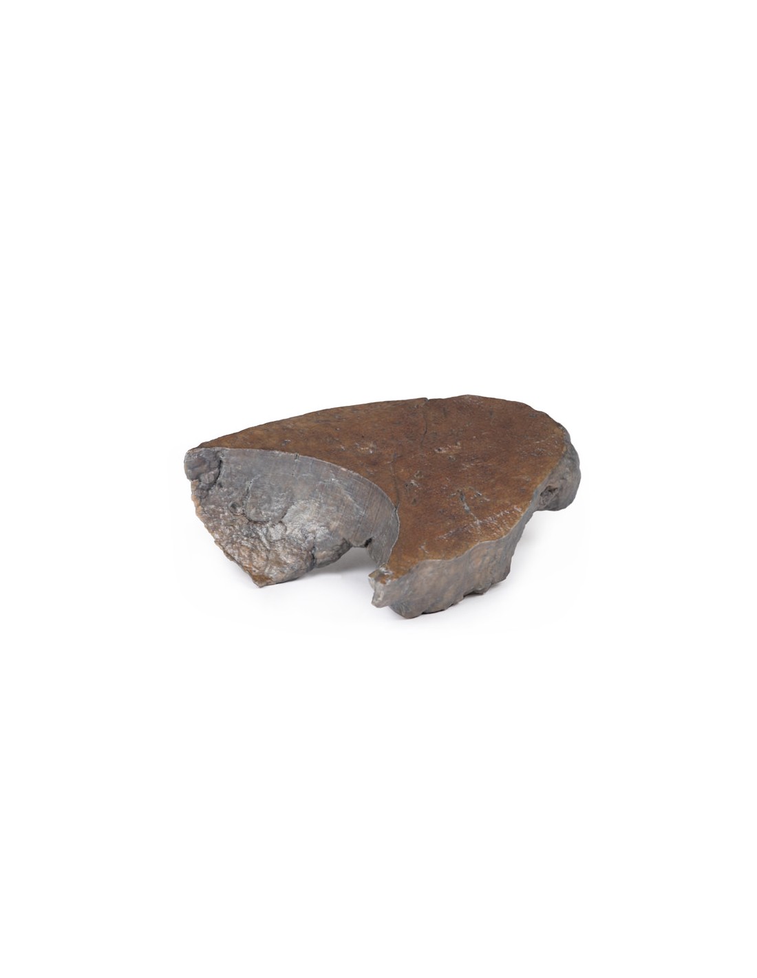
Dissection of the hilum of the right lung - Erler Zimmer 3D anatomy Series MP1127
erler zimmer
EZ-MP1127
€1,391.65
Tax included
Made in ultra-high resolution 3D printing in full color.
Dissection of the hilum of the right lung - Erler Zimmer 3D anatomy Series MP1127
This dissection model of the hilum of the right lung is part of the exclusive Monash 3D anatomy series, a comprehensive series of human dissections reproduced with ultra-high resolution color 3D printing.
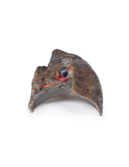
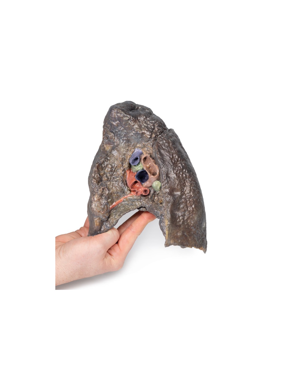
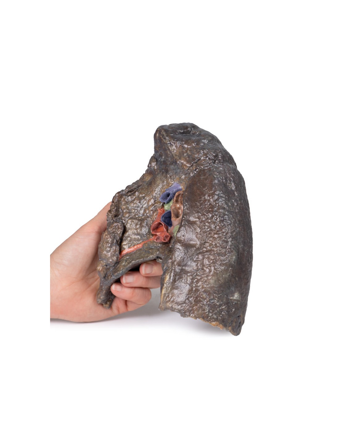
The hilum of a lung is where the visceral and parietal pleura meet and functions with the pulmonary ligament because the lungs are the only connection to the rest of the body. This connection includes the pulmonary artery, upper and lower pulmonary veins, main bronchi, nerves and lymphatics.
Since the definition of an artery involves transporting blood AWAY from the heart, this will be deoxygenated blood in the pulmonary system, as opposed to systemic circulation. Similarly, veins transport blood TOWARD the heart, which means it will be oxygenated in the pulmonary system.
With the specimen cut in a sagittal plane in line with the cardiac impression, the nerves and lymphatic vessels are difficult to identify, however, it is possible to see the sulcus from the esophagus as it descends posteriorly to perforate the diaphragm next to the cardiac impression (of the right atrium) is noticeably anterior to the hilum of the right lung; the right main bronchi and its subsequent divisions into lobar bronchi, which are located more posteriorly in the hilum in this specimen; the pulmonary artery and its divisions, located higher up within the hilum; the superior and inferior pulmonary veins and their divisions, which are more inferior and anterior in the specimen. the oblique and horizontal slits along the lateral surface of the specimen and the hilar lymph nodes around the hilum on the medial surface of the lung.
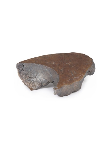
The diaphragmatic surface is located inferiorly and the visceral costal surface is located at the back of the specimen.
What advantages does the Monash University anatomical dissection collection offer over plastic models or plastinated human specimens?
- Each body replica has been carefully created from selected patient X-ray data or human cadaver specimens selected by a highly trained team of anatomists at the Monash University Center for Human Anatomy Education to illustrate a range of clinically important areas of anatomy with a quality and fidelity that cannot be achieved with conventional anatomical models-this is real anatomy, not stylized anatomy.
- Each body replica has been rigorously checked by a team of highly trained anatomists at the Center for Human Anatomy Education, Monash University, to ensure the anatomical accuracy of the final product.
- The body replicas are not real human tissue and therefore not subject to any barriers of transportation, import, or use in educational facilities that do not hold an anatomy license. The Monash 3D Anatomy dissection series avoids these and other ethical issues that are raised when dealing with plastinated human remains.
No reviews
Tap to zoom