Your cart
There are no more items in your cart


Section of lung, hilum removed - Erler Zimmer 3D anatomy Series MP1125
erler zimmer
EZ-MP1125
€1,099.10
Tax included
Made in ultra-high resolution 3D printing in full color.
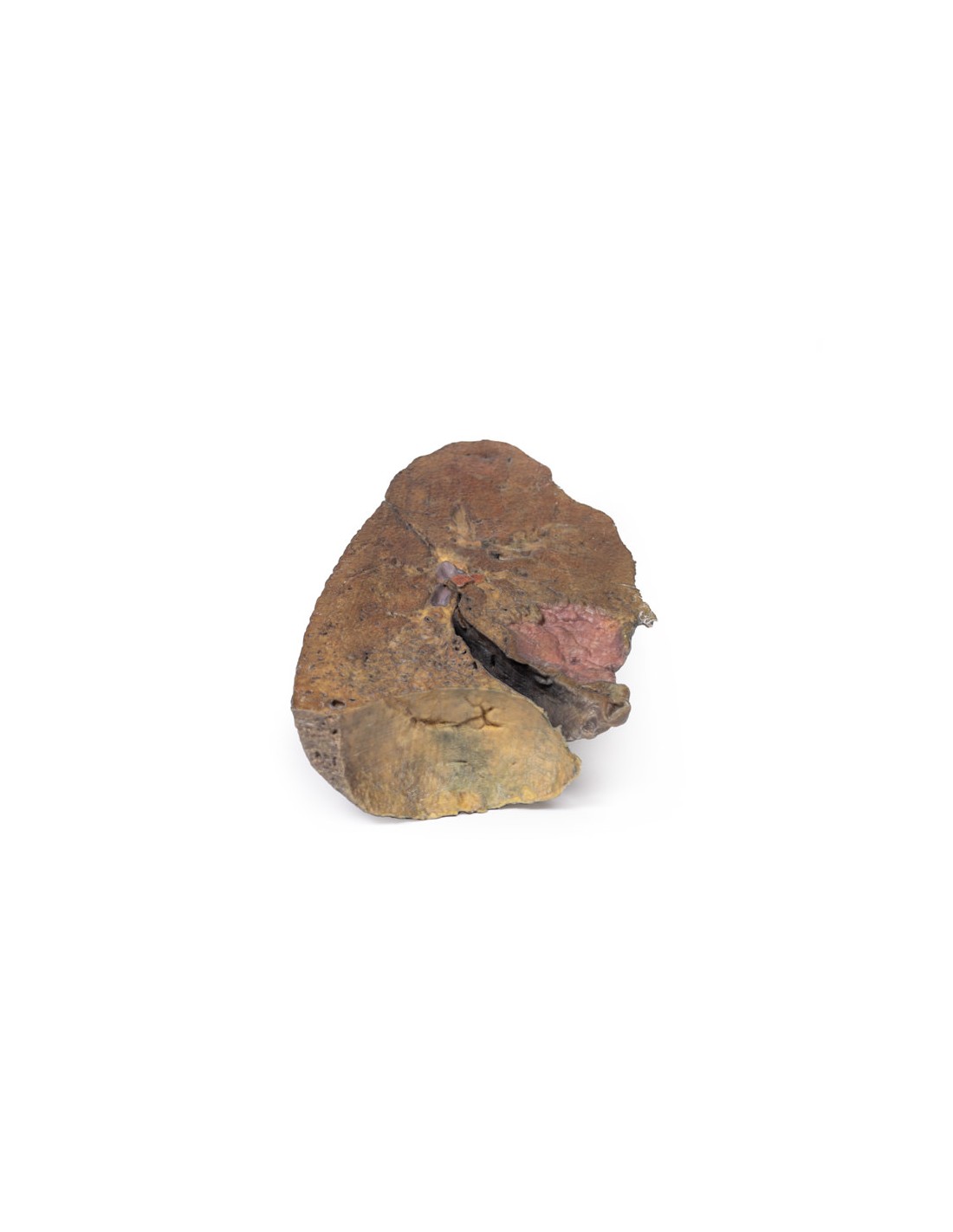
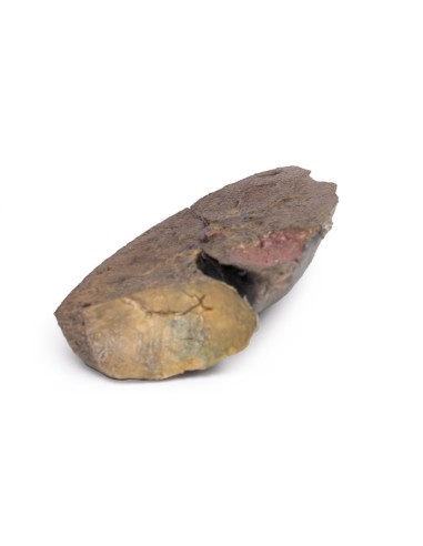
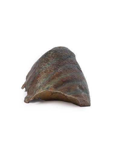
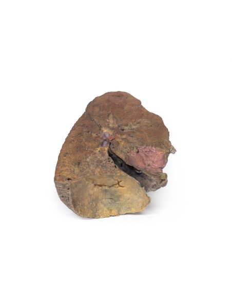
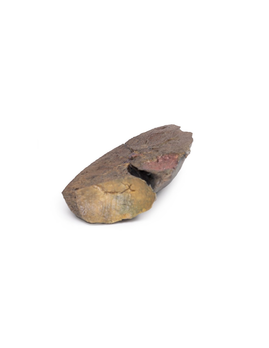
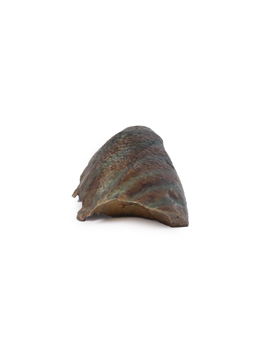
Lung section, hilum removed - Erler Zimmer 3D anatomy Series MP1125
This model of a section of the left lung is part of the exclusive Monash 3D anatomy series, a comprehensive series of human dissections reproduced with ultra-high resolution color 3D printing.
The lung was dissected following a parasagittal plane, removing the mediastinal surface. Usually, pulmonary arteries, veins, and bronchi can be observed entering the lung at the hilum, but the primary bronchi cannot be seen in this specimen because they have already divided substantially. It is unclear to what extent laterally the specimen has been sectioned, so the level of division of the bronchi (secondary or tertiary) cannot be determined.
The cardiac impression is formed by the left ventricle of the heart resting on the mediastinal surface of the lung. Although the lung was sectioned following a parasagittal plane, the cardiac footprint can still be observed as it is the most concave area of the medial surface of the lung.
The lung lies above the diaphragm, forming the concave diaphragmatic surface. The pleura was not preserved in this specimen, but normally there is a diaphragmatic recess bounded by the costal and diaphragmatic pleura. This would be located between the diaphragmatic imprint of the lung and the diaphragm.
What advantages does the Monash University anatomical dissection collection offer over plastic models or plastinated human specimens?
- Each body replica has been carefully created from selected patient X-ray data or human cadaver specimens selected by a highly trained team of anatomists at the Monash University Center for Human Anatomy Education to illustrate a range of clinically important areas of anatomy with a quality and fidelity that cannot be achieved with conventional anatomical models-this is real anatomy, not stylized anatomy.
- Each body replica has been rigorously checked by a team of highly trained anatomists at the Center for Human Anatomy Education, Monash University, to ensure the anatomical accuracy of the final product.
- The body replicas are not real human tissue and therefore not subject to any barriers of transportation, import, or use in educational facilities that do not hold an anatomy license. The Monash 3D Anatomy dissection series avoids these and other ethical issues that are raised when dealing with plastinated human remains.
No reviews
Tap to zoom