Your cart
There are no more items in your cart


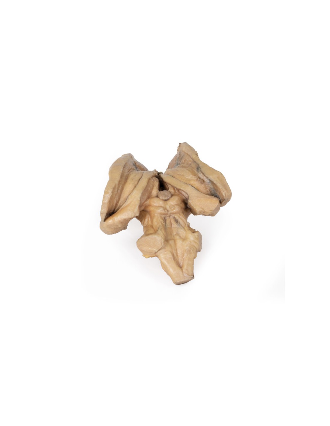
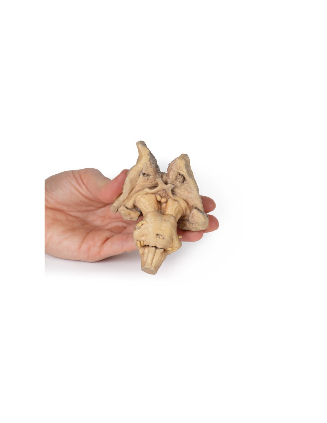
Encephalic trunk, deep brain and diencephalic structures - Erler Zimmer 3D anatomy Series MP1100
erler zimmer
EZ-MP1100
€602.56
Tax included
Made in ultra-high resolution 3D printing in full color.
Encephalic trunk, deep brain and diencephalic structures - Erler Zimmer 3D anatomy Series MP1100
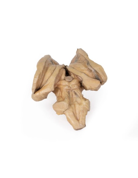
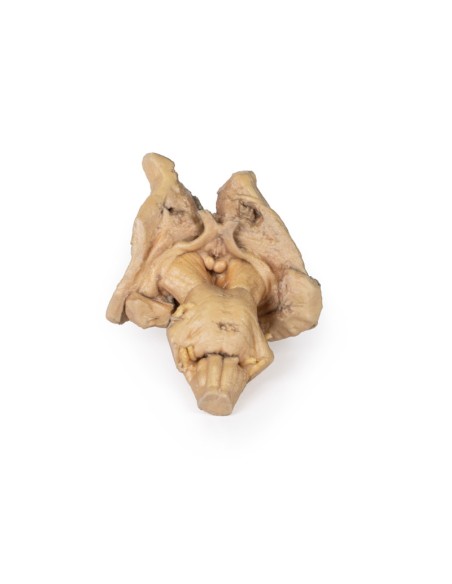
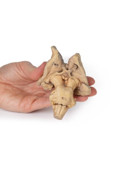
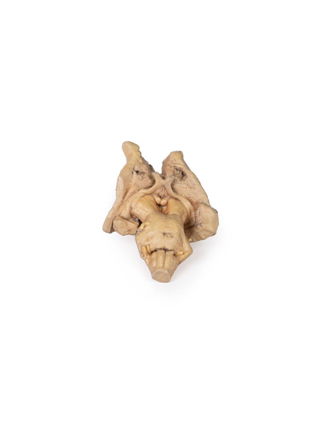
This model is part of the exclusive Monash 3D anatomy series, a comprehensive series of human dissections reproduced with ultra-high resolution color 3D printing.
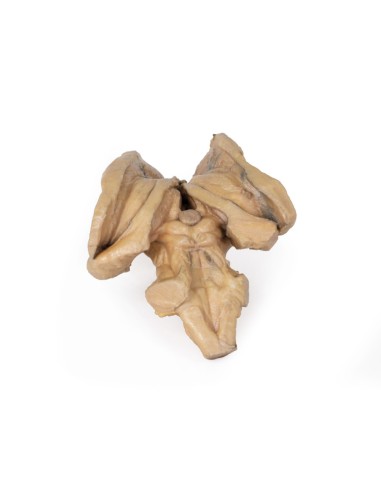
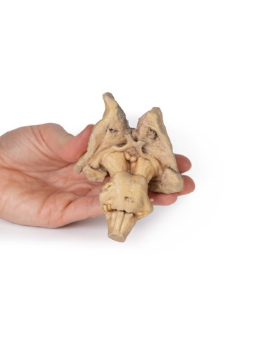
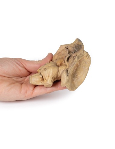
This 3D model preserves the many deep brain and diencephalic structures down to the proximal medulla oblongata and complements the other isolated brainstem (BRW10) in our series.
Above, on the right side of the 3D model, is the lentiform (lenticular) nucleus and the corona radiata of the internal capsule can be seen emerging around it. On the left side, the lentiform nucleus is absent, but the head and body of the caudate nucleus are present medially on both sides, wrapping medially to the preserved inner capsule margins and leading to the amygdaloid bodies on each side. The thalami are present bilaterally, and the third ventricle is slightly open in the midline inferior to the epithalamus (pineal gland).
Anteriorly, cerebral peduncles are present, with optic nerves extending from the chiasm and preserved tracts. The interpeduncular region is exposed with both the mammillary bodies and the dissected infundibulum visible. Caudal to the interpeduncular region is the bridge that preserves the origins of the middle cerebellar peduncles and the origins of cranial nerves V, VII and VIII. The preserved portion of the medulla oblongata possesses prominent pyramids and olives.
Posteriorly, the superior and inferior colliculi lie just above the dissected superior cerebellar peduncles, and the fourth ventricle is open to expose the rhomboid fossa and floor features: the medial eminence, facial colliculus, hypoglossal triangle, vestibular triangle, and vagal triangle.
What advantages does the Monash University anatomical dissection collection offer over plastic models or plastinated human specimens?
? Each body replica is carefully created from selected radiographic data of the patient or from
human cadaver specimens selected by a highly trained team of anatomists at the Monash University Center for Human Anatomy Education to illustrate a range of clinically important areas of anatomy with a quality
and fidelity that cannot be achieved with conventional anatomical models-this is real anatomy, not stylized anatomy.
? Each body replica has been rigorously checked by a team of highly trained anatomists at the Center for Human Anatomy Education, Monash
University, to ensure the anatomical accuracy of the final product.
? The body replicas are not real human tissue and therefore not subject to any barriers of transportation, import, or use in educational facilities
that do not hold an anatomy license. The Monash 3D Anatomy dissection series avoids these and other ethical issues that are raised when dealing with plastinated human remains.
No reviews
Tap to zoom