Your cart
There are no more items in your cart


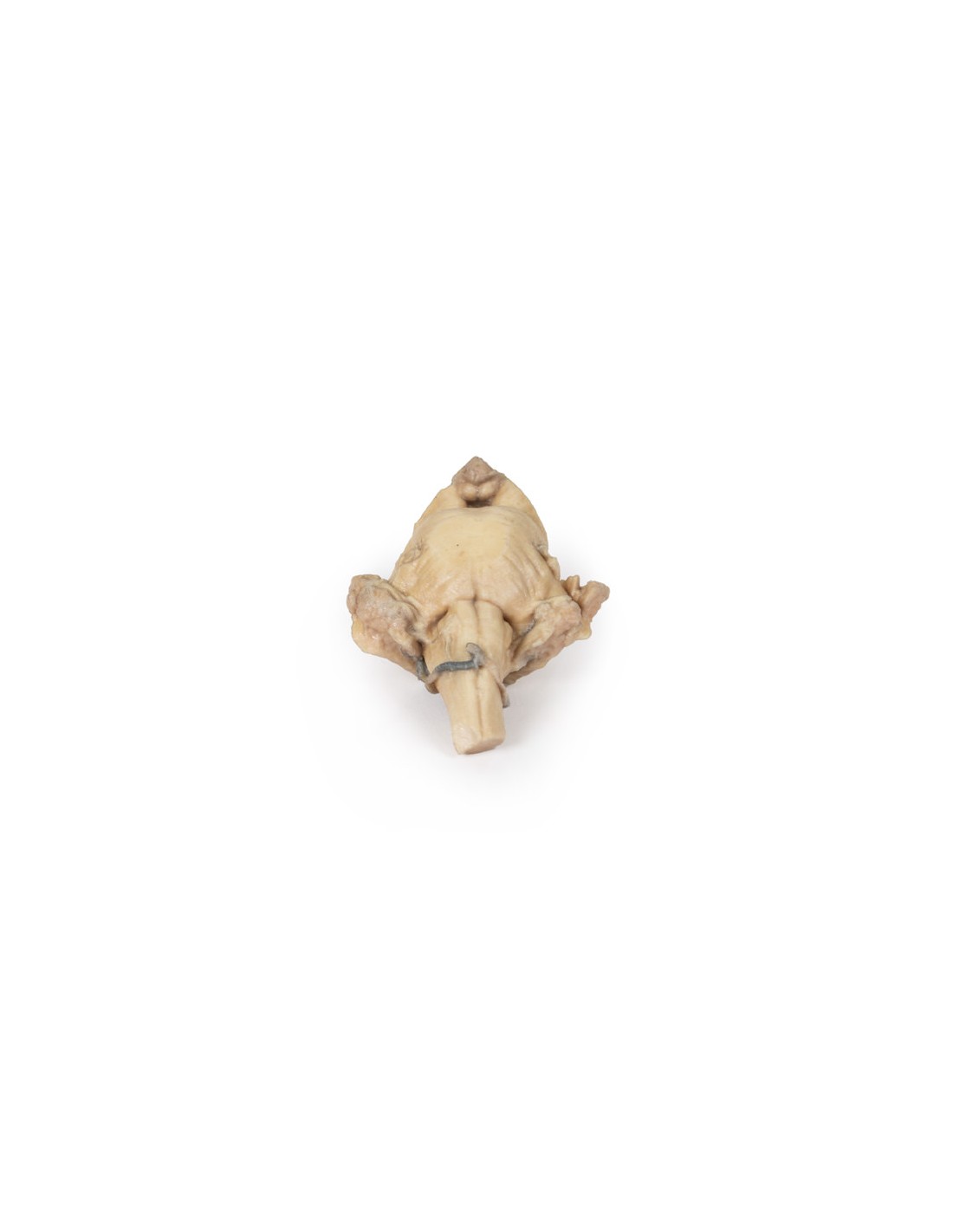
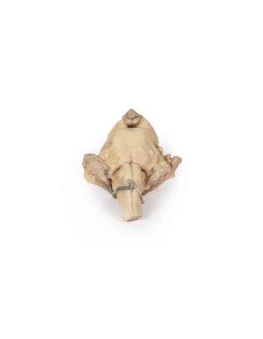
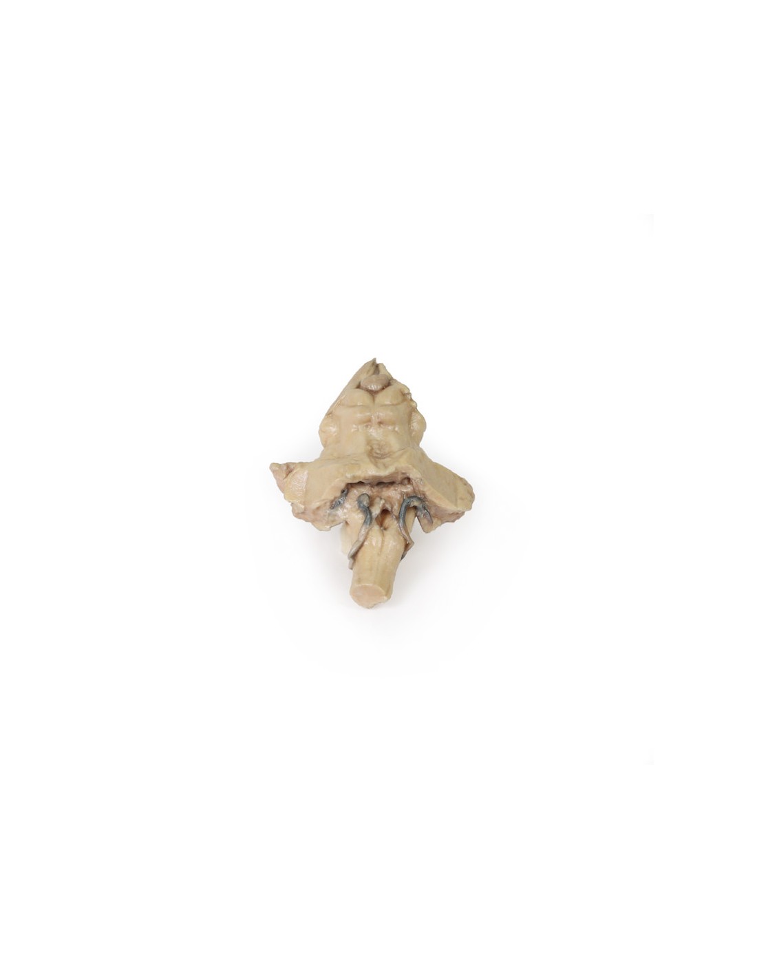
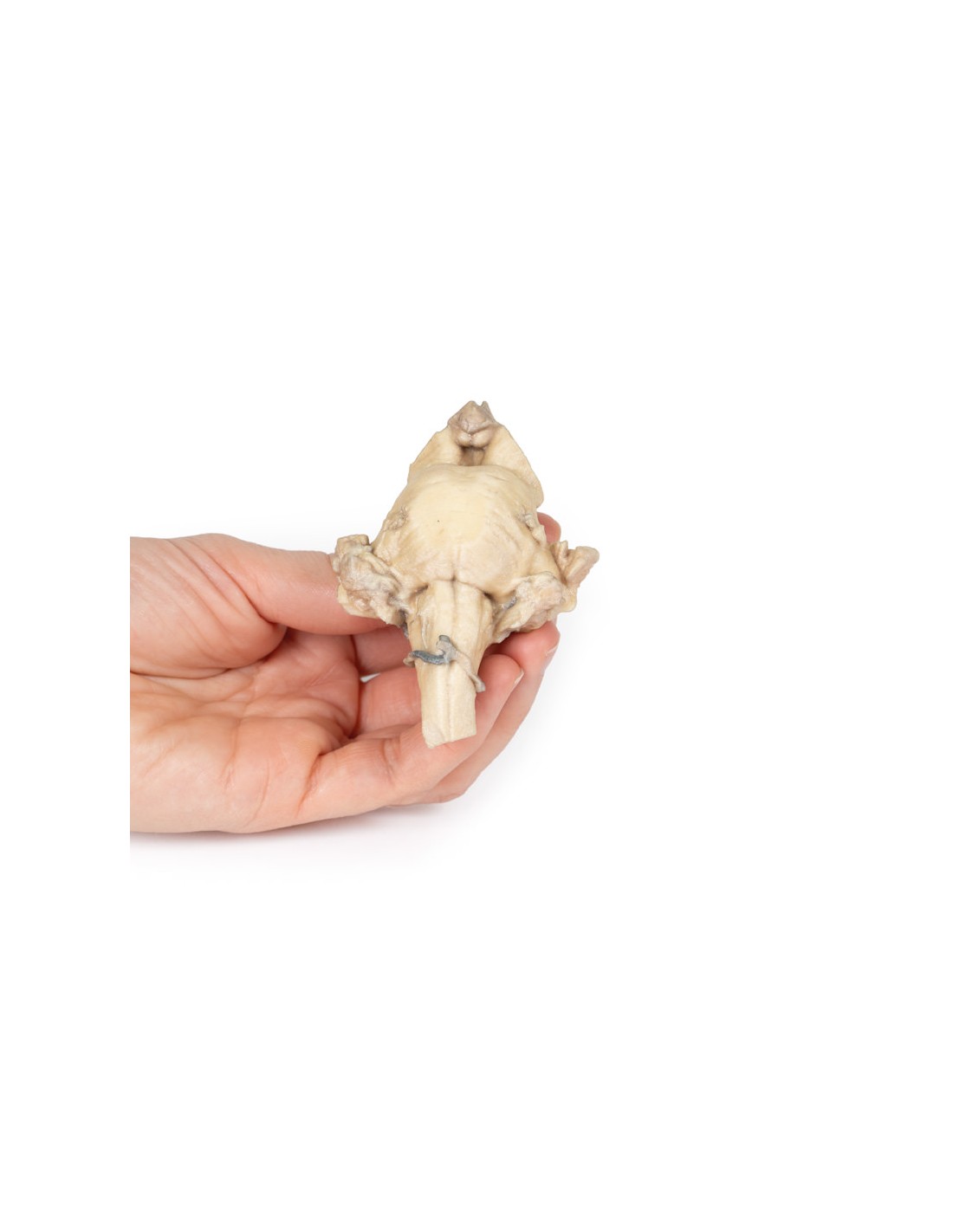
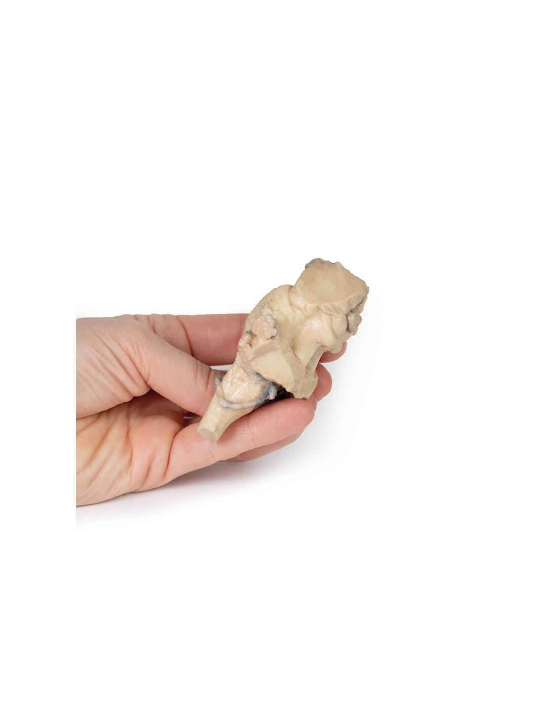
Brain stem, isolated anatomy from midbrain to medulla oblongata - Erler Zimmer 3D anatomy Series MP1101
erler zimmer
EZ-MP1101
€390.52
Tax included
Made in ultra-high resolution 3D printing in full color.
Brain stem, isolated anatomy from midbrain to medulla oblongata - Erler Zimmer 3D anatomy Series MP1101
This model is part of the exclusive Monash 3D anatomy series, a comprehensive series of human dissections reproduced with ultra-high resolution color 3D printing.
This 3D model provides a view of isolated brainstem anatomy from midbrain to medulla oblongata and complements the other 3D diencephalon/brainstem model (BR 10) in our series.
Rostrally, the 3D model was sectioned at an angle to the overlying diencephalon, while retaining the mamillary bodies of the hypothalamus between the brain peduncles (anteriorly) and the pineal gland/epithalamus (posteriorly). Posteriorly, the quadrigeminal bodies (the superior and inferior collective colliculi) of the midbrain are prominently adjacent to the superior cerebellar peduncles. The cerebellum itself has been removed, leaving the cross section of the middle and lower cerebellar peduncles on each side. Inferior to the dissected peduncles is the partially open fourth ventricle and the remains of the posterior inferior cerebellar arteries.
In the ventral aspect of the 3D model the pons with the origin of the trigeminal nerve (CN V) preserved (particularly on the left side). Inferior to the pons on the medulla oblongata, both pyramids and olives are visible on both sides (particularly sharp on the right).
What advantages does the Monash University anatomical dissection collection offer over plastic models or plastinated human specimens?
- Each body replica has been carefully created from selected patient X-ray data or human cadaver specimens selected by a highly trained team of anatomists at the Monash University Center for Human Anatomy Education to illustrate a range of clinically important areas of anatomy with a quality and fidelity that cannot be achieved with conventional anatomical models-this is real anatomy, not stylized anatomy.
- Each body replica has been rigorously checked by a team of highly trained anatomists at the Center for Human Anatomy Education, Monash University, to ensure the anatomical accuracy of the final product.
- The body replicas are not real human tissue and therefore not subject to any barriers of transportation, import, or use in educational facilities that do not hold an anatomy license. The Monash 3D Anatomy dissection series avoids these and other ethical issues that are raised when dealing with plastinated human remains.
No reviews
Tap to zoom