Your cart
There are no more items in your cart


Pathways of the paranasal sinus - Erler Zimmer 3D anatomy Series MP1106
erler zimmer
EZ-MP1106
Last items in stock
€1,011.87
Tax included
Made in ultra-high resolution 3D printing in full color.
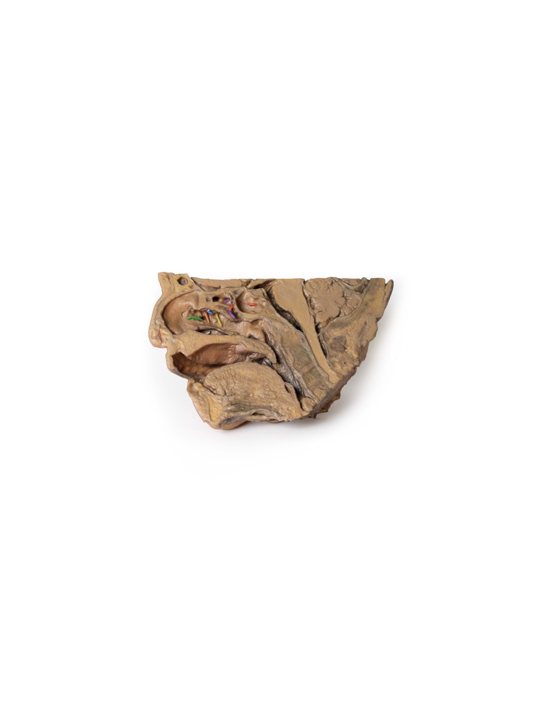
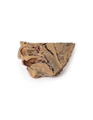
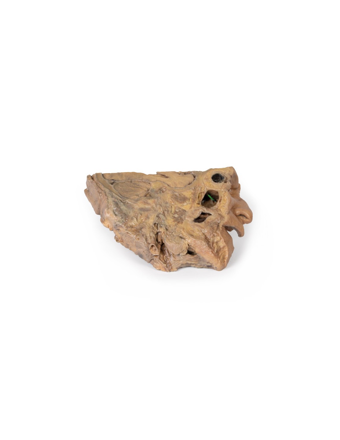
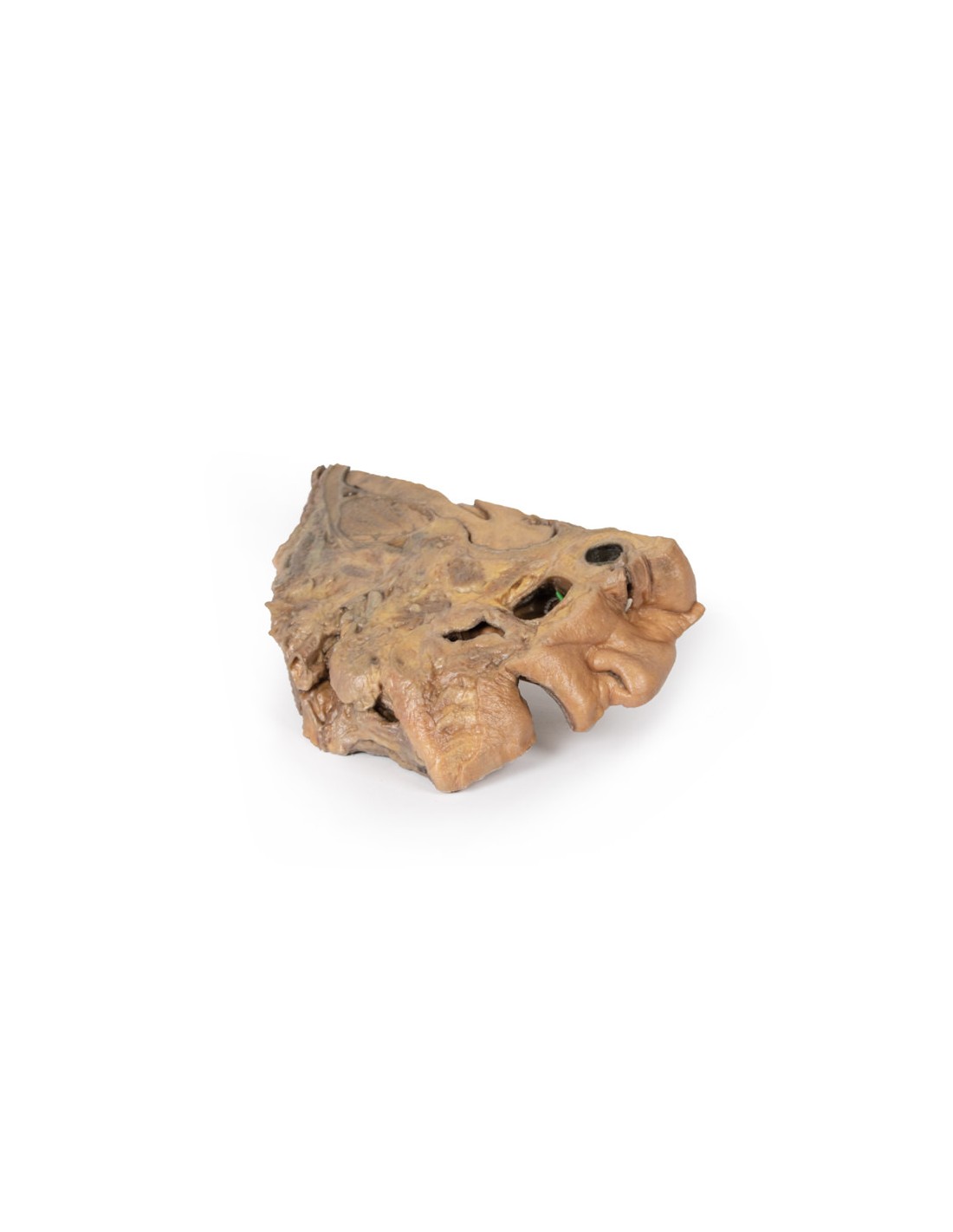
Pathways of the paranasal sinus - Erler Zimmer 3D anatomy Series MP1106
This model is part of the exclusive Monash 3D anatomy series, a comprehensive series of human dissections reproduced with ultra-high resolution color 3D printing.
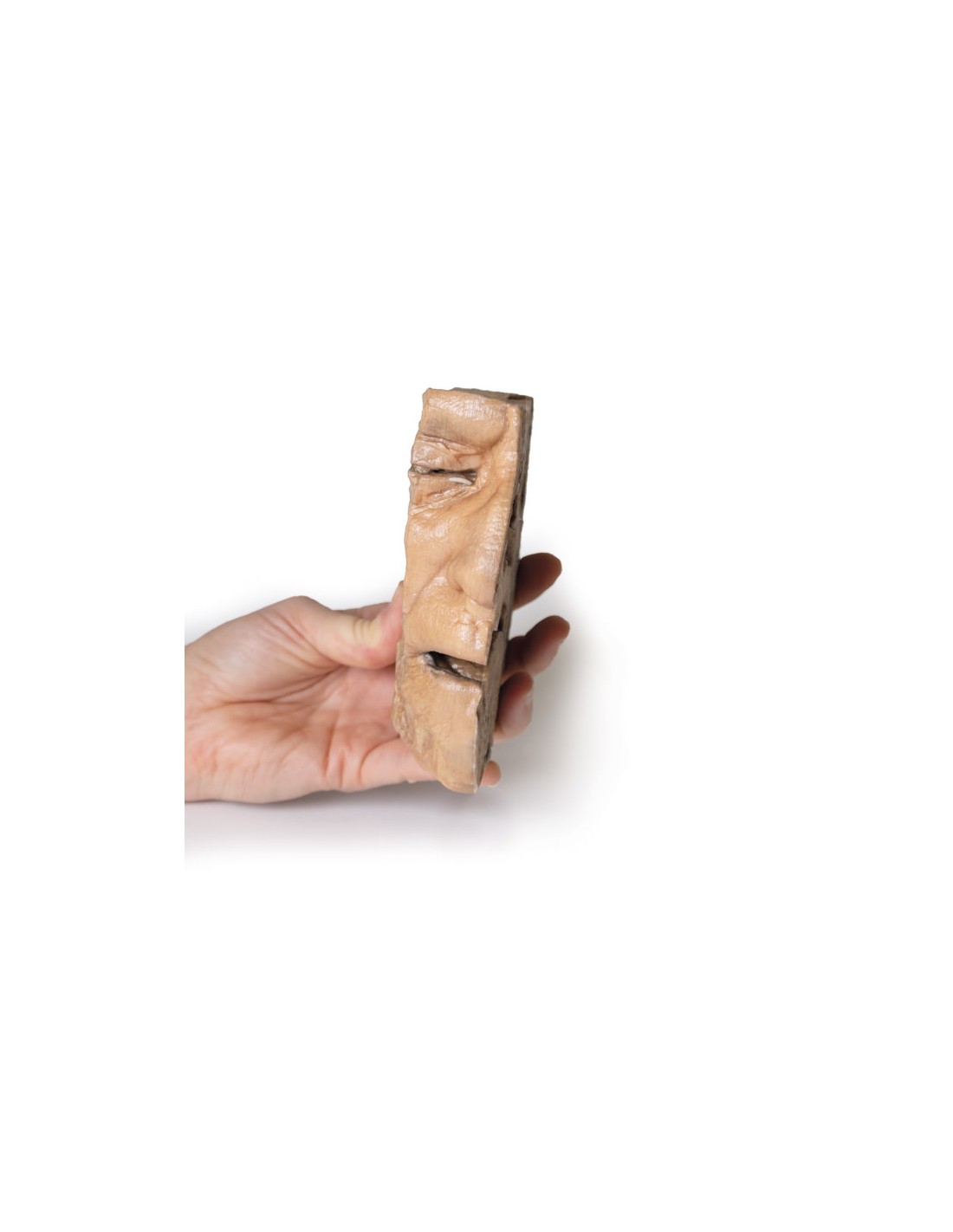
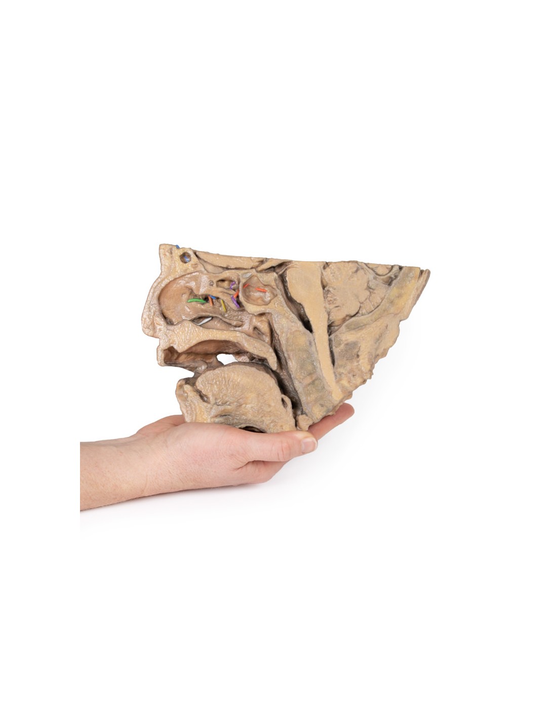
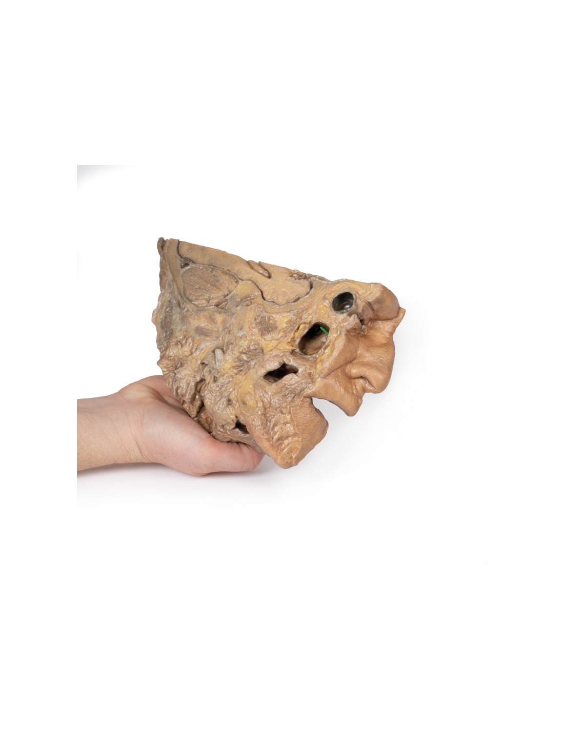
This 3D model provides a mid-sagittal to parasagittal segment of a right head to demonstrate the relationships and transitions of the sinuses. These passages were highlighted with thin colored markers to indicate the relationship of these pathways between the sinuses and the nasal cavity.
Starting anteriorly in the nasal cavity, the opening of the nasolacrimal duct (white) is present just deep to the inferior nasal concha. The middle nasal concha was dissected to allow a clear view of the sinus opening (visible in the parasagittal plane) through the semilunar hiatus (green), as well as the frontal sinus drainage (blue; with the sinus visible superiorly in the section and transverse cut of the specimen) and the anterior (orange) and middle (yellow) ethmoidal cells. The opening of the posterior ethmoidal cells in the superior meatus is shown through the purple marker, which is visible within a small open window in the ethmoid just above the nasal cavity. Finally,
In addition to these pathways, this 3D model also captures some of the surrounding anatomy within the section. In the mediosagittal view, the other primary structures of the nasal cavity are visible from the nostril to the opening of the ear tube posteriorly. The soft palate and uvula are preserved, as well as the rest of the pharynx just to the level of the epiglottis and the collapsed laryngeal region in the lower part of the preserved specimen. The oral cavity is visualized in cross section, with distinct genioglossus and geniohyoid muscles. Parts of the brain are preserved in the cranial cavity including the lower parts of the frontal lobe and the optic nerve/chiasma/right tract. The pituitary gland is visible in cross section just above the sphenoid sinus. The ??ponte, medulla oblongata, and most of the cerebellum are present, with a small part of the tentorium cerebellum separating the cerebellum from the right occipital lobe of the brain. On the parasagittal side of the specimen is a continuation of the tentorium cerebelli separating these parts of the brain, with clear cross sections of the transverse sinus and part of the sigmoid sinus on either side of the cerebellum. Overlying this is a small part of the medial temporal lobe of the cerebellum with part of the anterior horn of the lateral ventricle deep within the lobe. with clear transverse sections of the transverse sinus and part of the sigmoid sinus on either side of the cerebellum. Overlying this is a small part of the medial temporal lobe of the cerebellum with part of the anterior horn of the lateral ventricle deep within the lobe. with clear cross sections of the transverse sinus and part of the sigmoid sinus on both sides of the cerebellum. Overlying this is a small part of the medial temporal lobe of the cerebellum with part of the anterior horn of the lateral ventricle deep within the lobe.
What advantages does the Monash University anatomical dissection collection offer over plastic models or plastinated human specimens?
- Each body replica has been carefully created from selected patient X-ray data or human cadaver specimens selected by a highly trained team of anatomists at the Monash University Center for Human Anatomy Education to illustrate a range of clinically important areas of anatomy with a quality and fidelity that cannot be achieved with conventional anatomical models-this is real anatomy, not stylized anatomy.
- Each body replica has been rigorously checked by a team of highly trained anatomists at the Center for Human Anatomy Education, Monash University, to ensure the anatomical accuracy of the final product.
- The body replicas are not real human tissue and therefore not subject to any barriers of transportation, import, or use in educational facilities that do not hold an anatomy license. The Monash 3D Anatomy dissection series avoids these and other ethical issues that are raised when dealing with plastinated human remains.
No reviews
Tap to zoom