Your cart
There are no more items in your cart


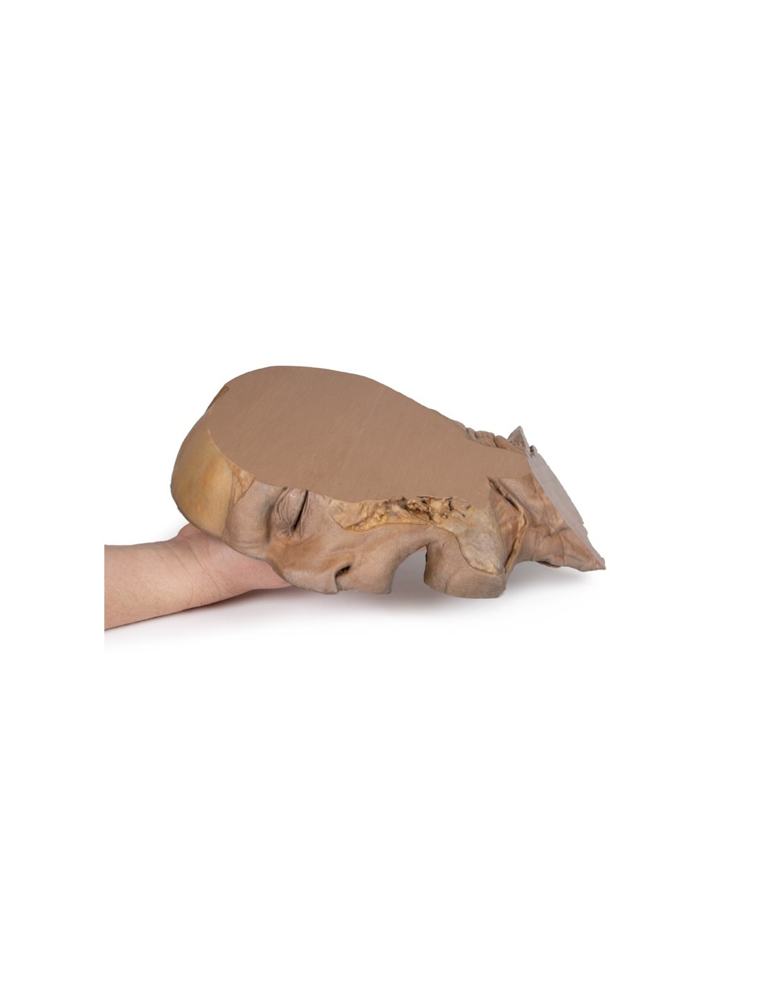
Parasagittal section of the head and neck - Erler Zimmer 3D anatomy Series MP1107
erler zimmer
EZ-MP1107
€2,434.39
Tax included
Made in ultra-high resolution 3D printing in full color.
Parasagittal section of the head and neck - Erler Zimmer 3D anatomy Series MP1107
This model is part of the exclusive Monash 3D anatomy series, a comprehensive series of human dissections reproduced with ultra-high resolution color 3D printing.
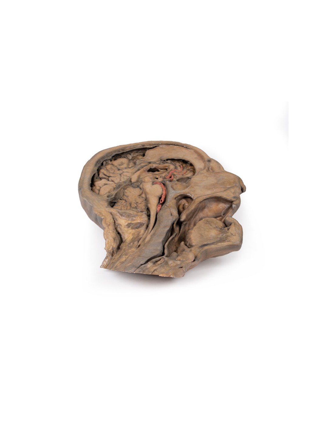
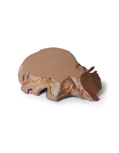
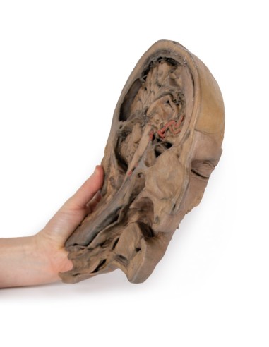
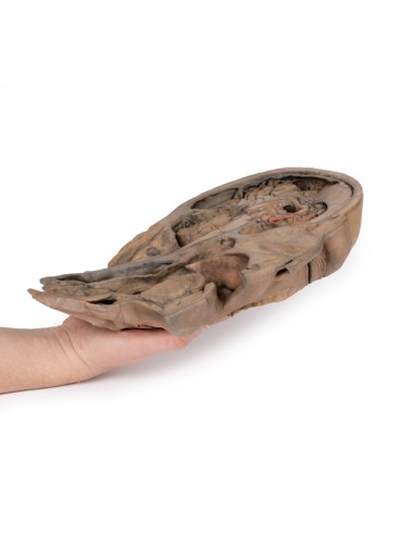
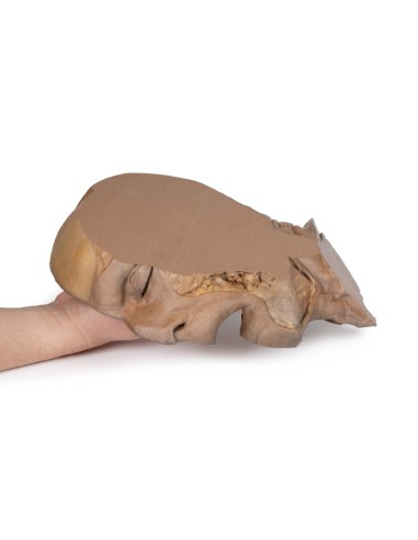
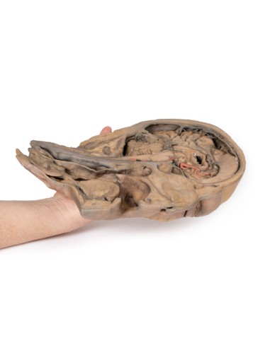
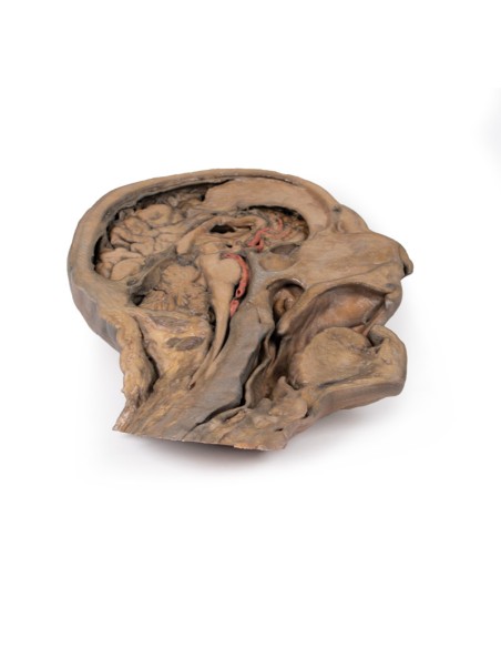
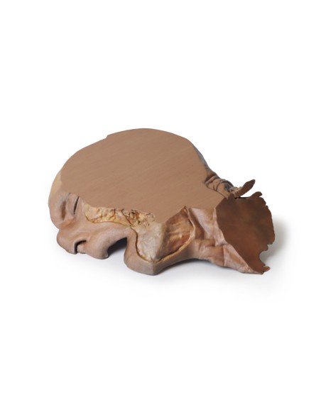
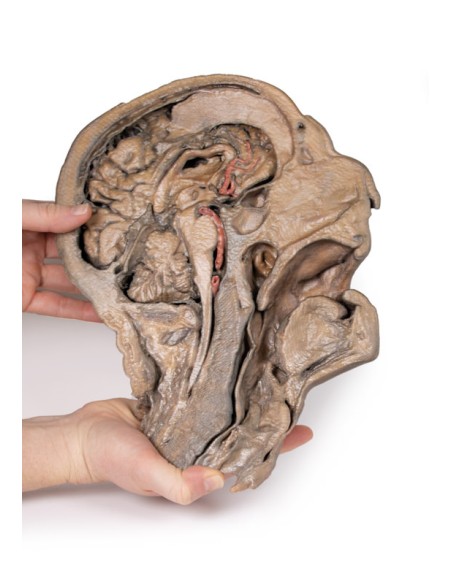
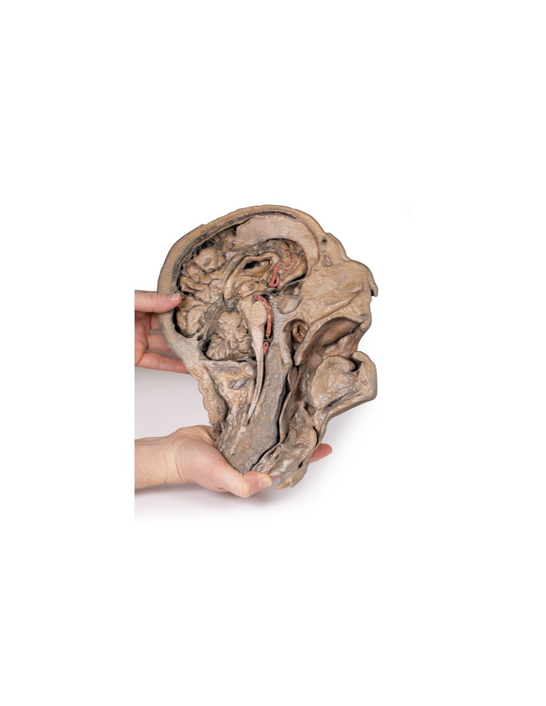
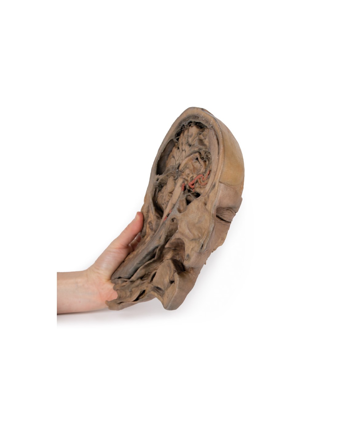
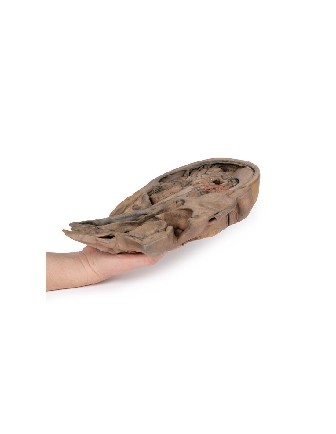
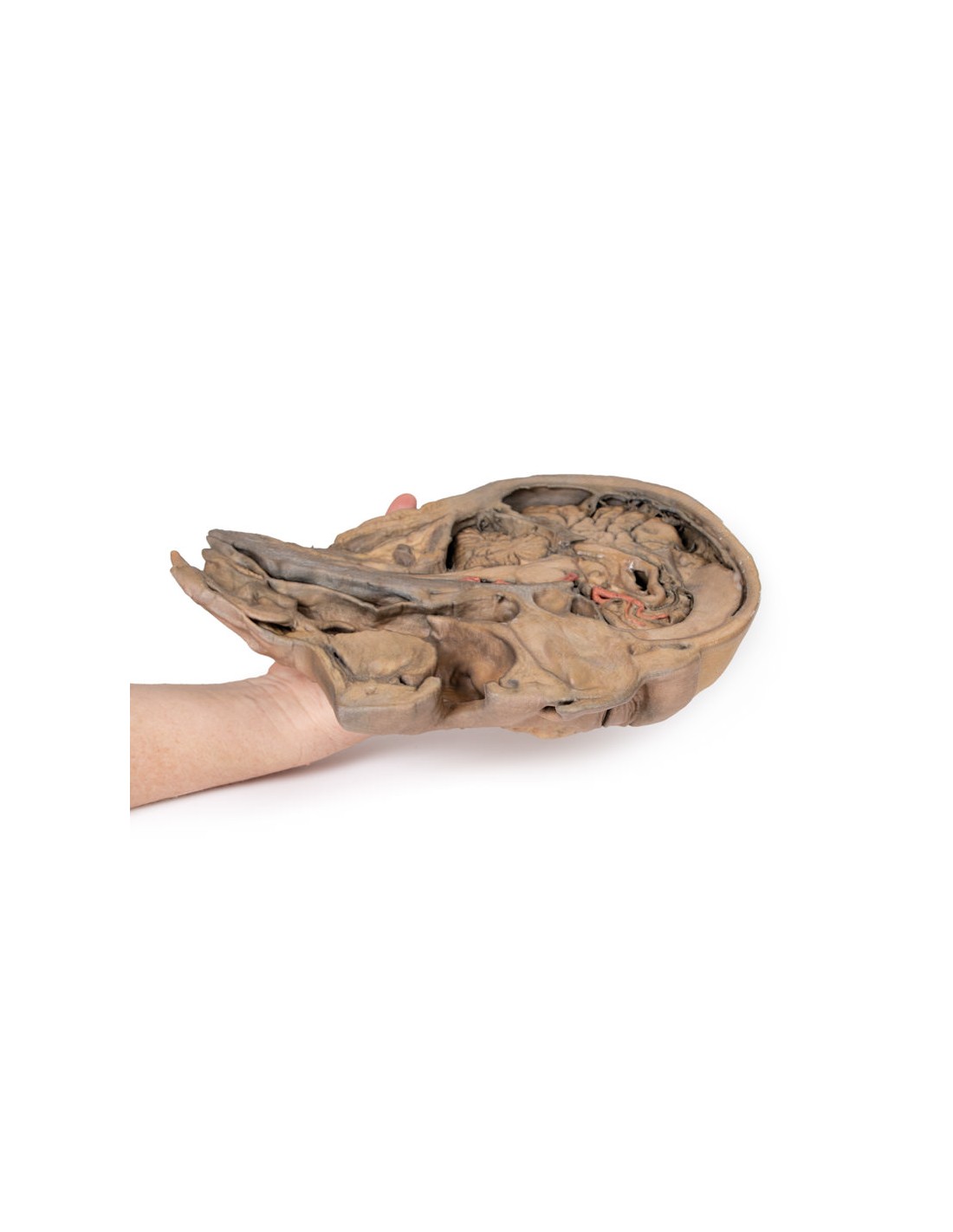
This 3D model of the head and neck represents a specimen dissected just outside the mid sagittal plane to preserve some midline anatomical structures (e.g., the cerebral sickle, septum pellucidum, nasal septum) that are absent from other specimens in the series. There was also a fixative-induced shrinkage of neural tissue. This volume reduction has the advantage of exaggerating the space between the brain and the contours and endocranial structures that are normally in close approximation. The non-sectioned side of the specimen was digitally removed.
The anterior part of the falx cerebri was retained from its anterior attachment to the galli crest until about the midpoint of its extension toward the tentorium cerebelli. At the attachment of the falx, the dura portion was removed to demonstrate the extension of the superior sagittal sinus within the retained portion of the dural fold. The brain itself was dissected preserving the septum pellucidum and the interventricular foramen (of Monro) defining the transition between the deep lateral ventricle and the dissected third ventricle. This plane of section also captures the infundibulum extending from the hypothalamus to the pituitary gland, which is located adjacent to a well-developed sphenoidal sinus. Both the cerebral aqueduct and the fourth ventricle are preserved, as are parts of the left vertebral artery,
The retention of the nasal septum in this specimen (and in contrast to other 3D models of the head and neck in the series) allows us to appreciate the relationship between the septum and the hard and soft palate, the entrance to the auditory tube, and the overall nasopharynx with respect to the nasal cavity and the oropharyngeal region below it. The muscular wall of the pharynx was isolated to demonstrate position relative to the cervical spine. Inferiorly, tracheal cartilages including the epiglottis, arytenoid, and thyroid were maintained to demonstrate the position of these cartilages relative to the hyoid bone, as well as the vestibule, vestibular fold, and vocal cord in cross section.
What advantages does the Monash University anatomical dissection collection offer over plastic models or plastinated human specimens?
- Each body replica has been carefully created from selected patient X-ray data or human cadaver specimens selected by a highly trained team of anatomists at the Monash University Center for Human Anatomy Education to illustrate a range of clinically important areas of anatomy with a quality and fidelity that cannot be achieved with conventional anatomical models-this is real anatomy, not stylized anatomy.
- Each body replica has been rigorously checked by a team of highly trained anatomists at the Center for Human Anatomy Education, Monash University, to ensure the anatomical accuracy of the final product.
- The body replicas are not real human tissue and therefore not subject to any barriers of transportation, import, or use in educational facilities that do not hold an anatomy license. The Monash 3D Anatomy dissection series avoids these and other ethical issues that are raised when dealing with plastinated human remains.
No reviews
Tap to zoom