Your cart
There are no more items in your cart


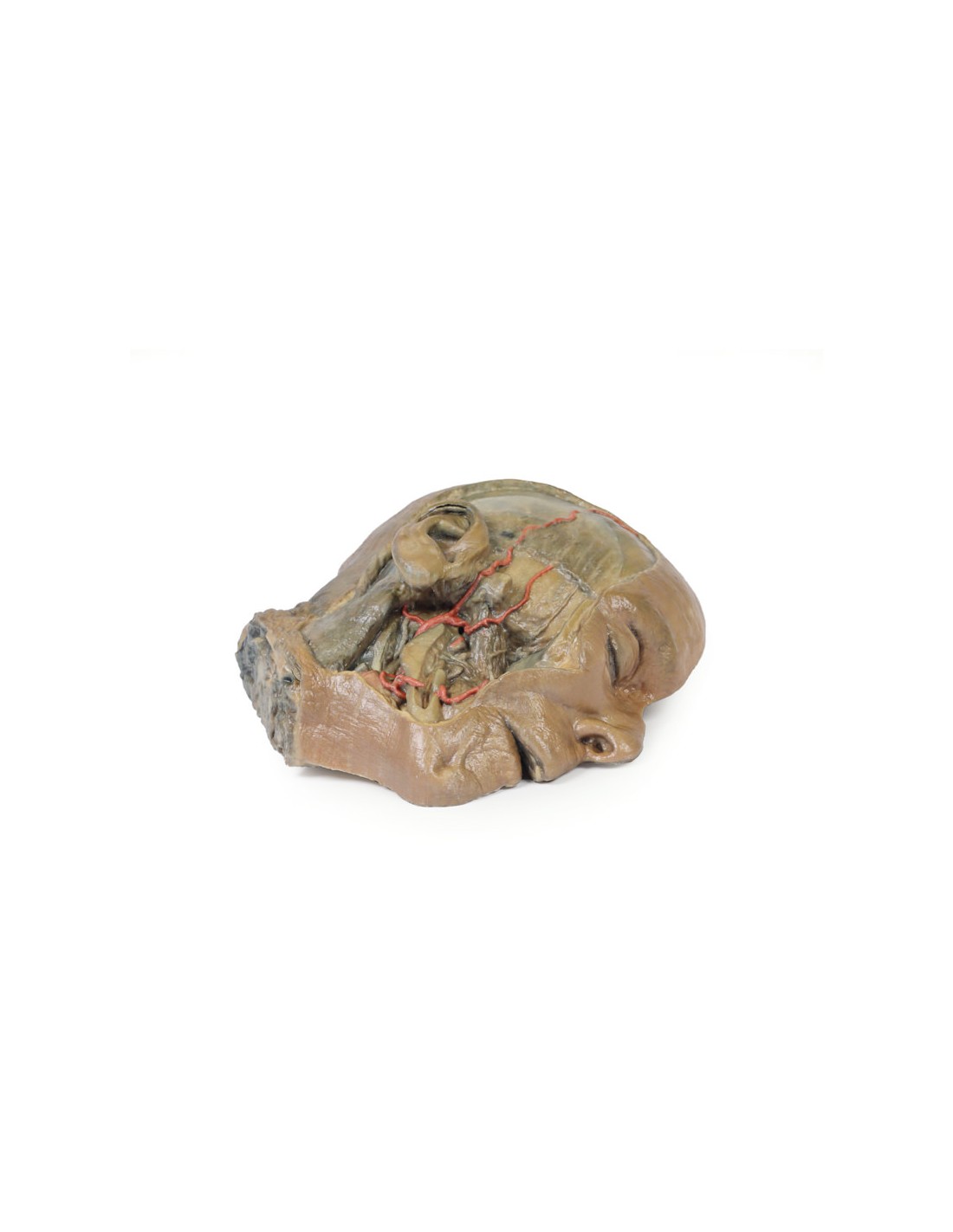
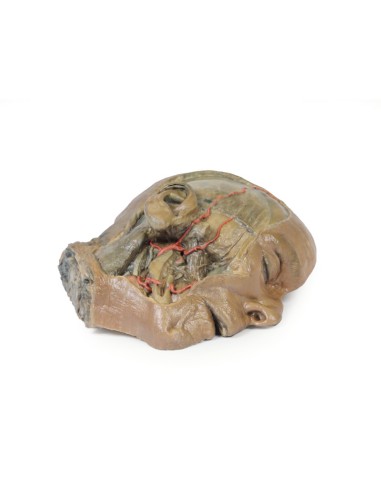
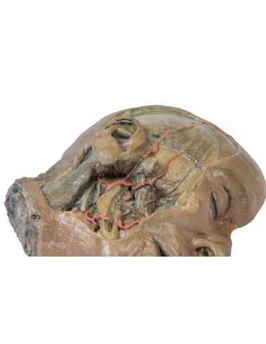
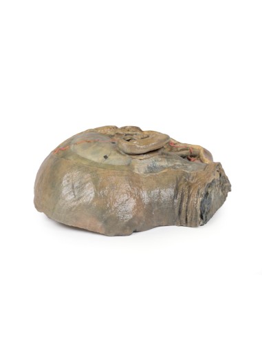
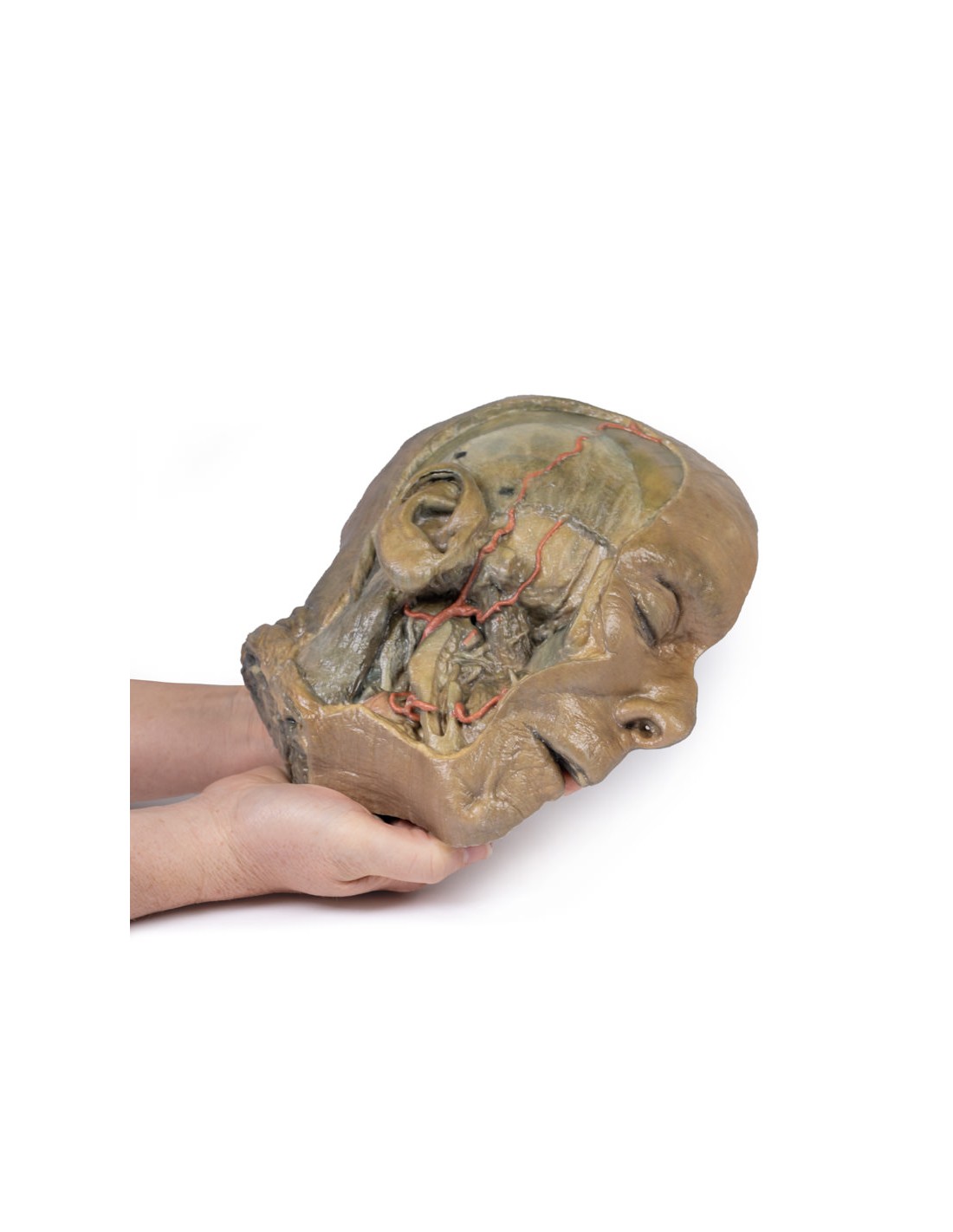
Sagittal section of the head with infratemporal fossa dissection - Erler Zimmer 3D anatomy Series MP1104
erler zimmer
EZ-MP1104
€2,422.31
Tax included
Made in ultra-high resolution 3D printing in full color.
Sagittal section of the head with infratemporal fossa dissection - Erler Zimmer 3D anatomy Series MP1104
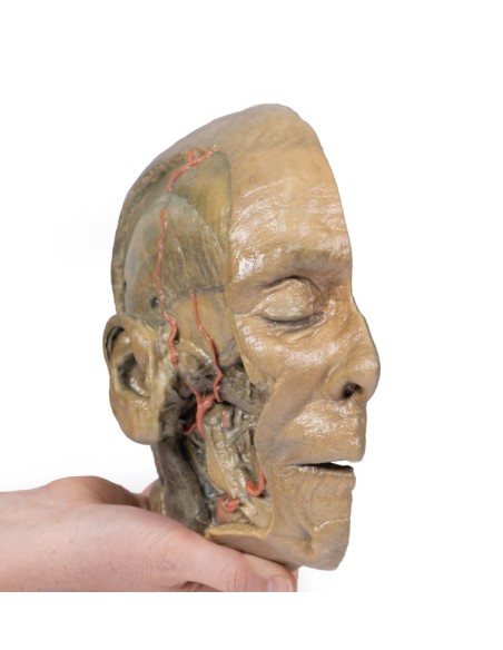
This model is part of the exclusive Monash 3D anatomy series, a comprehensive series of human dissections reproduced with ultra-high resolution color 3D printing.
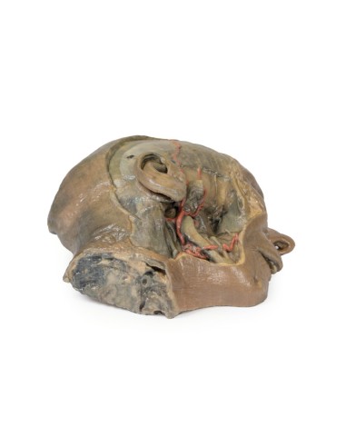
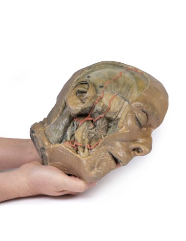
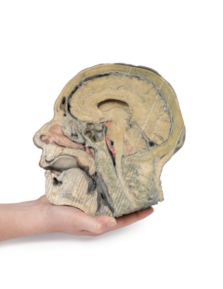
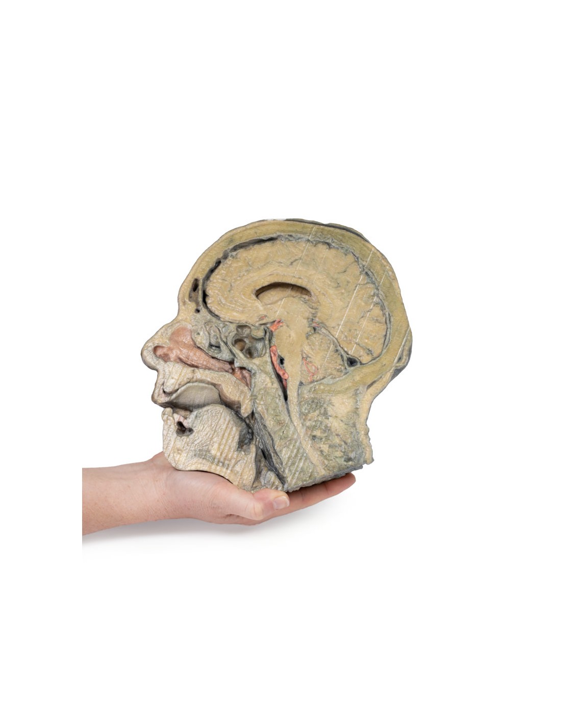
This 3D model provides a combined mediosagittal section through the head and upper neck coupled with a deep dissection in the infratemporal fossa region and a superficial scalp dissection.
In the preserved mediosagittal section there is preservation of the endocranial contents, nasal and oral cavities, and pharynx at the level of the laryngeal cartilages. The nasal cavity is preserved almost intact except for a small window excised in the middle nasal concha to expose the ethmoidal air cells. A very large sphenoidal sinus exists in the individual just superior to the torus of the ear tube in the nasopharynx. The oral cavity and laryngopharynx are not dissected, with the larynx being preserved only distally at the level of the arytenoid cartilages and not including a clear set of vocal cords.
Within the endocranial cavity, the dissected brain is slightly out of the medioagittal plane, so that neither the superior sagittal sinus nor the third ventricle are clearly defined, but the lateral ventricle is open and part of the fourth ventricle is preserved between the bridge and the cerebellum. The gyrus and sulcus of the brain are not well separated, but the cingulate gyrus and corpus callosum can be separated. Cross sections of the optic tract, pituitary gland, superior and inferior colliculi, superior cerebellar peduncle, and the transition between the medulla oblongata and spinal cord are visible. The cerebellar tentorium and transverse confluence/sinus is located between the cerebellar hemisphere and occipital lobe. Small portions of the posterior inferior cerebellar artery, vertebral arteries, basilar artery,
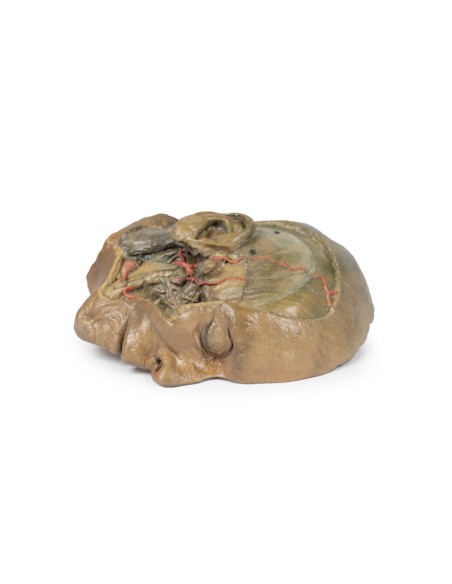
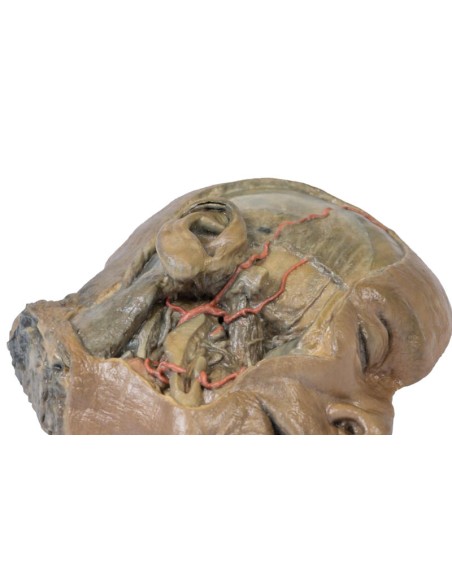
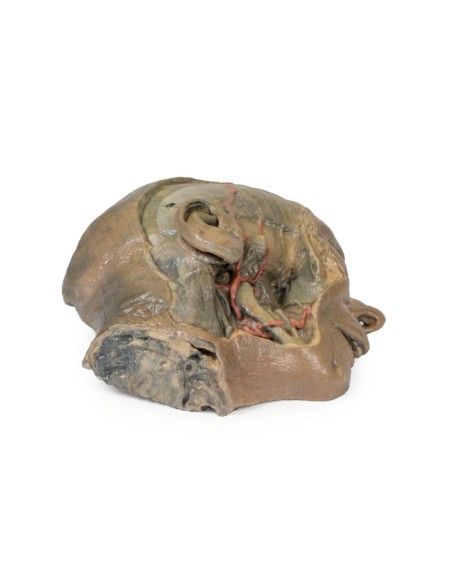
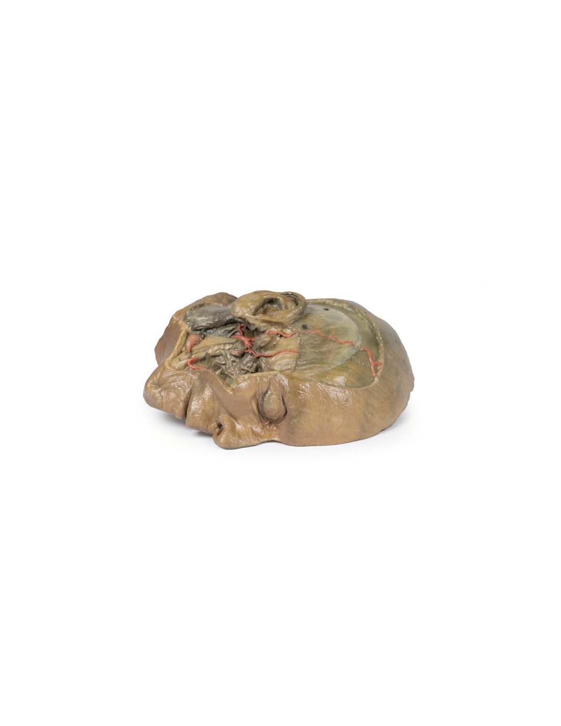
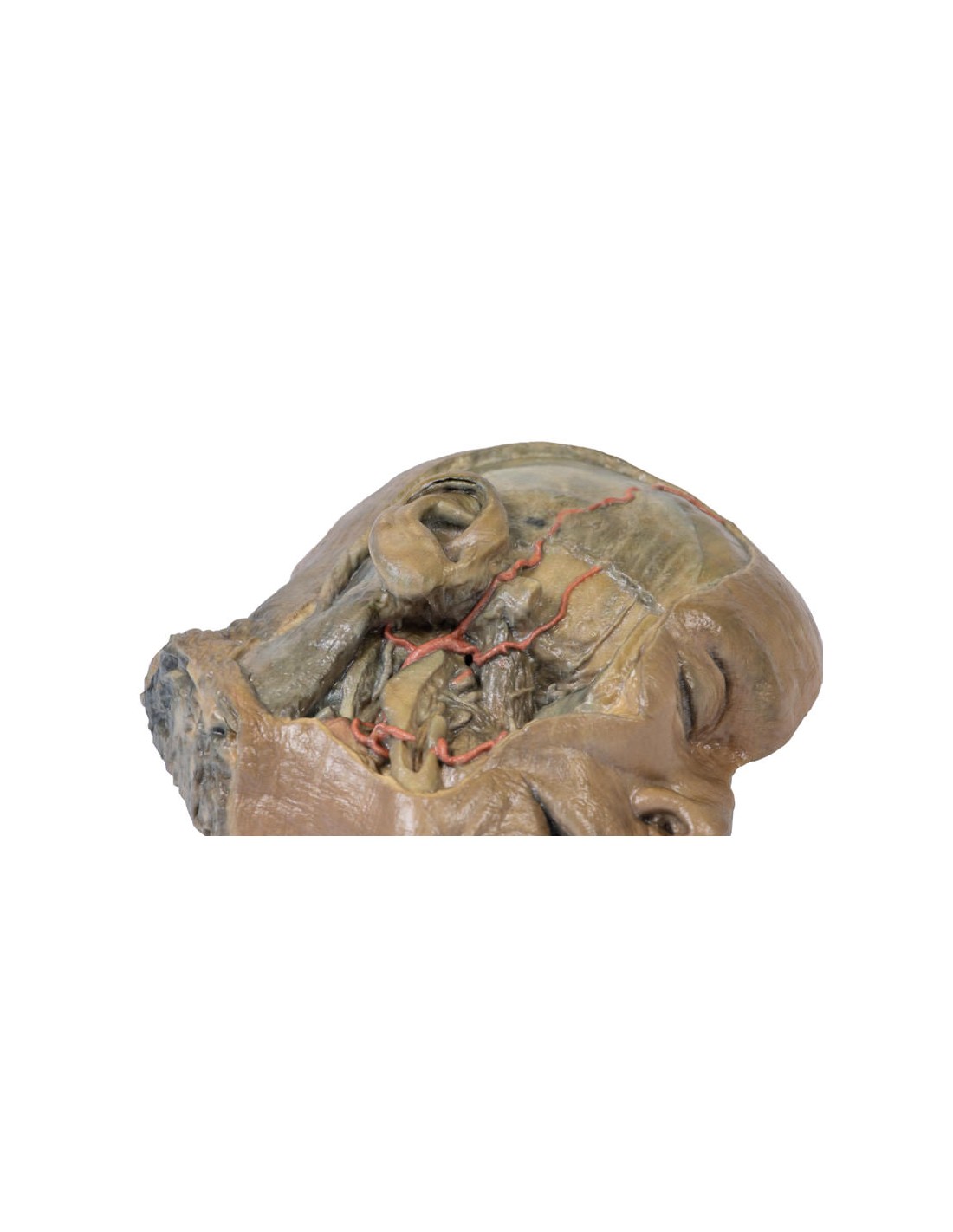
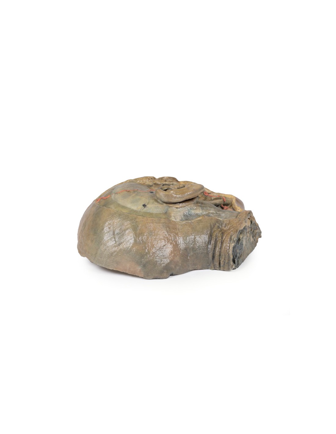
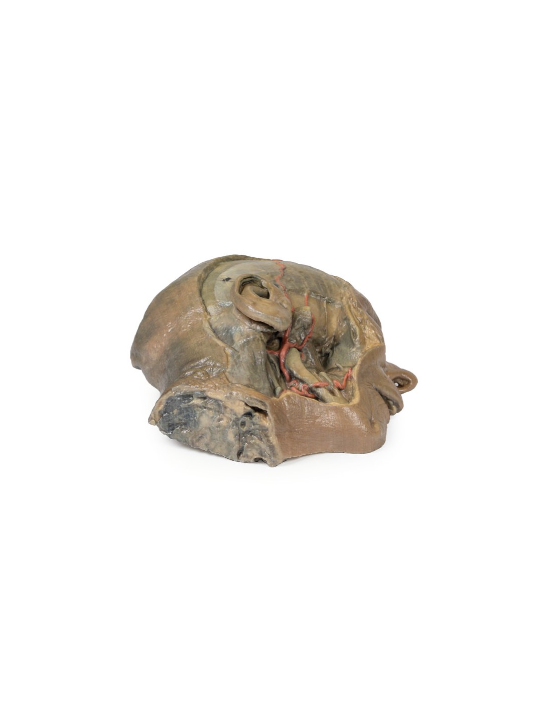
On the opposite side of the model, superficial and deep dissection opened a wide window into the anatomy of the lateral scalp and infratemporal fossa. Across the scalp there is a well-preserved posterior auricular nerve and a superficial temporal artery highlighted on the superficial surface of the temporalis muscle. Anteriorly, the temporalis was dissected to expose the deep temporal arteries originating on the other side of the maxillary artery.
The deep level of dissection exposed parts of the infratemporal fossa (through partial removal of the ramus and mandibular body) and dissection of the retromandibular tissues. At the lower margin of the dissection window, the cut edge of the retromandibular vein lies adjacent to the submandibular gland and the ascending path of the facial artery as it crosses toward the corner of the mouth. Just superior to the cut retromandibular vein is the posterior belly of the digastric muscle, overlying a small exposure of the deeper internal jugular vein.
Just behind the retained ascending branch of the mandible are the external carotid artery and the occipital artery (which run in parallel before passing posteriorly). Tracing the external carotid artery superiorly, the posterior auricular artery, superficial temporal artery, and maxillary artery are visible. The maxillary artery passes deep into the lateral pterygoid muscle and infratemporal fossa, reappearing superior to the lateral pterygoid as it passes into the pterygomaxillary fissure. Along its course, it gives rise to the posterior deep temporal artery, the inferior alveolar artery (which is exposed in the dissected mandibular body), the anterior deep temporal artery, and the posterior superior alveolar artery. Finally, the inferior alveolar nerve can be seen running inside the open mandibular body, and the lingual nerve resting on the medial pterygoid. The buccinator muscle is also retained, with the distal part of the parotid duct preserved as it enters the muscle toward the oral mucosa.
What advantages does the Monash University anatomical dissection collection offer over plastic models or plastinated human specimens?
- Each body replica has been carefully created from selected patient X-ray data or human cadaver specimens selected by a highly trained team of anatomists at the Monash University Center for Human Anatomy Education to illustrate a range of clinically important areas of anatomy with a quality and fidelity that cannot be achieved with conventional anatomical models-this is real anatomy, not stylized anatomy.
- Each body replica has been rigorously checked by a team of highly trained anatomists at the Center for Human Anatomy Education, Monash University, to ensure the anatomical accuracy of the final product.
- The body replicas are not real human tissue and therefore not subject to any barriers of transportation, import, or use in educational facilities that do not hold an anatomy license. The Monash 3D Anatomy dissection series avoids these and other ethical issues that are raised when dealing with plastinated human remains.
No reviews
Tap to zoom