Your cart
There are no more items in your cart


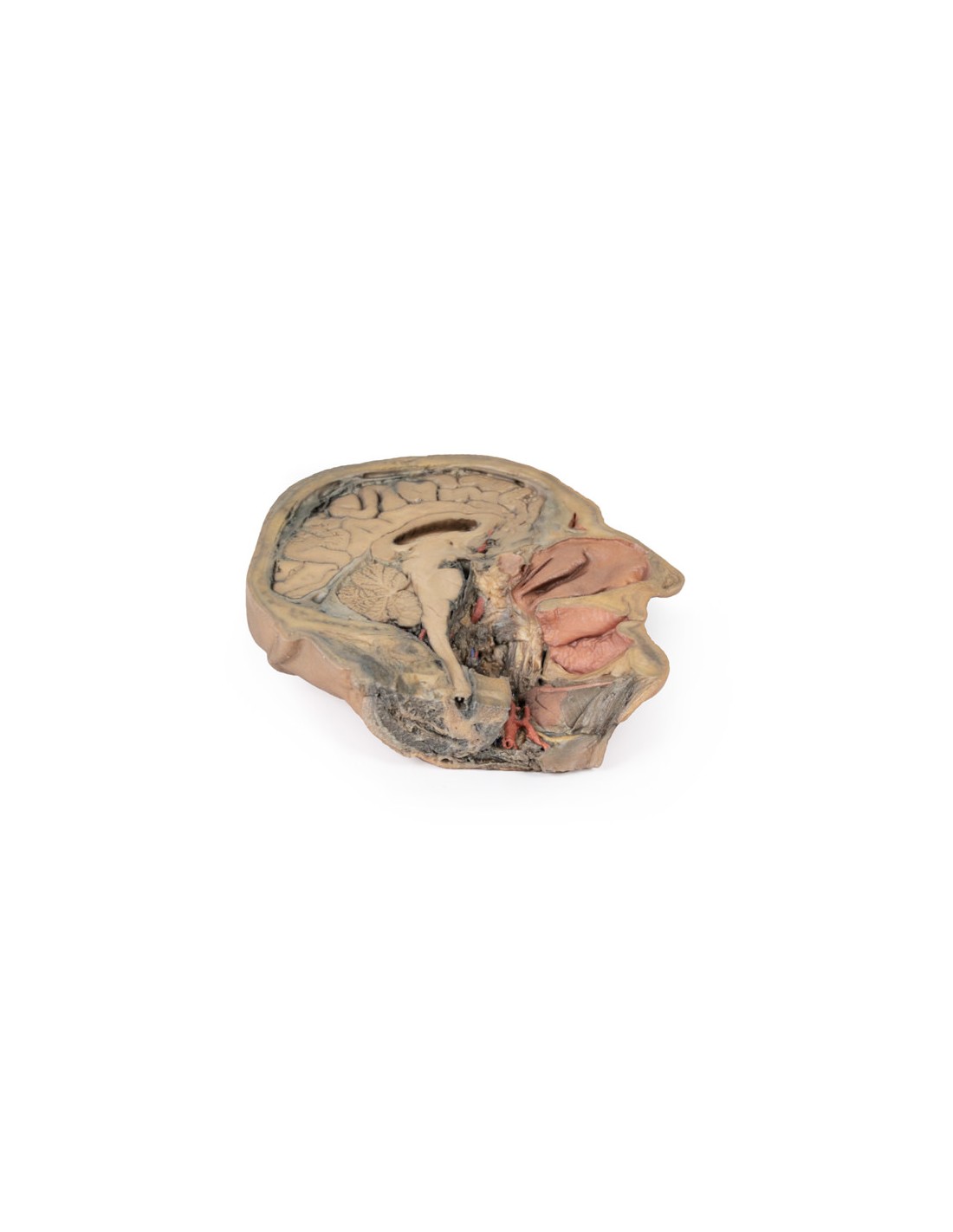
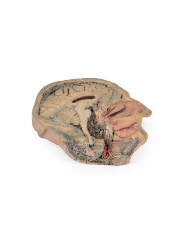
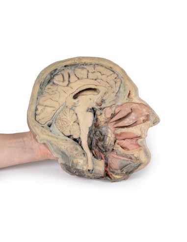
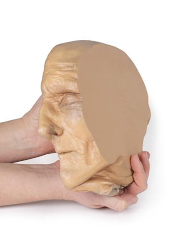
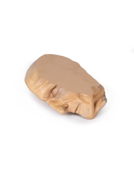
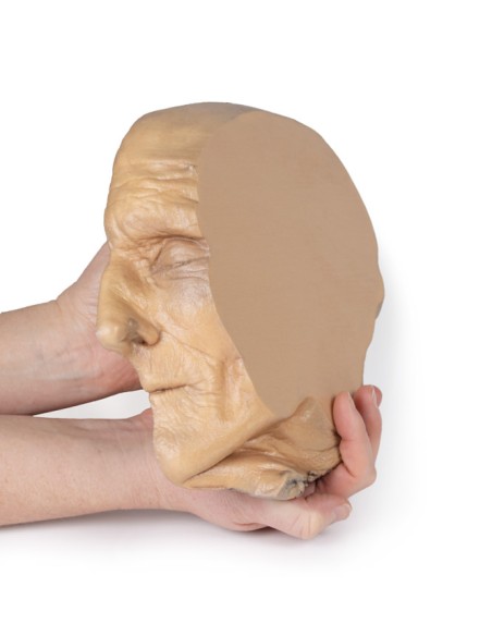
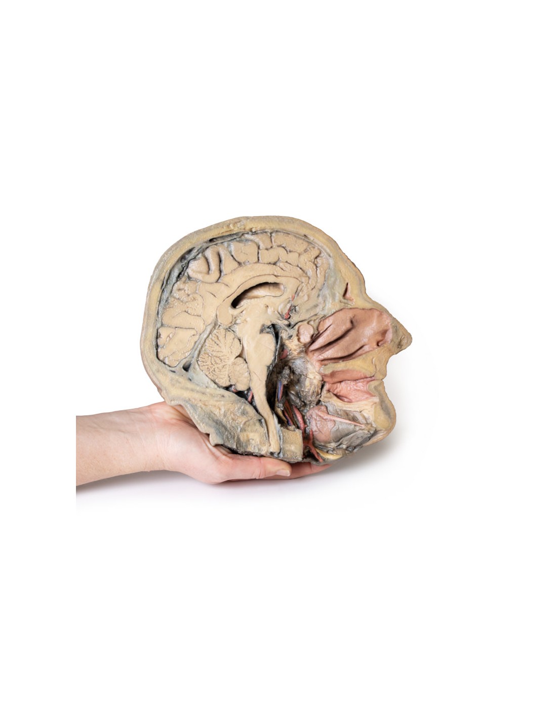
Sagittal section of the head with deep dissection - Erler Zimmer 3D anatomy Series MP1105
erler zimmer
EZ-MP1105
€1,898.93
Tax included
Made in ultra-high resolution 3D printing in full color.
Sagittal section of the head with infratemporal fossa dissection - Erler Zimmer 3D anatomy Series MP1104
This model is part of the exclusive Monash 3D anatomy series, a comprehensive series of human dissections reproduced with ultra-high resolution color 3D printing.
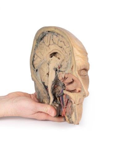
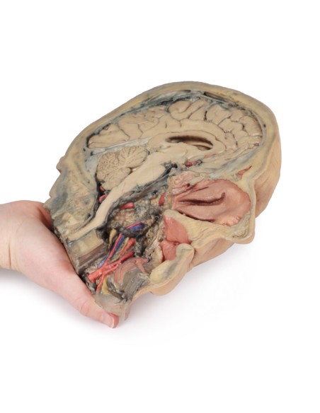
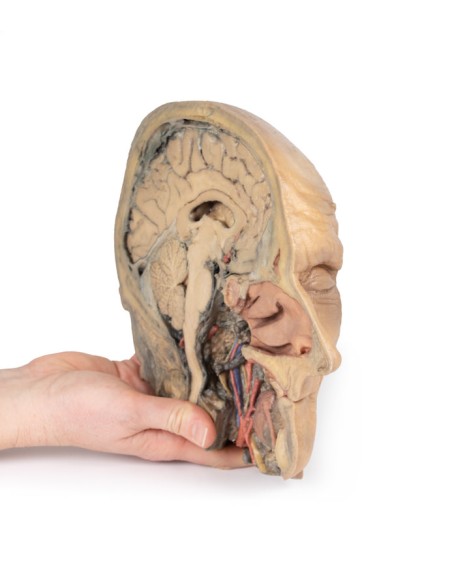
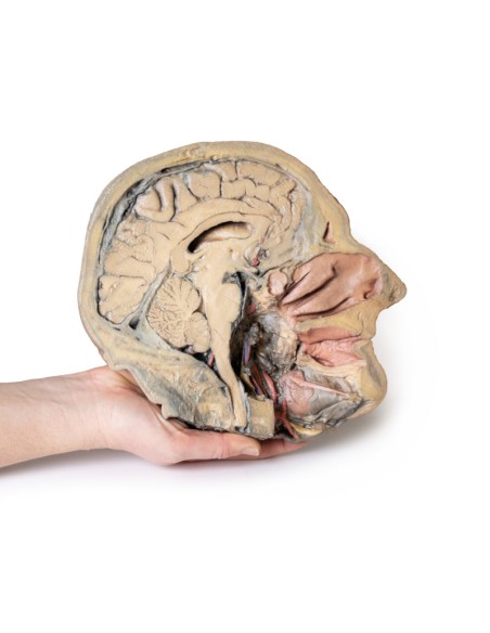
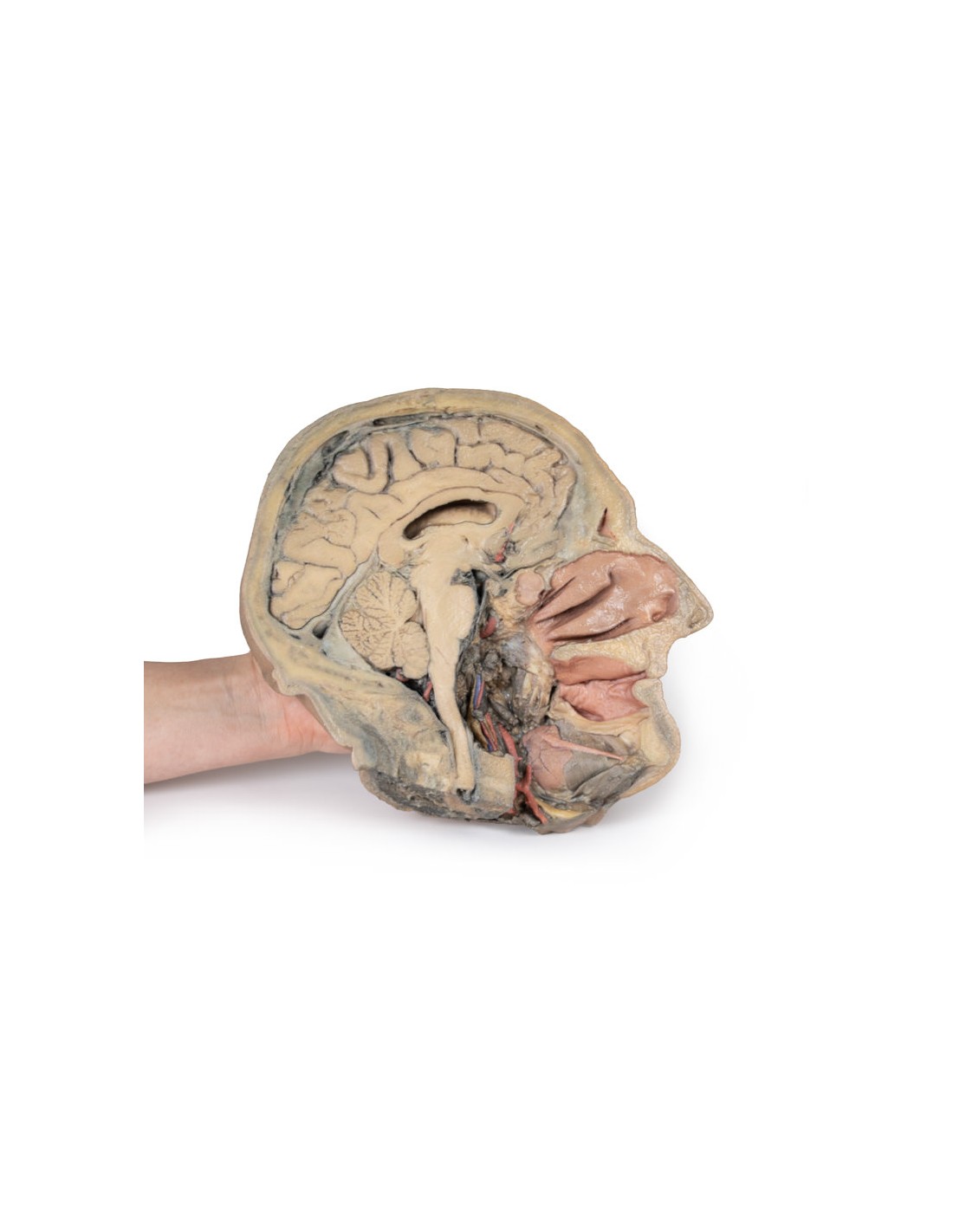
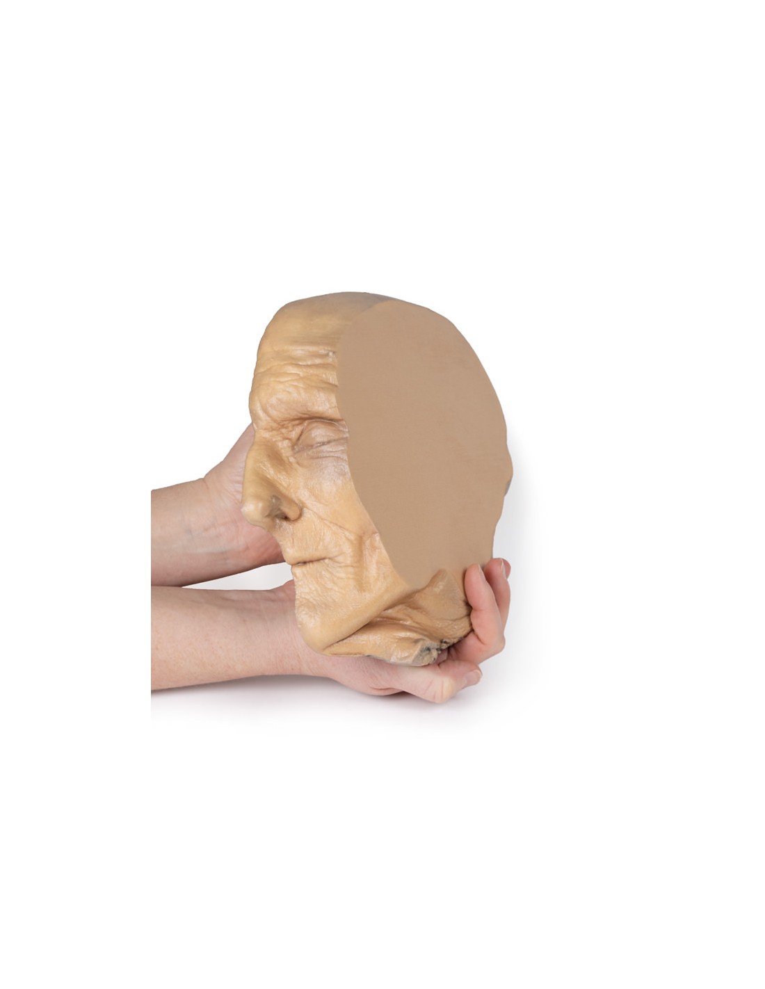
This 3D model combines a mediosagittal section of the head with preservation of the anatomy of the brain and cranial cavity, with a unique deep dissection of the pharyngeal region by removal of the basicranial bone and anterior parts of the atlas and axis. Since the opposite side is not dissected, it was digitally removed from the model.
Within the endocranial cavity, preservation of the dura preserves the superior sagittal sinus for most of its course from anterior to posterior, reaching the confluence of sinuses visible in cross section. Both tentorium cerebelli and falx cerebelli are preserved. The brain is well reserved with retention of the cingulate gyrus and sulcus and removal of the septum pellucidum inferior to the corpus callosum providing a view into the lateral ventricle (with retention of the interventricular foramen at the inferior margin of the septum). The structures of the diencephalon and midbrain (epithalamus, colliculi, mammillary body, infundibulum) are all appreciable in cross section, as are the cerebellar hemisphere and fourth ventricle. Small views of the inferior anterior and posterior cerebellar arteries are visible (and in false color).
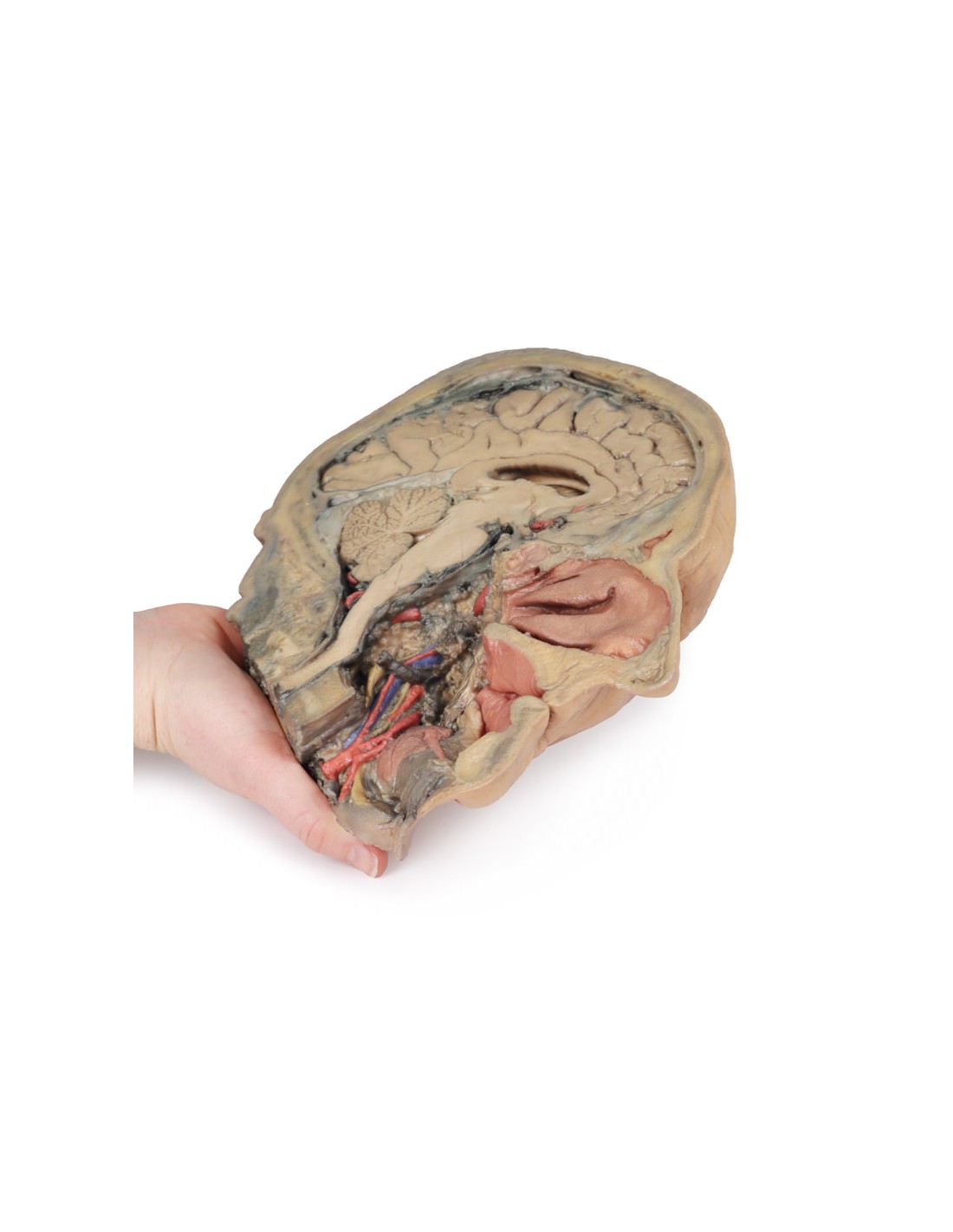
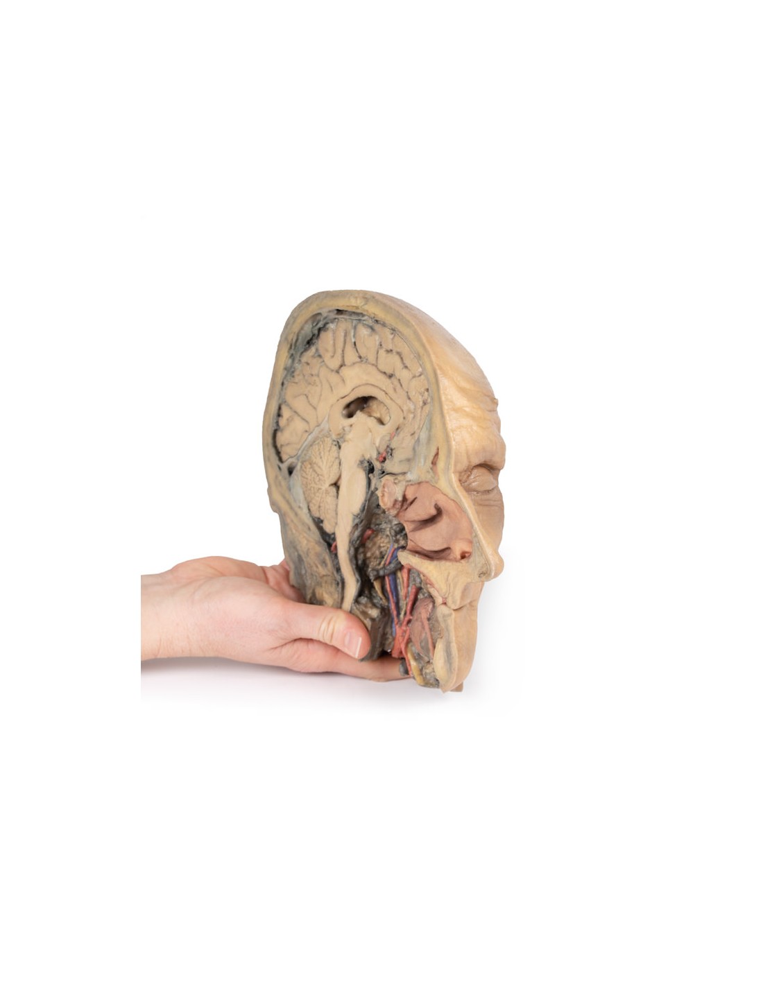
Outside the endocranium, removal of parts of the occipital, temporal, and sphenoid bones (along with the atlas and axis) was coupled with removal of the pharyngeal constrictors, carotid sheath, and oral mucosa to demonstrate a single view of several key neurovascular and glandular structures. Within the area of the removed tissue is a partial exposure of the right common carotid artery within the dissected petrous portion of the temporalis, as well as a partial exposure of the left vertebral artery through the interruption of the occipital and dural lining.
The medial and lateral pterygoids are exposed near the posterior margin of the largely intact nasal cavity. Between the exposed dura and medulla and the pterygoids (and trapped deep within the dissected and reflexed stylohyoid muscle) the dissected carotid sheath has exposed the internal jugular vein, vagus nerve, internal carotid artery (with predominantly ascending pharyngeal artery from the external carotid artery), and sympathetic trunk (with superior cervical ganglion and internal carotid nerve). Immediately anterior to this bundle of neurovascular structures is the external carotid artery, which gives rise to the ascending pharyngeal artery, a common trunk for the lingual and facial arteries, and then continues superiorly out of the plane of dissection. The submandibular gland can be seen resting on the mylohyoid muscle near the lingual artery (which passes deeper than the gland), with the ductus passing toward the genu of the mandible and the origin of the reflex genioglossus muscle. At the lower edge of the specimen, the reflex margin of the dissected tongue, the hypoglossal nerve can be seen deep down to the lingual artery.
What advantages does the Monash University anatomical dissection collection offer over plastic models or plastinated human specimens?
- Each body replica has been carefully created from selected patient X-ray data or human cadaver specimens selected by a highly trained team of anatomists at the Monash University Center for Human Anatomy Education to illustrate a range of clinically important areas of anatomy with a quality and fidelity that cannot be achieved with conventional anatomical models-this is real anatomy, not stylized anatomy.
- Each body replica has been rigorously checked by a team of highly trained anatomists at the Center for Human Anatomy Education, Monash University, to ensure the anatomical accuracy of the final product.
- The body replicas are not real human tissue and therefore not subject to any barriers of transportation, import, or use in educational facilities that do not hold an anatomy license. The Monash 3D Anatomy dissection series avoids these and other ethical issues that are raised when dealing with plastinated human remains.
No reviews
Tap to zoom