Your cart
There are no more items in your cart


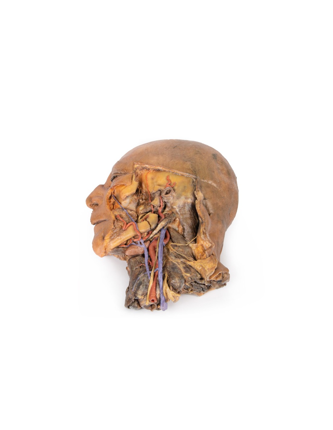
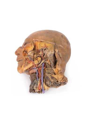
Sagittal section of head and neck with intratemporal fossa and carotid sheath - Erler Zimmer 3D anatomy Series MP1111
erler zimmer
EZ-MP1111
€2,535.04
Tax included
Made in ultra-high resolution 3D printing in full color.
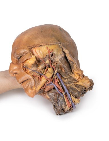
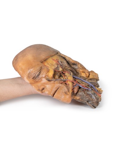
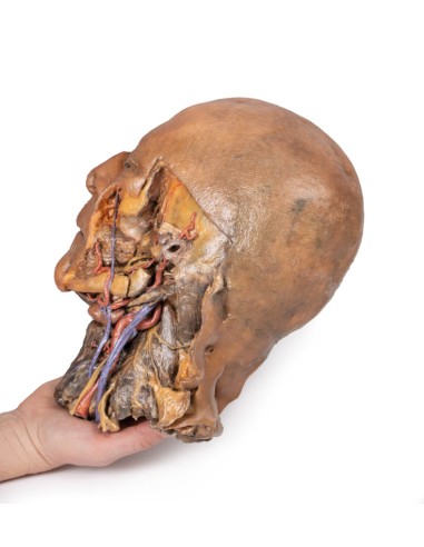
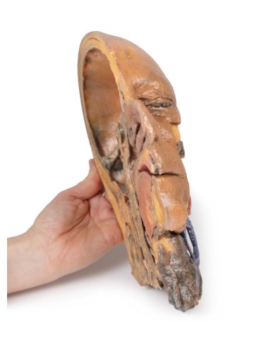
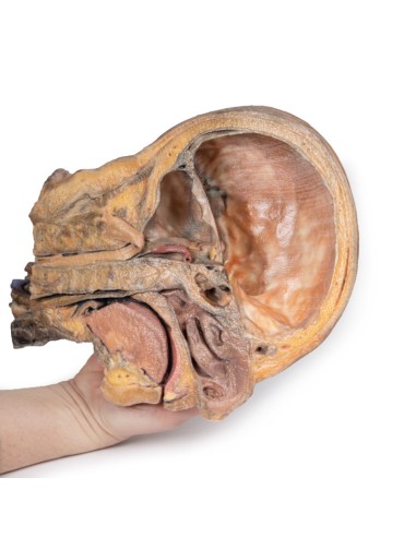
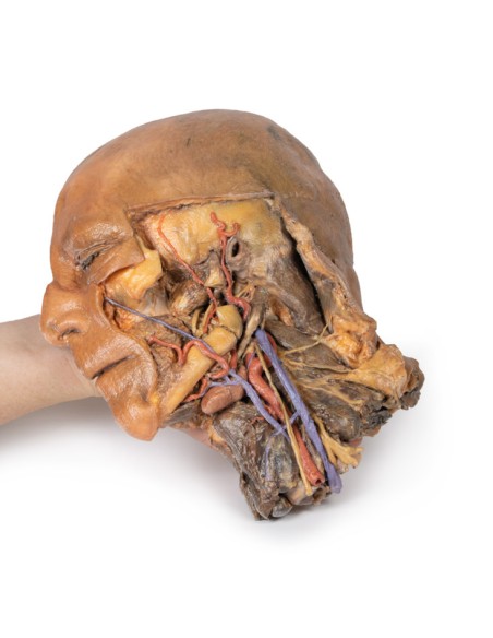
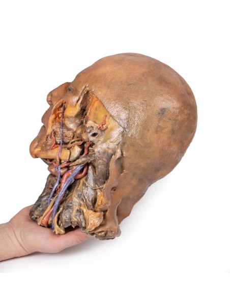
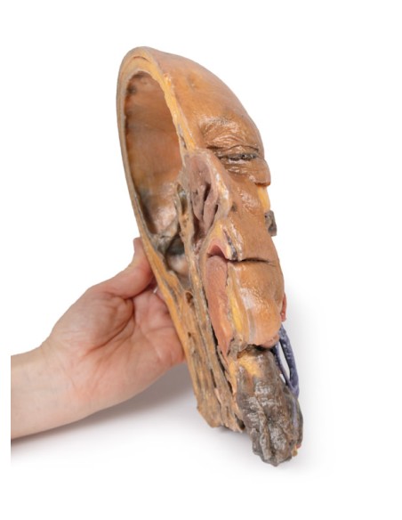
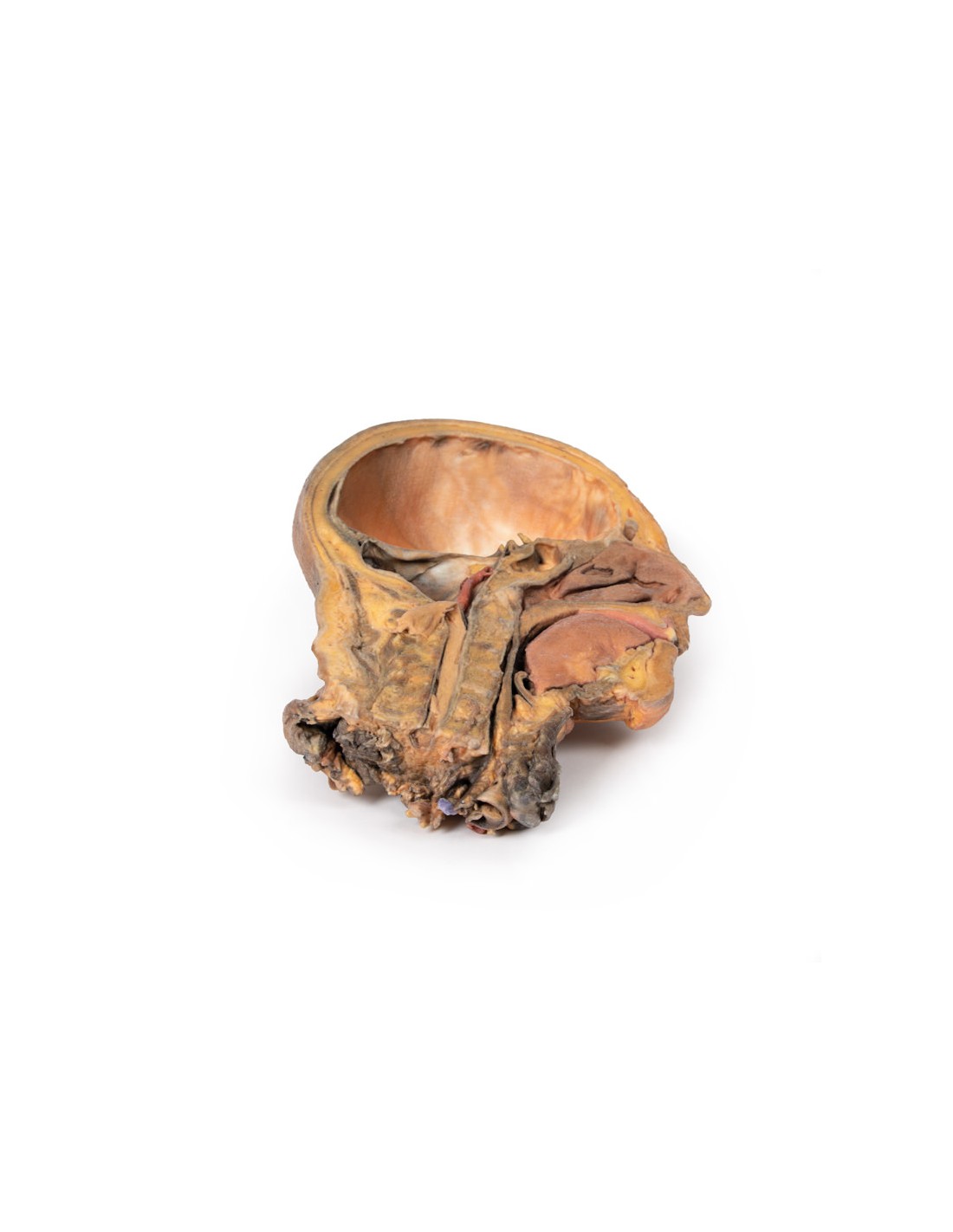
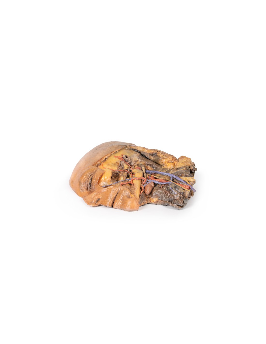
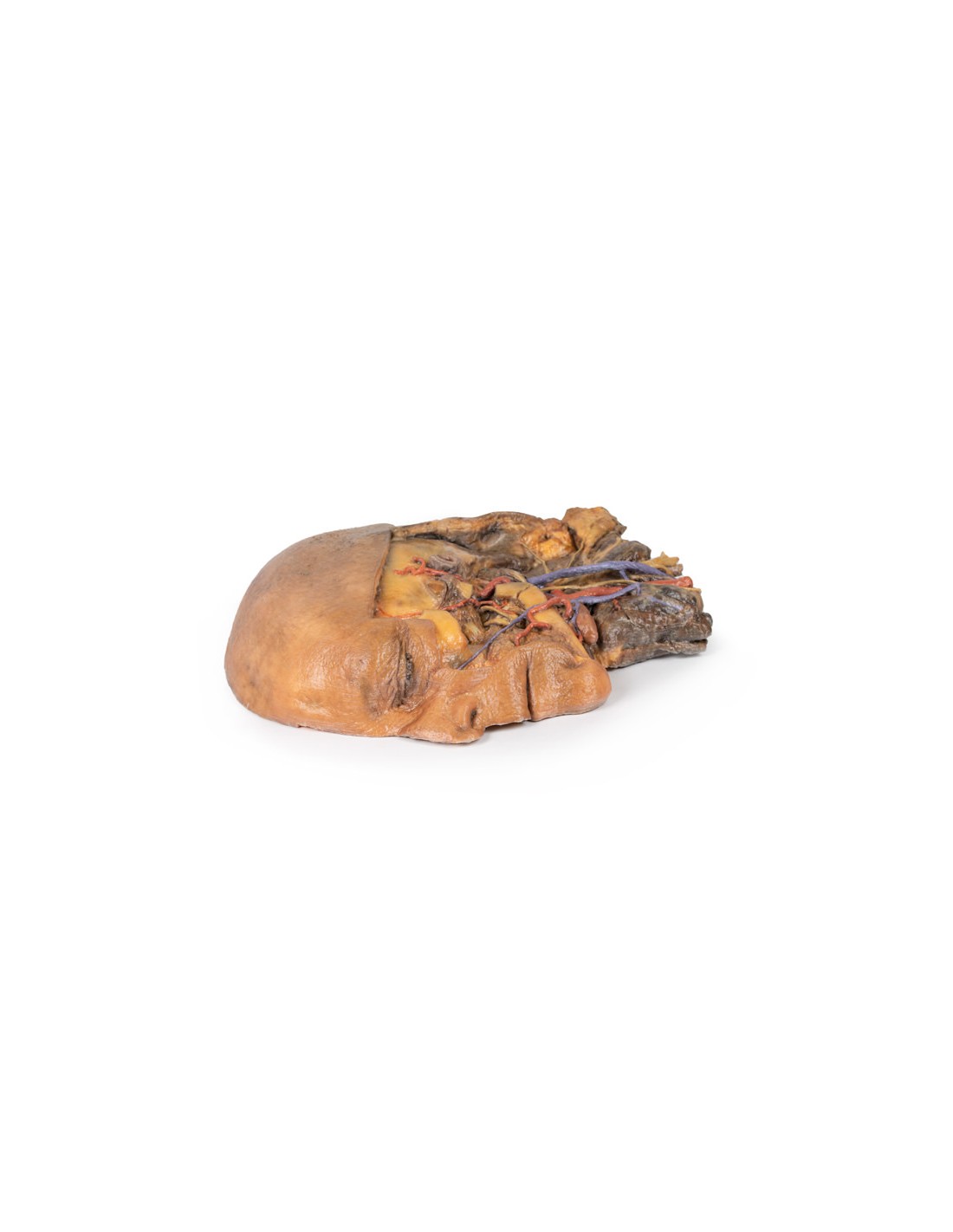
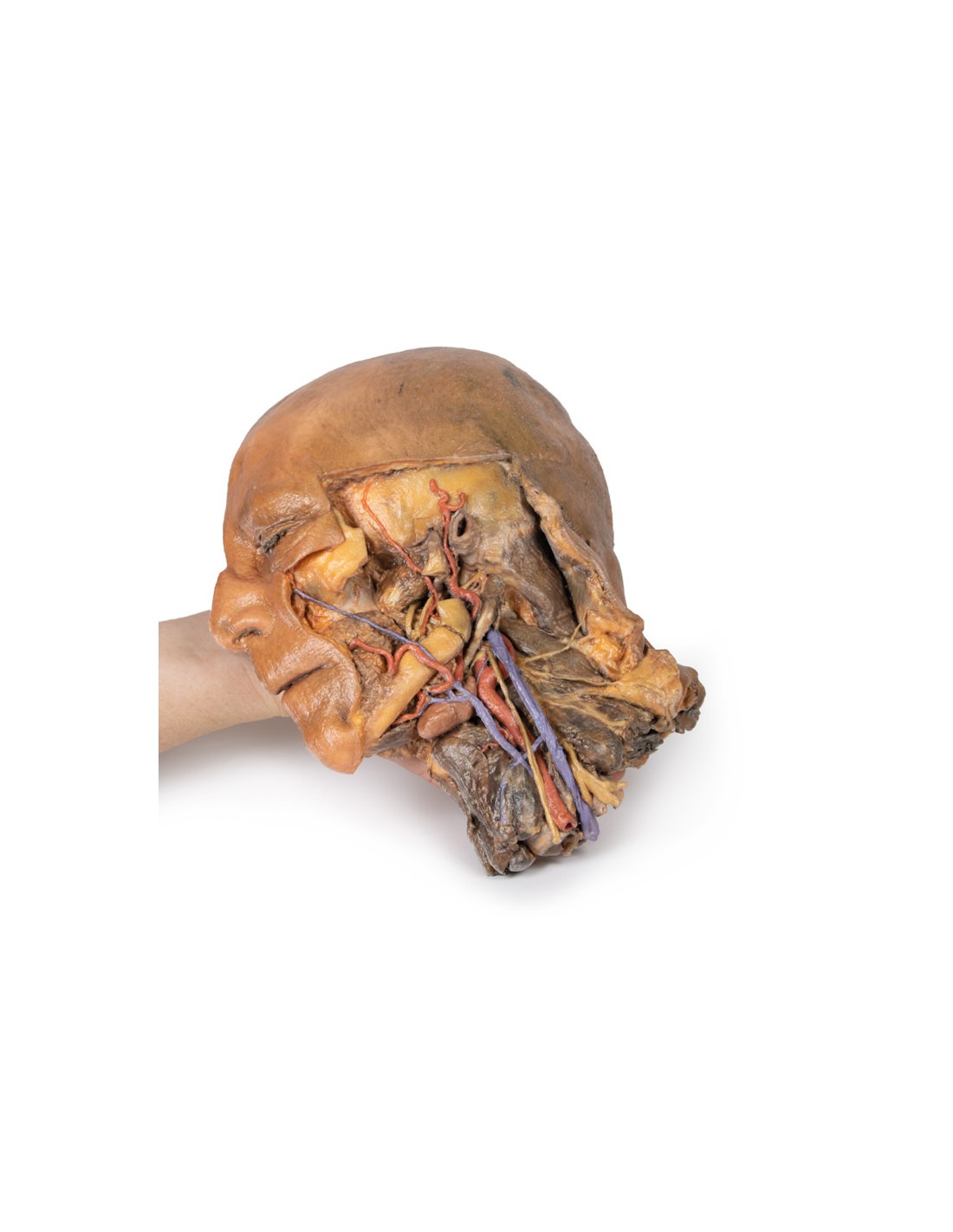
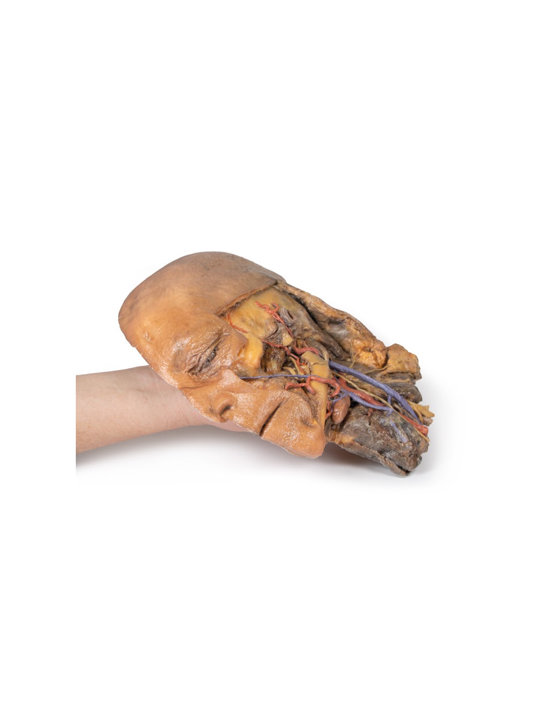
Sagittal section of the head and neck with intratemporal fossa and carotid sheath dissection - Erler Zimmer 3D anatomy Series MP1111
This model is part of the exclusive Monash 3D anatomy series, a comprehensive series of human dissections reproduced with ultra-high resolution color 3D printing.
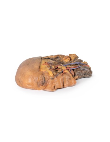
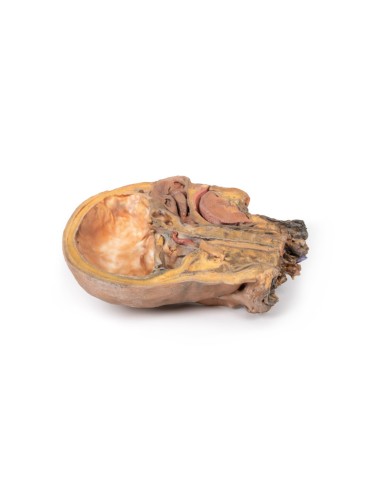
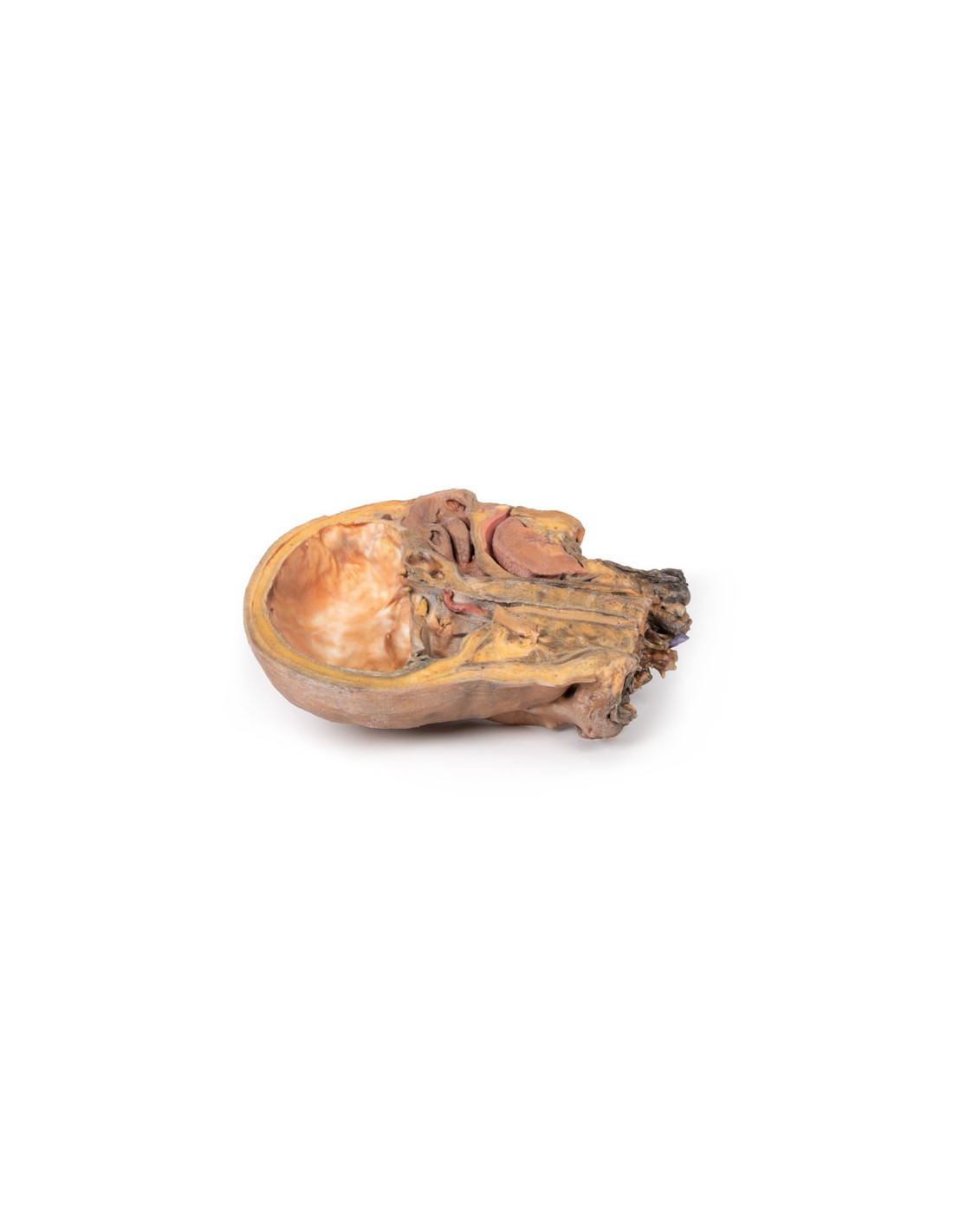
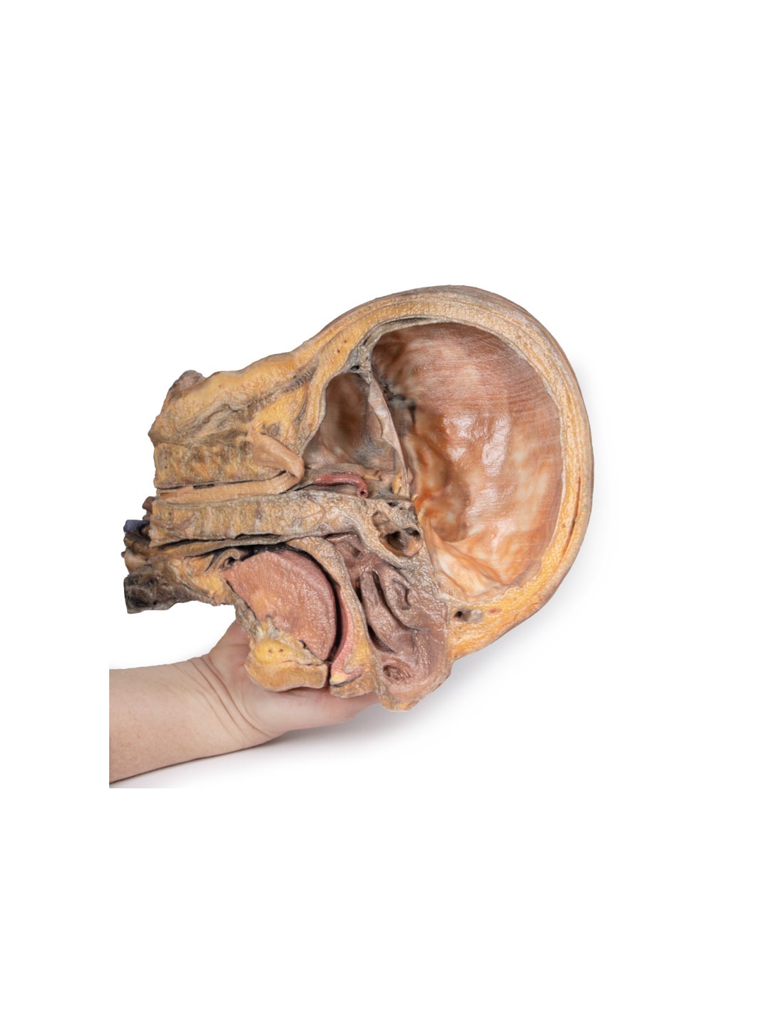
This 3D model provides a complementary specimen to the H 11 and H 12 head and neck specimens by providing a perspective of the endocranial cavity without the brain and a lateral dissection including neck anatomy.
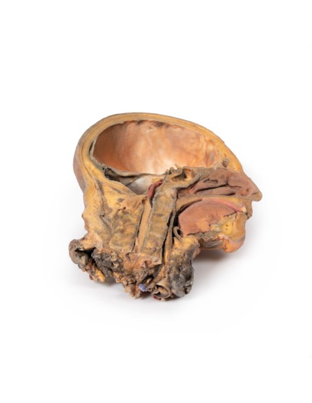
In the mediosagittal section, removal of the brain (and reflection of the medulla inferiorly) provides a complete view of the dura mater lining the endocranial cavity, including the cerebral tentorium extending from the transverse sinus to the attachment to the clinoid process of the sphenoid. A number of cranial nerves can be seen, including the optic (CN II), oculomotor (CN III), trigeminal (CN V), abducens (CN VI), and combined facial (CN VII) and vestibulocochlear (CN VIII) nerves. piercing the dura. The pituitary gland can be seen in cross section within the sella turcica, and the left vertebral artery can be seen ascending into the posterior cranial fossa.
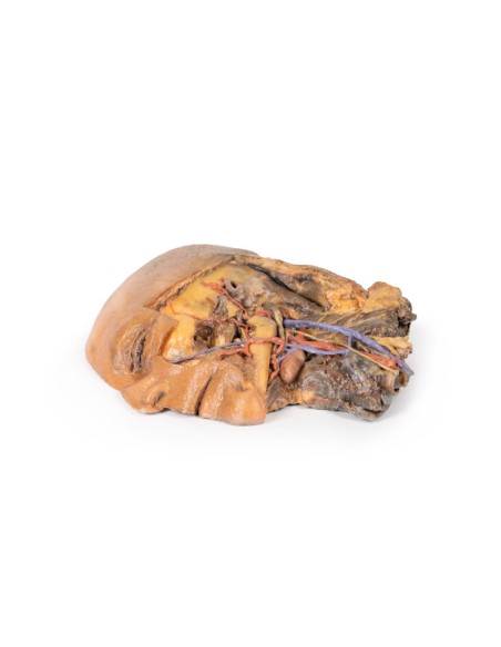
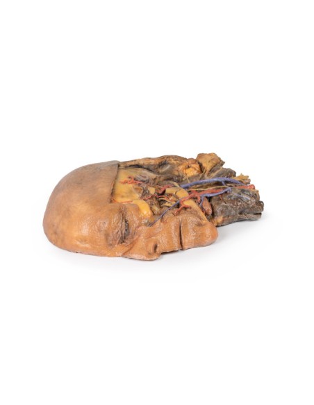
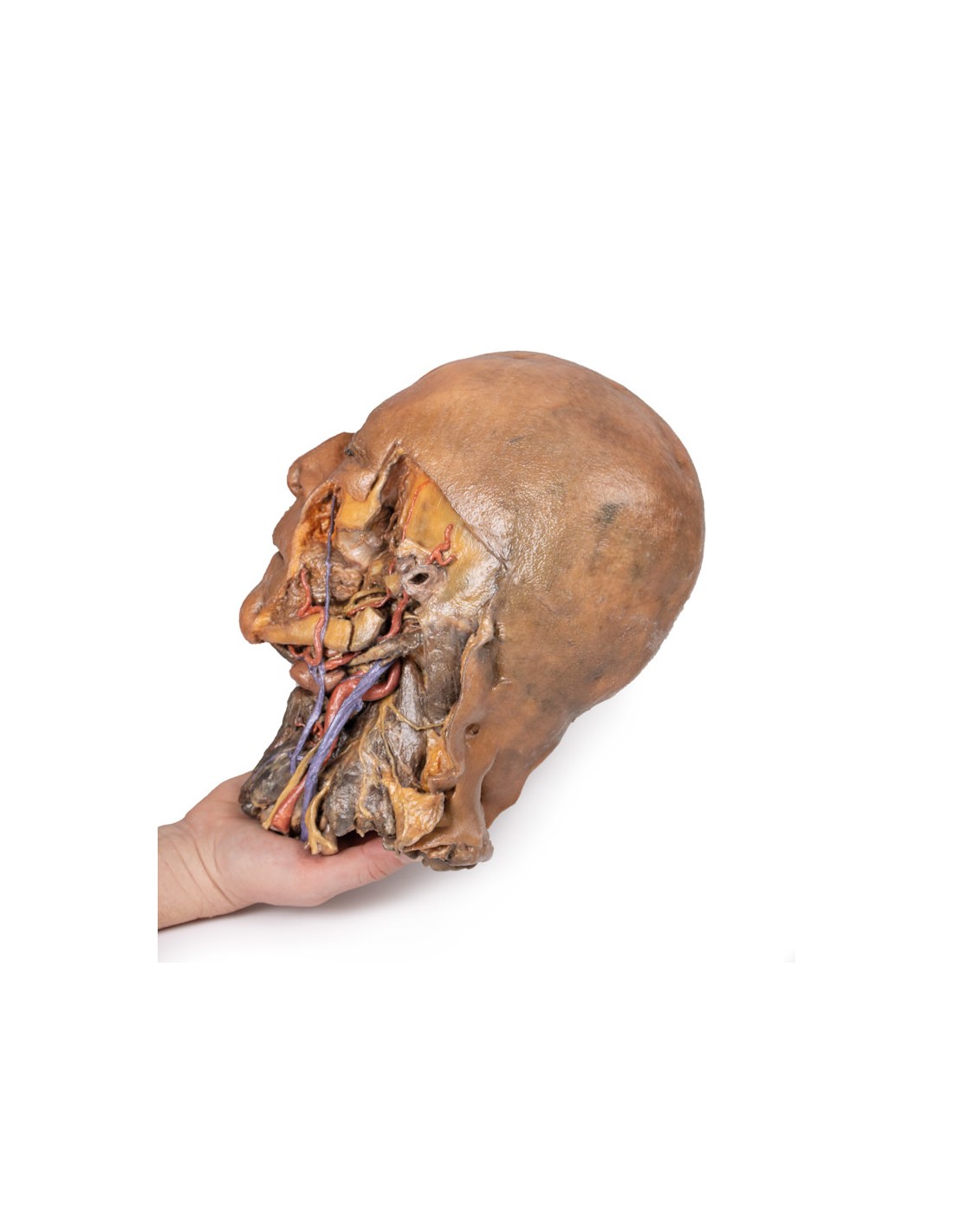
Lateral dissection to the face preserved some superficial structures while simultaneously exposing the anatomy within the infratemporal fossa. The facial vein and facial artery were preserved but are dissected away from any superficial fascia or facial expression muscles and are located through the body of the mandible and buccinator muscle. Most of the ascending branch of the mandible and zygomatic arch has been removed to demonstrate some of the anatomy of the infratemporal fossa, including the inferior alveolar artery and lingual nerve (resting on the medial pterygoid), the posterior deep temporal artery (resting on the lateral pterygoid), and the articulation of the mandibular condyle with the glenoid fossa. The terminal part of the external carotid artery is visible,
Posterior to the infratemporal region, the facial nerve (CN VII) can be seen briefly adjacent to the posterior belly of the digastic muscle. The posterior belly of the digastric angles slopes superficially to obscure the internal and external carotid arteries and the internal jugular vein, which have been dissected by the carotid sheath (next to the vagus nerve [CN X]). At the angle of the mandible, and along the lower margin of the body, the hypoglossal nerve (CN XII) rests just adjacent to the central tendon of the digastric and external carotid arteries. Anteriorly, the facial artery is integrated into the submandibular gland before ascending through the mandibular body, where the lingual artery and anterior belly of the digastric can be seen.
In the neck region of the specimen, the hyoid bone is immediately deep relative to the submandibular gland and receives infrahyoid muscles just superficial to a robust thyroid gland. In the cut section of the dissection inferiorly, the underlying larynx can also be seen. Posterior to the carotid sheath structures, radiating cutaneous branches from the cervical plexus rest on the scalene muscles, and near the lower margin of the specimen the superior roots of the brachial plexus adjacent to the exposed internal jugular vein are preserved.
What advantages does the Monash University anatomical dissection collection offer over plastic models or plastinated human specimens?
- Each body replica has been carefully created from selected patient X-ray data or human cadaver specimens selected by a highly trained team of anatomists at the Monash University Center for Human Anatomy Education to illustrate a range of clinically important areas of anatomy with a quality and fidelity that cannot be achieved with conventional anatomical models-this is real anatomy, not stylized anatomy.
- Each body replica has been rigorously checked by a team of highly trained anatomists at the Center for Human Anatomy Education, Monash University, to ensure the anatomical accuracy of the final product.
- The body replicas are not real human tissue and therefore not subject to any barriers of transportation, import, or use in educational facilities that do not hold an anatomy license. The Monash 3D Anatomy dissection series avoids these and other ethical issues that are raised when dealing with plastinated human remains.
No reviews
Tap to zoom