Your cart
There are no more items in your cart


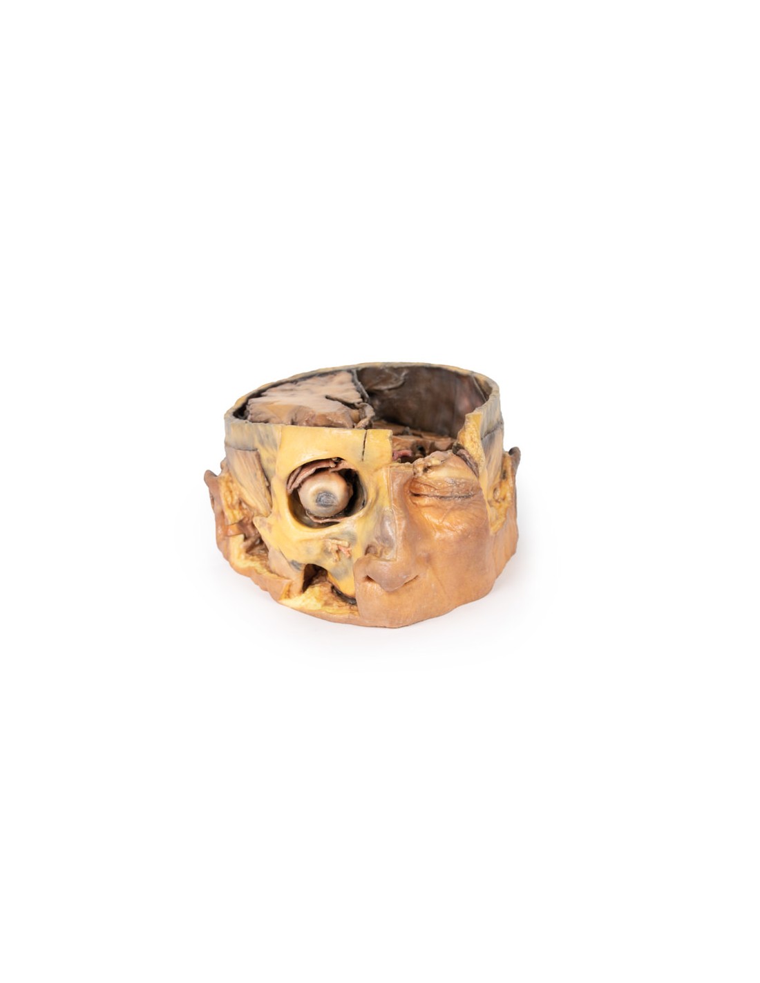
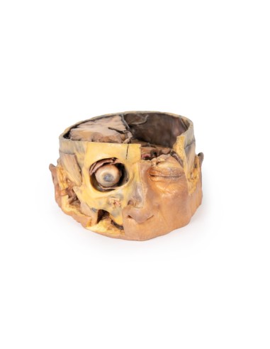
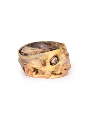
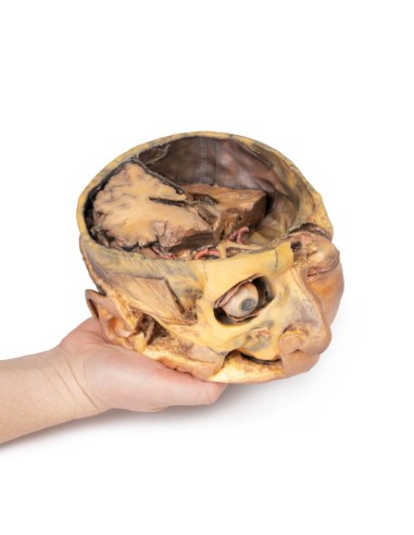
Cross section of the head - Erler Zimmer 3D anatomy Series MP1110
erler zimmer
EZ-MP1110
€2,712.18
Tax included
Made in ultra-high resolution 3D printing in full color.
Cross section of the head - Erler Zimmer 3D anatomy Series MP1110
This model is part of the exclusive Monash 3D anatomy series, a comprehensive series of human dissections reproduced with ultra-high resolution color 3D printing.
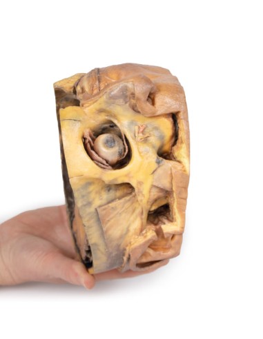
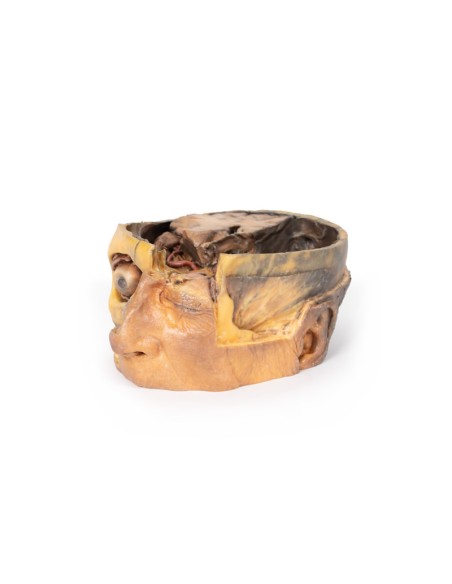
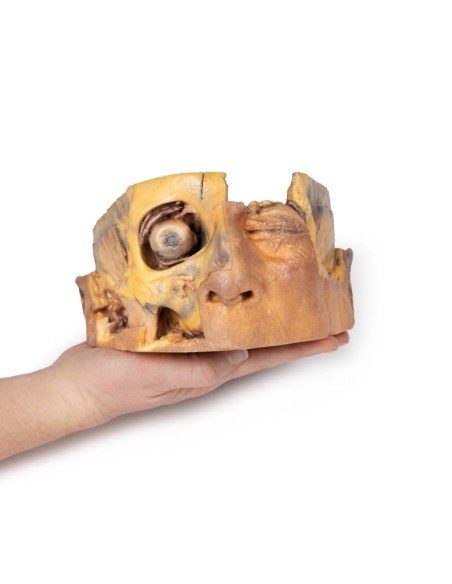
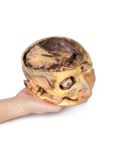
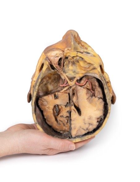
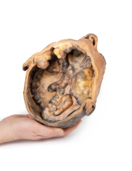
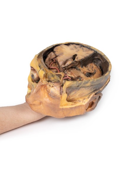
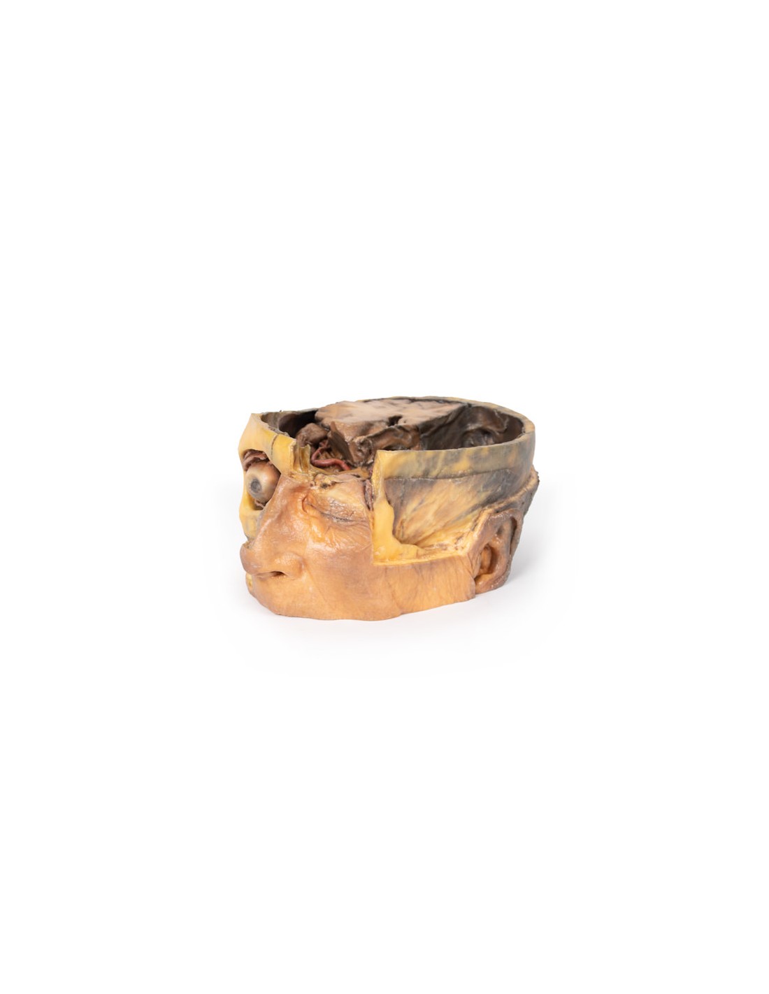
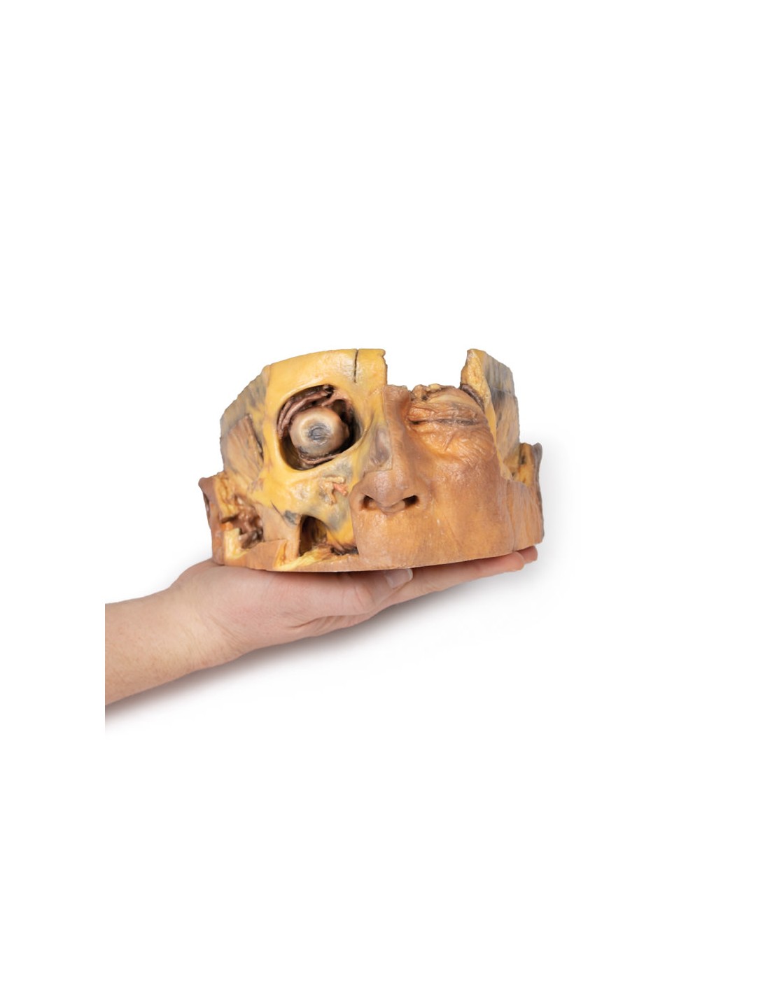
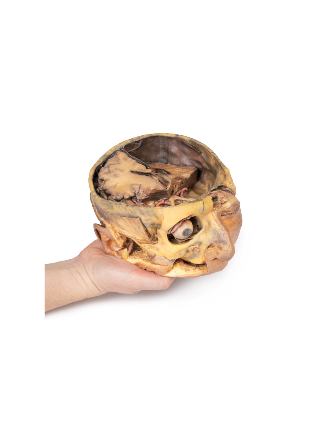
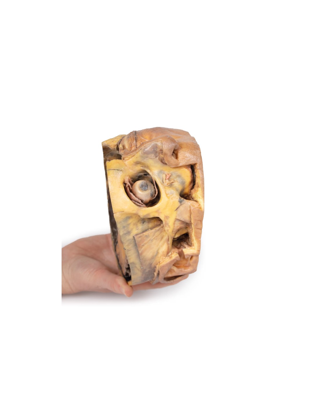
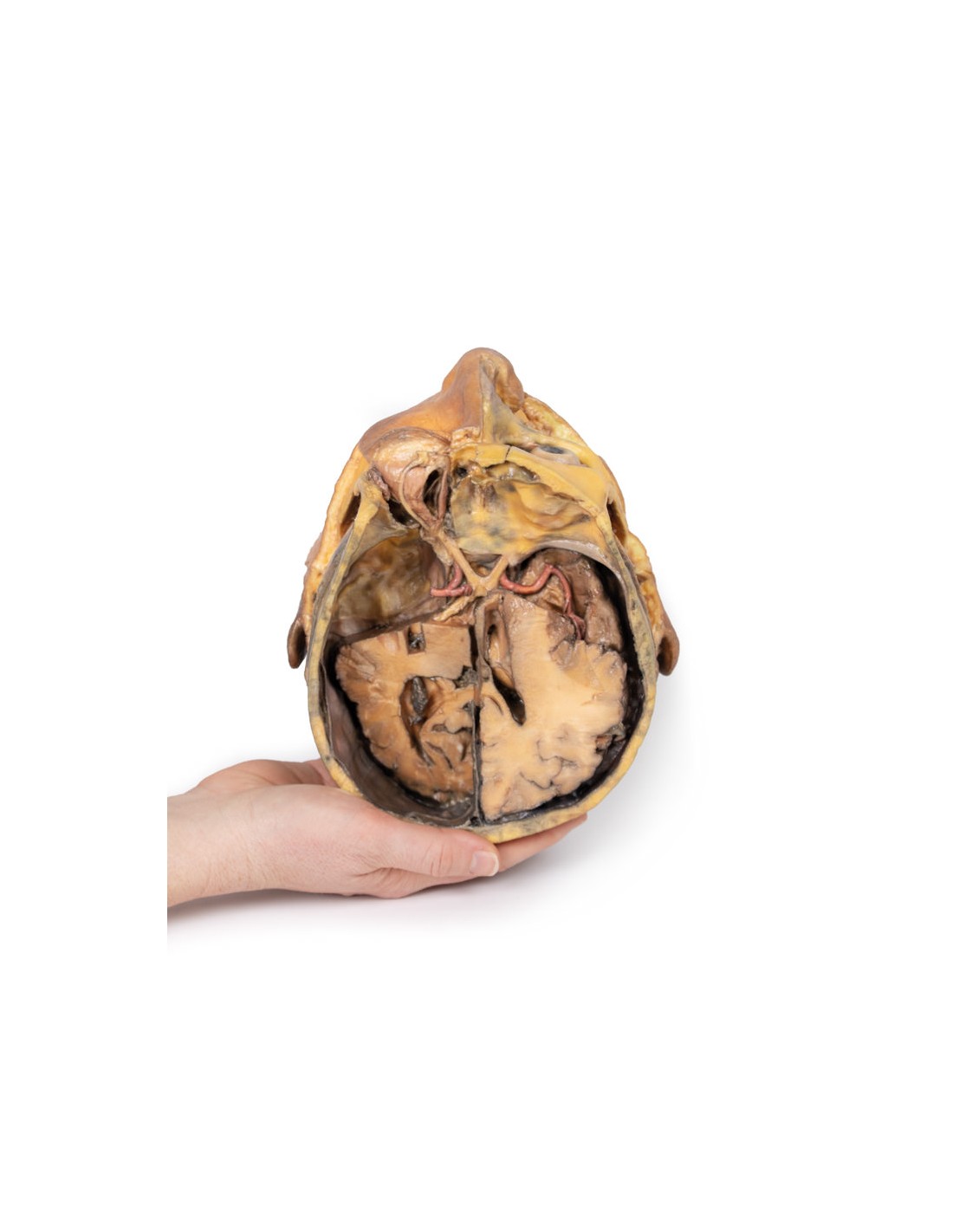
This 3D model preserves a cross section of the cranial cavity with partial dissection of the brain and exposure of the left orbital roof, along with a deep dissection of the face and temporomandibular joint region.
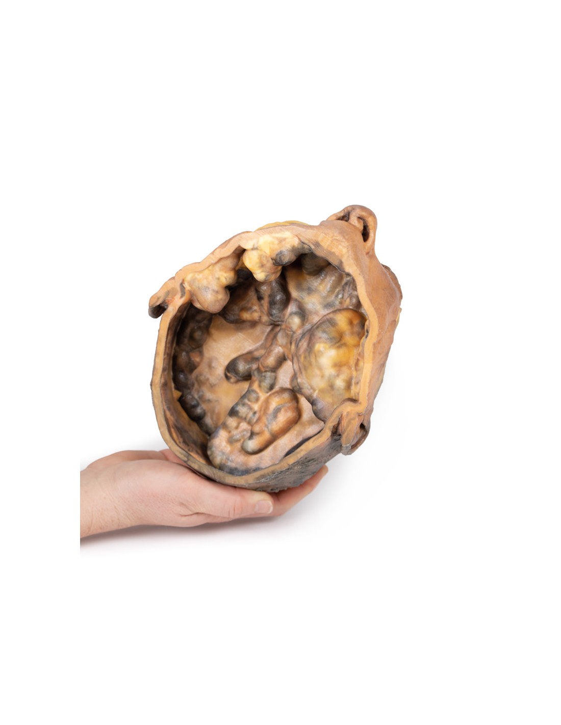
Inside the cranial cavity, the dura mater was largely removed from the anterior cranial fossa, with retention of the layer partly through the middle and posterior cranial fossae. On the right side, the brain was dissected to expose the lateral ventricle and to open the lateral fissure to demonstrate the course of the middle cerebral artery between the frontal, parietal and temporal lobes. A more significant dissection of the brain on the left side allows an appreciation of the midline third ventricle and the retained septum pellucidum on the right side, the cerebral sickle (with the superior sagittal sinus visible in cross section), and parts of the anterior and posterior horns of the lateral ventricle with choroid plexus. This differential dissection of the brain also provides an excellent view of the optic nerves, chiasm and tracts,
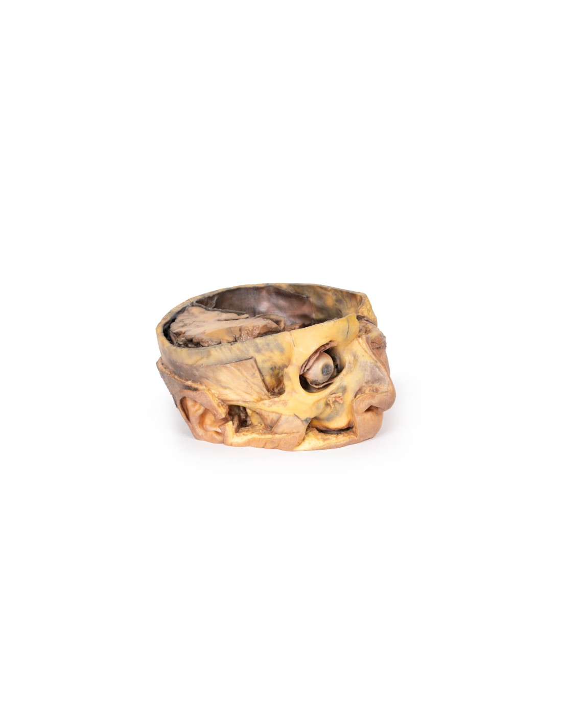
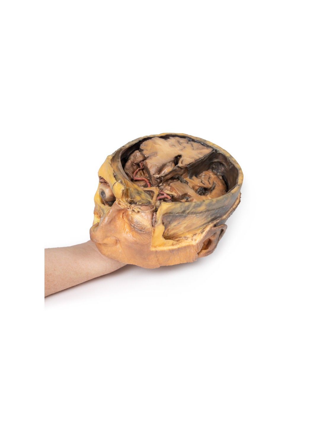
Anteriorly in the cranial cavity, the left optic nerve can be followed into the left orbit, which has been opened to expose several key orbital structures. Centrally, the frontal nerve is well preserved on the elevator muscle of the upper eyelid. Laterally, the lacrimal gland rests in the upper quadrant, while medially the partial dissection in the frontal bone and sinuses provides a clear view of the superior oblique muscle passing through the trochlea. Deep to these superficial structures, extraocular fat was removed to show the medial rectus muscle laterally and the nasociliary nerve and medial rectus muscles medially.
In the face, the skin, superficial tissue, orbicularis oculi, and extraocular fat were removed from the right orbit to expose the extraocular muscles and lacrimal gland. The elevator of the upper eyelid is well defined despite being detached from the superior tarsal plate. The reflex of the superior oblique muscle from the trochlea on the eye can be seen, as well as the insertions of the medial and lateral rectus muscle and the entire course of the inferior oblique.
Through the rest of the right side of the face and temporal region a deep dissection revealed a number of structures. Inferior to the orbital margin, the infraorbital artery and nerve were exposed exiting through the infraorbital foramen. The superficial and deep heads of the masseter are well defined, and partial dissection of the temporalis muscle provides perspective on its broad fiber origin and depth near the pterion (and in contrast to the exposed, non-sectioned right side). Inferior to the zygomatic, the parotid gland was dissected to expose the mandibular condyle resting in the glenoid fossa and to demonstrate the relationship of the external ear to the external auditory meatus.
What advantages does the Monash University anatomical dissection collection offer over plastic models or plastinated human specimens?
- Each body replica has been carefully created from selected patient X-ray data or human cadaver specimens selected by a highly trained team of anatomists at the Monash University Center for Human Anatomy Education to illustrate a range of clinically important areas of anatomy with a quality and fidelity that cannot be achieved with conventional anatomical models-this is real anatomy, not stylized anatomy.
- Each body replica has been rigorously checked by a team of highly trained anatomists at the Center for Human Anatomy Education, Monash University, to ensure the anatomical accuracy of the final product.
- The body replicas are not real human tissue and therefore not subject to any barriers of transportation, import, or use in educational facilities that do not hold an anatomy license. The Monash 3D Anatomy dissection series avoids these and other ethical issues that are raised when dealing with plastinated human remains.
No reviews
Tap to zoom