Your cart
There are no more items in your cart


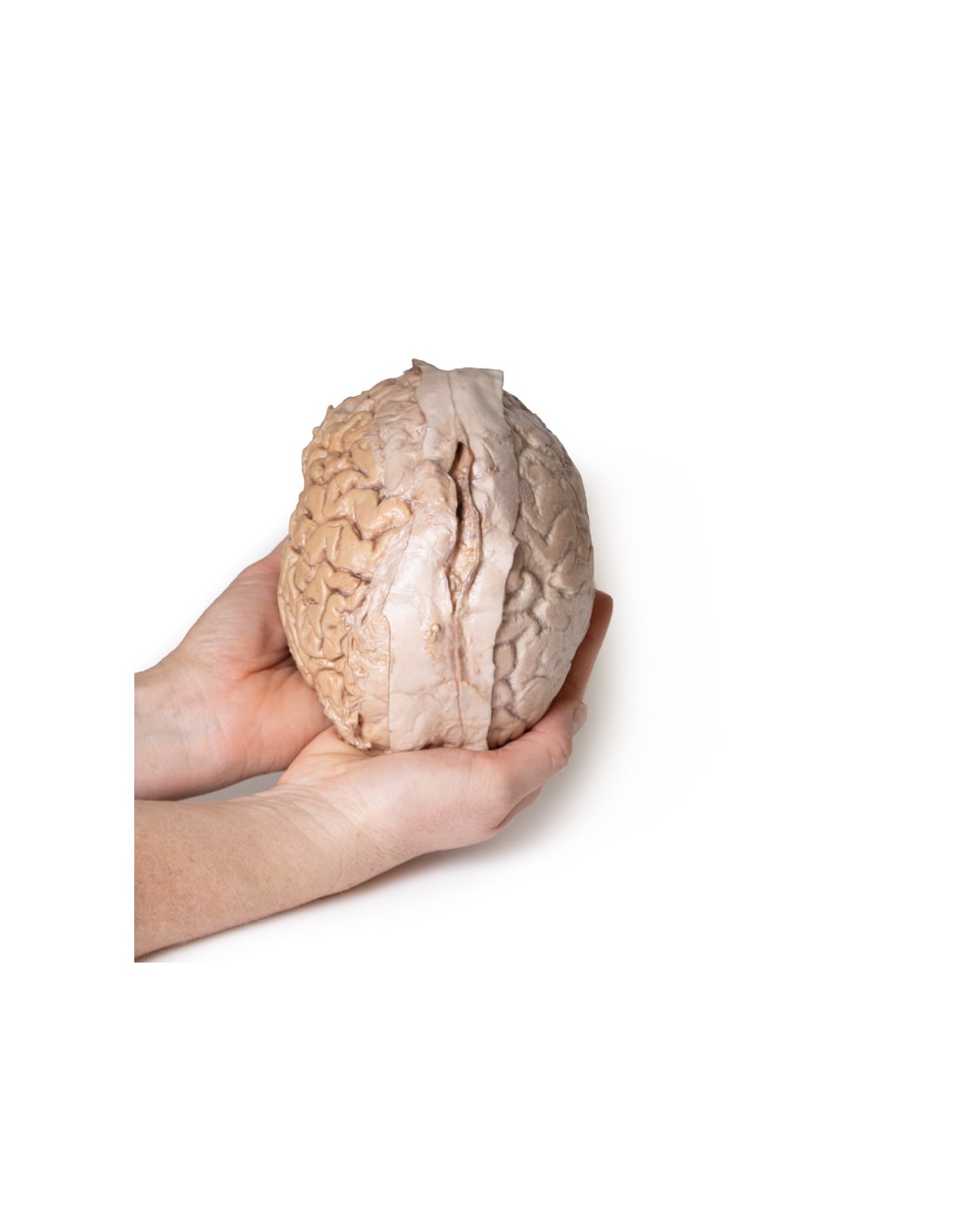
Made in ultra-high resolution 3D printing in full color.
Human Brain - Erler Zimmer 3D anatomy Series MP1103
This model is part of the exclusive Monash 3D anatomy series, a comprehensive series of human dissections reproduced with ultra-high resolution color 3D printing.
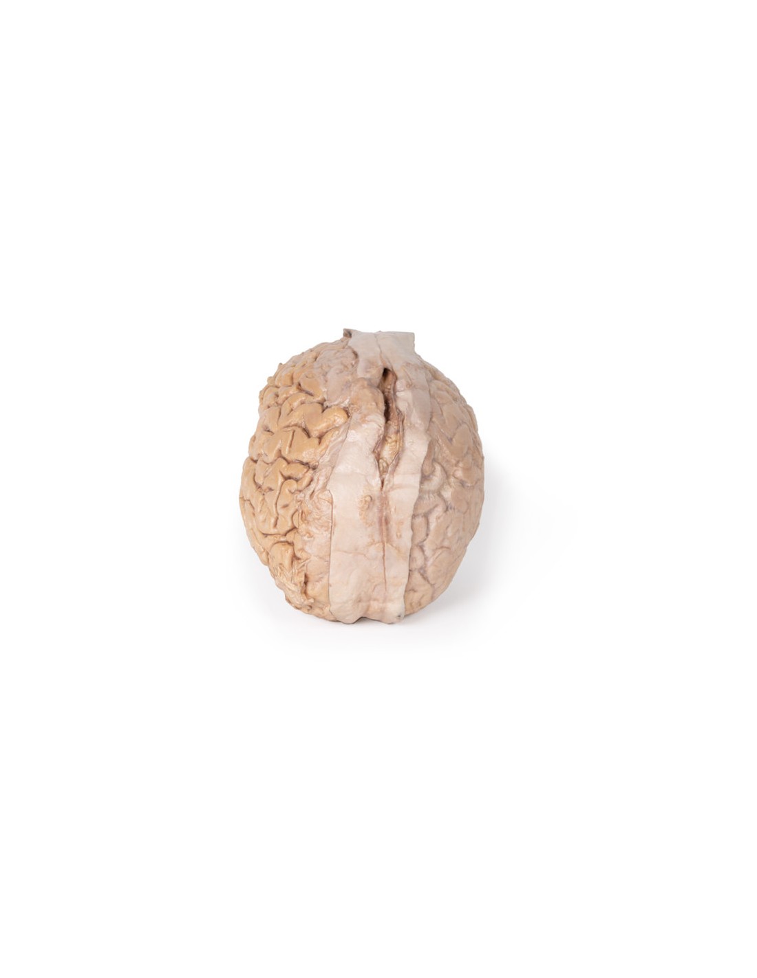
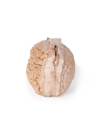
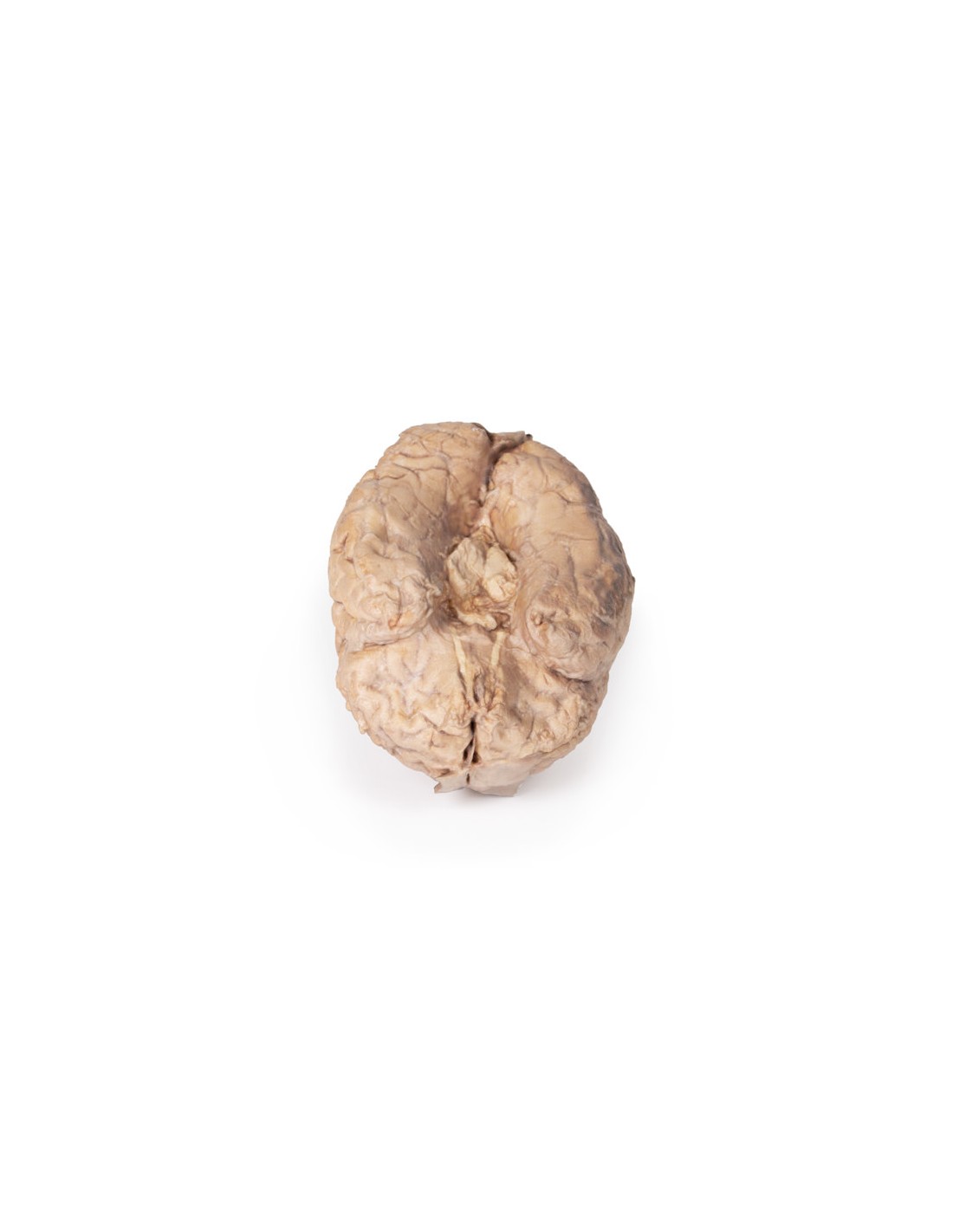
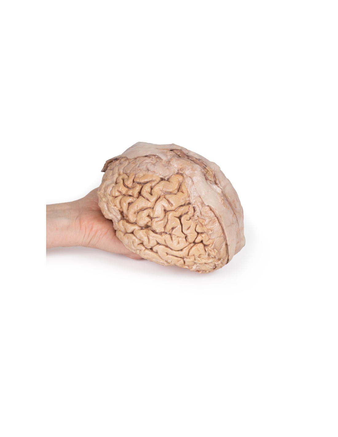
This 3D model provides a unique perspective on the anatomy of the brain with respect to the meninges. The brain has been separated from the brain stem and cerebellum, with only parts of the midbrain and cerebral peduncles visible on the lower surface.
Adjacent to the cut section can be seen the olfactory tracts and bulbs extending along the lower margin of the frontal lobes of the brain.
Variable dissection between the left and right cerebral hemispheres allows one to appreciate the organization of the brain and meninges as it normally appears within the cranial cavity.
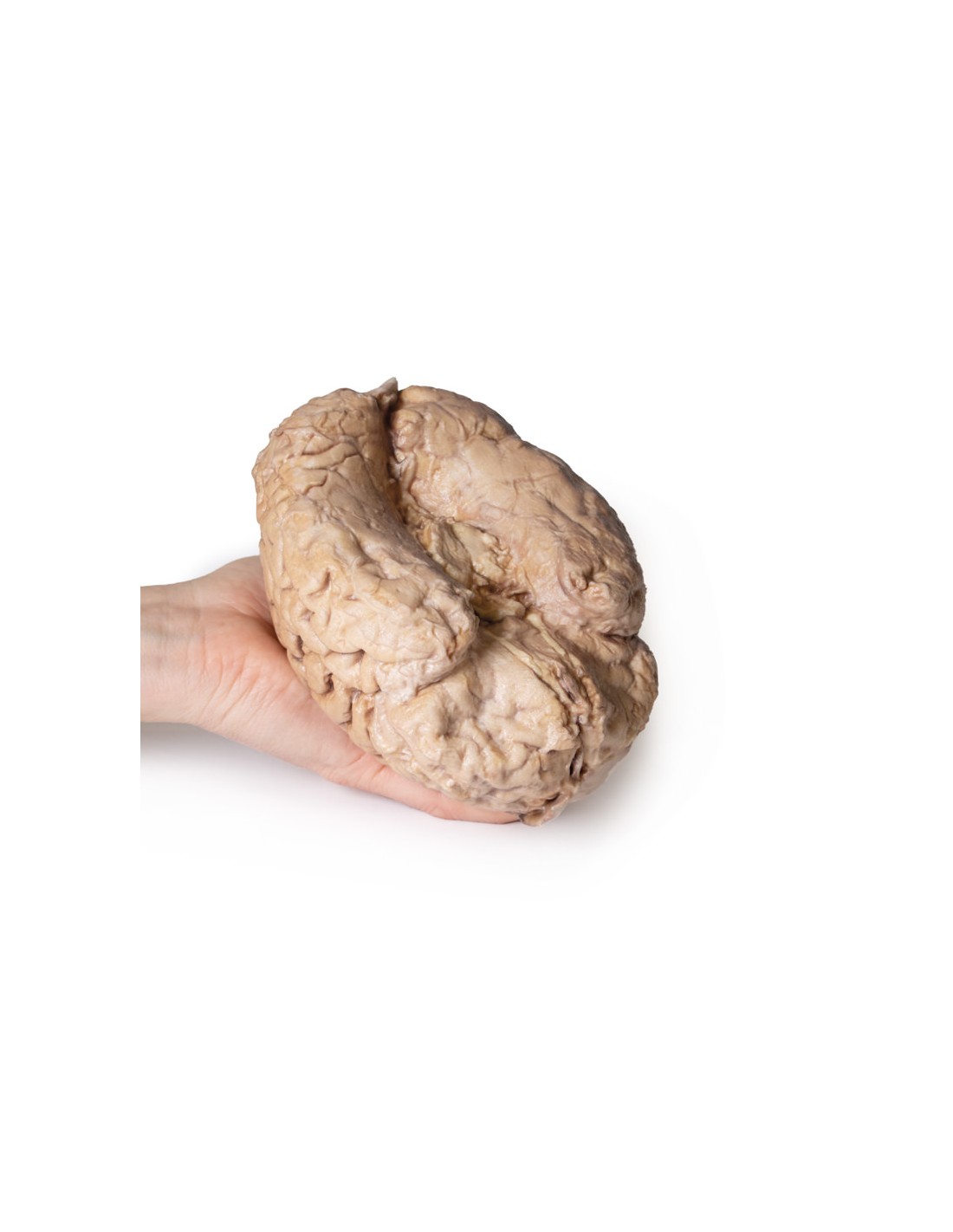
In the midline, the dura mater has been preserved from anterior (rostral) to posterior. The central portion of the true dura (endosteal) has opened to expose the superior sagittal sinus (between the endosteal and meningeal layers of the dura mater).
Numerous arachnoid granulations (clusters of arachnoid villi) are visible within the open superior sagittal sinus, as well as across the margins of the preserved dura.
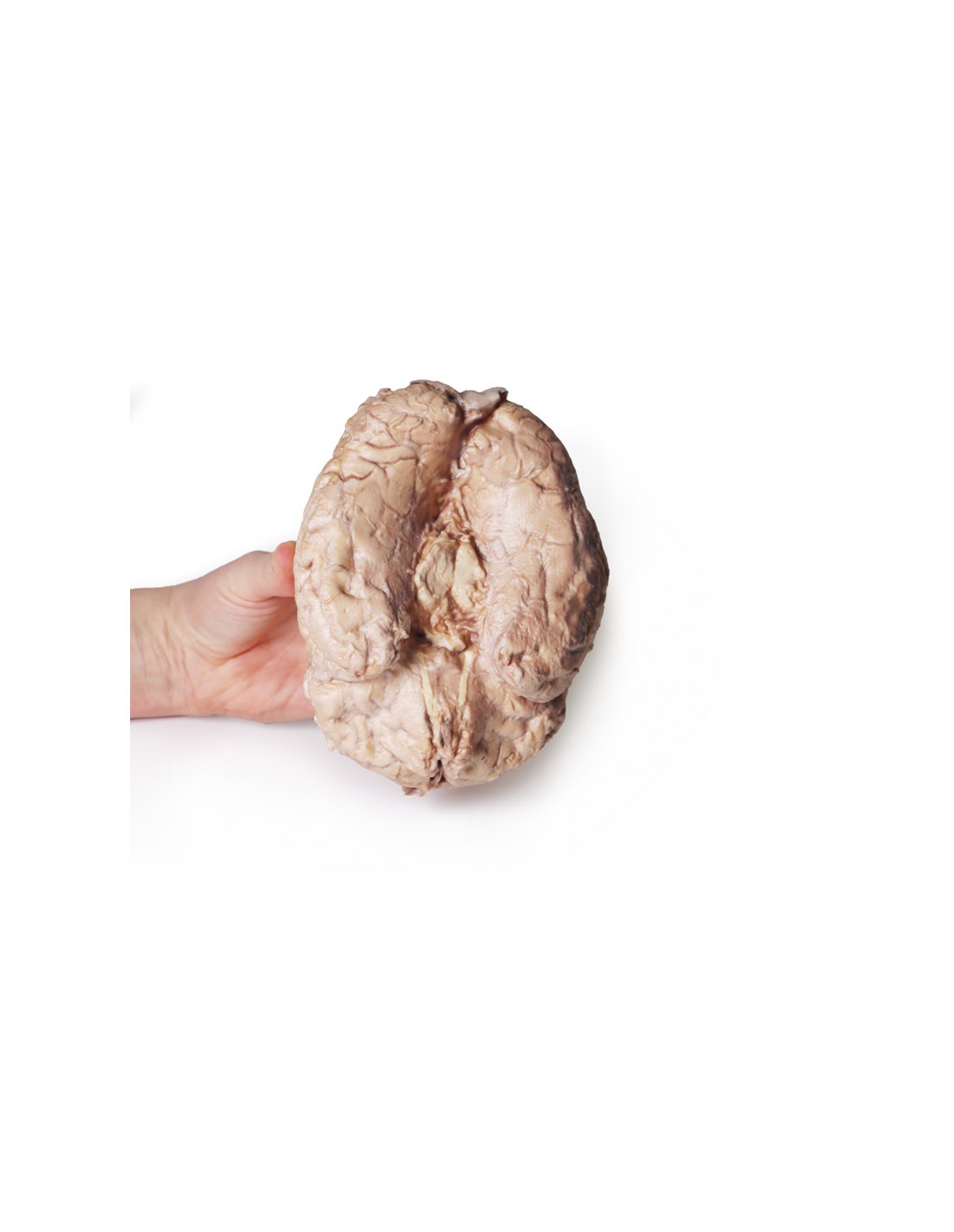
On the right cerebral hemisphere, the dura mater has been completely removed to expose the underlying arachnoid, which obscures the appearance of the underlying cerebral circumvolutions and furrows, as well as the terminal branches of the cerebral arteries. in contrast, the arachnoid mater has dissected most of the hemisphere (except for a reference margin) to expose the pia mater-covered circumvolutions and furrows.
This allows a clear view of the lateral sulcus and the central sulcus, with the latter defining the boundaries of the frontal and parietal lobes and separating the primary sensory and motor cortical areas on the gyrus on either side of the sulcus.
What advantages does the Monash University anatomical dissection collection offer over plastic models or plastinated human specimens?
- Each body replica has been carefully created from selected patient X-ray data or human cadaver specimens selected by a highly trained team of anatomists at the Monash University Center for Human Anatomy Education to illustrate a range of clinically important areas of anatomy with a quality and fidelity that cannot be achieved with conventional anatomical models-this is real anatomy, not stylized anatomy.
- Each body replica has been rigorously checked by a team of highly trained anatomists at the Center for Human Anatomy Education, Monash University, to ensure the anatomical accuracy of the final product.
- The body replicas are not real human tissue and therefore not subject to any barriers of transportation, import, or use in educational facilities that do not hold an anatomy license. The Monash 3D Anatomy dissection series avoids these and other ethical issues that are raised when dealing with plastinated human remains.
No reviews
Tap to zoom