Your cart
There are no more items in your cart


Dissection of hemipelvi and male thigh - Erler Zimmer 3D anatomy Series MP1142
erler zimmer
EZ-MP1142
€6,601.30
Tax included
Made in ultra-high resolution 3D printing in full color.
Dissection of hemipelvis and male thigh - Erler Zimmer 3D anatomy Series MP1142
This model is part of the exclusive Monash 3D anatomy series, a comprehensive series of human dissections reproduced with very high resolution color 3D printing.
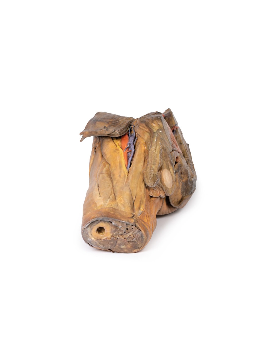
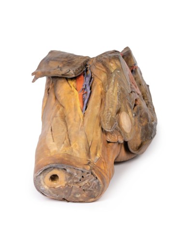
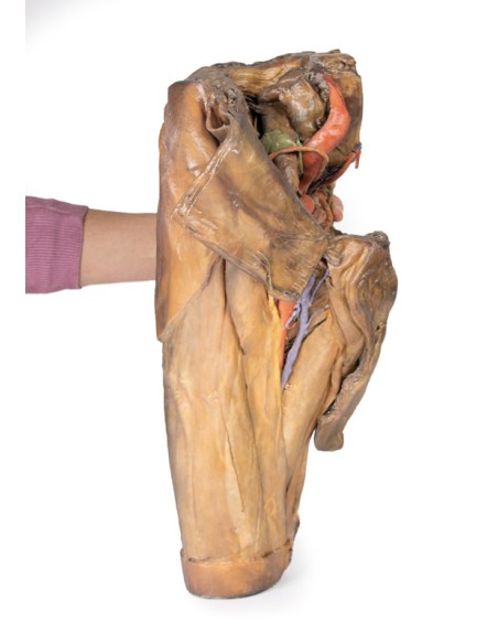
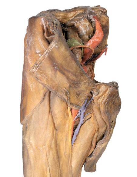
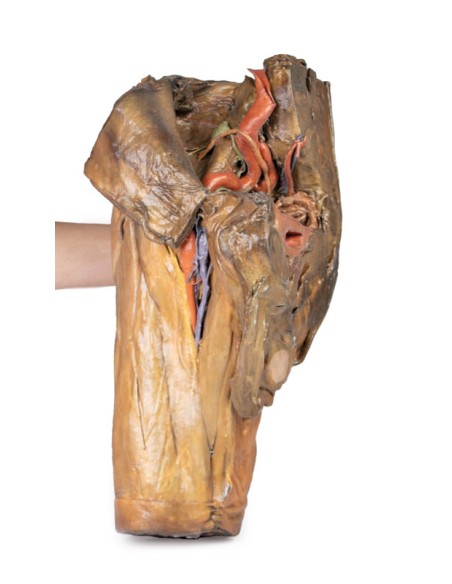
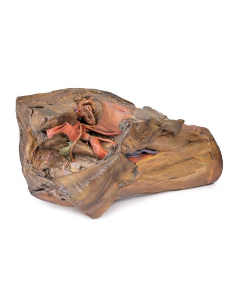
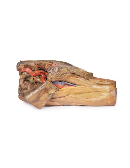
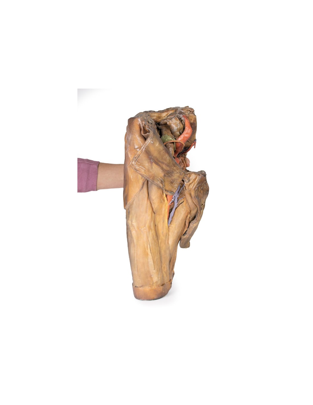
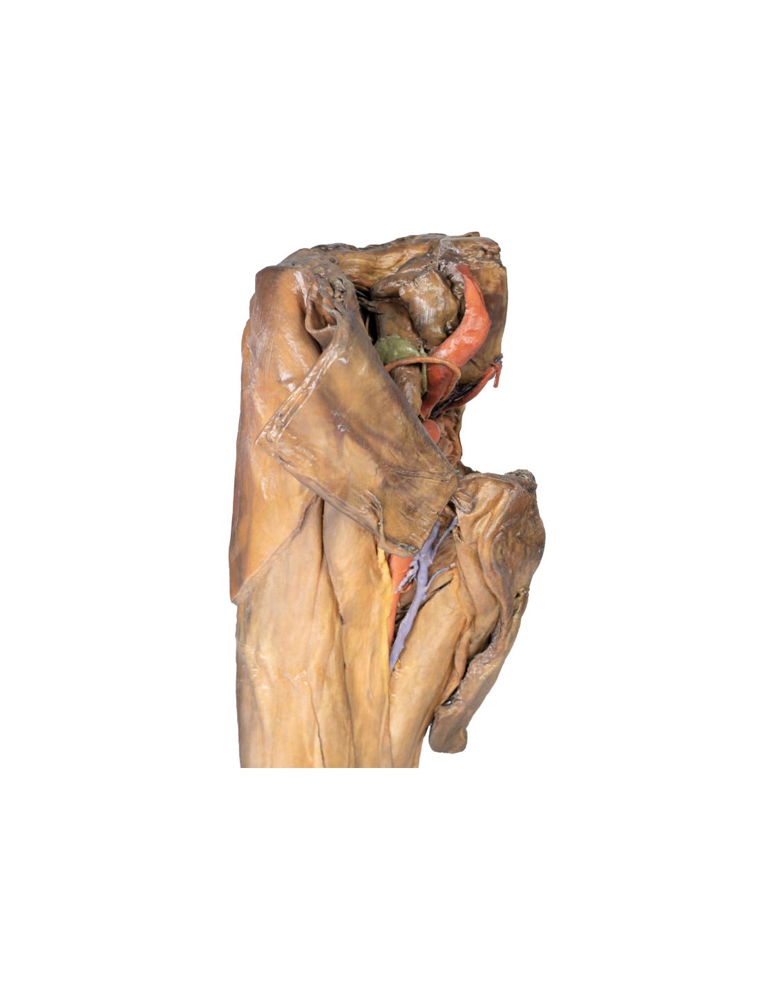
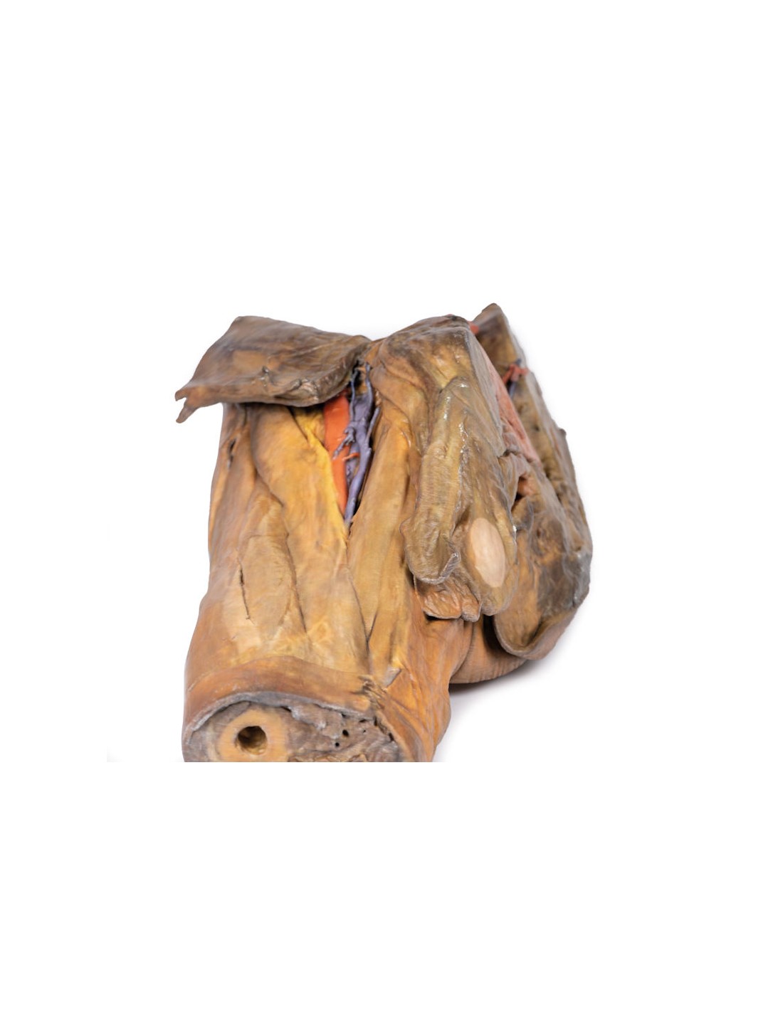
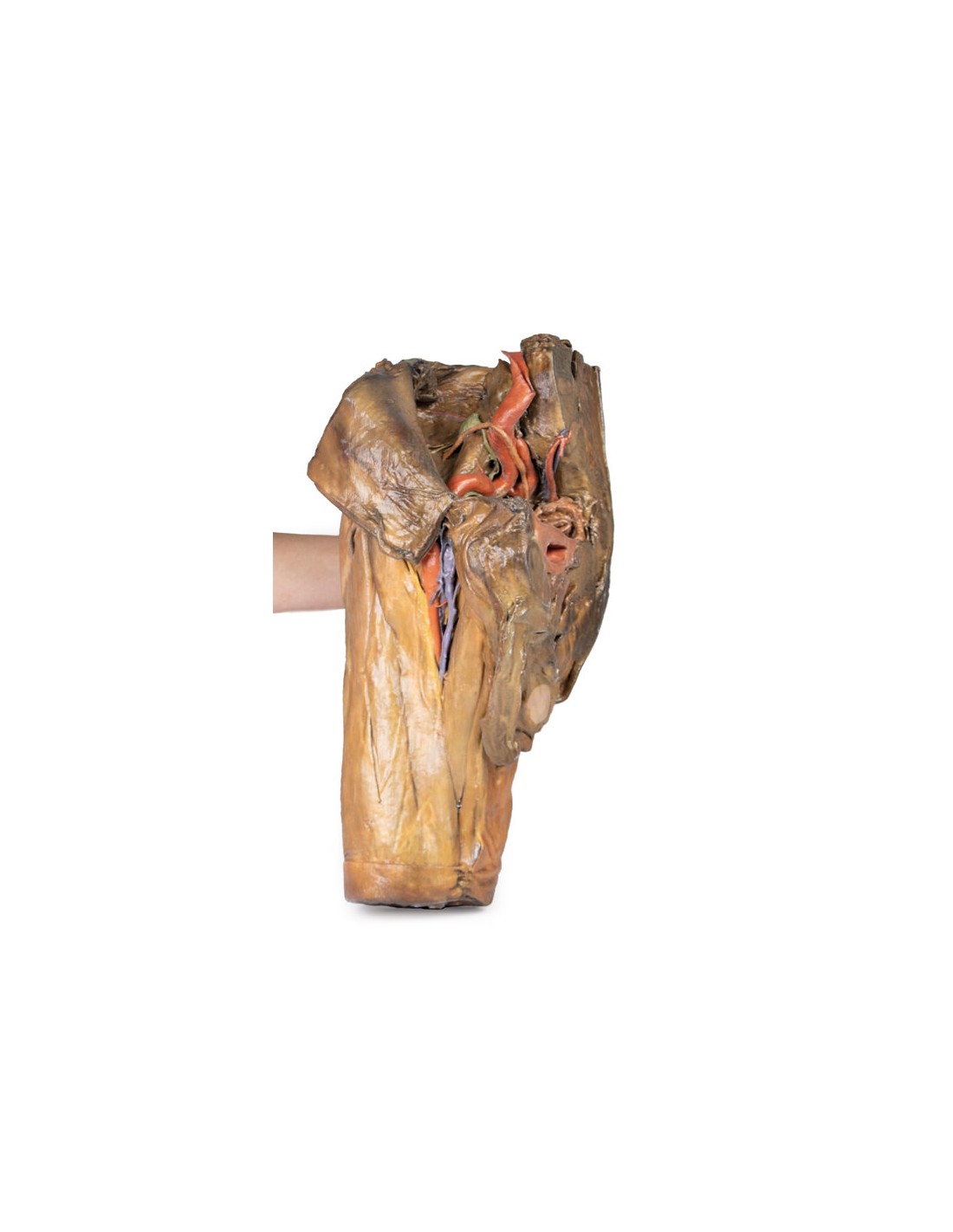
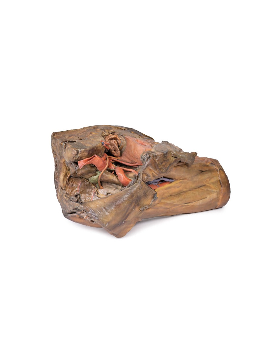
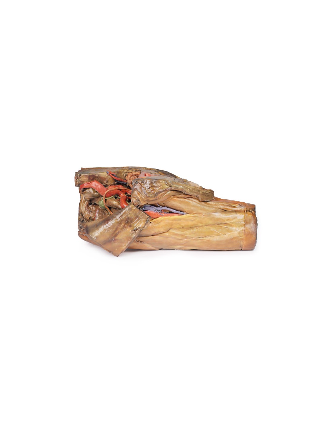
This 3D model preserves a right male pelvis dissected just above the L5 vertebra and dissected in the mid sagittal plane, with the thigh preserved near the midshaft of the femur.
This specimen matches our LW 91 female hemipelvic specimen and thigh.
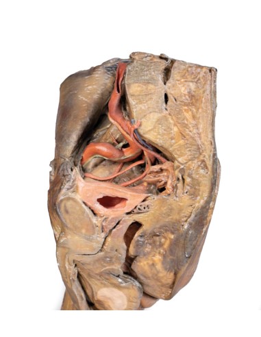
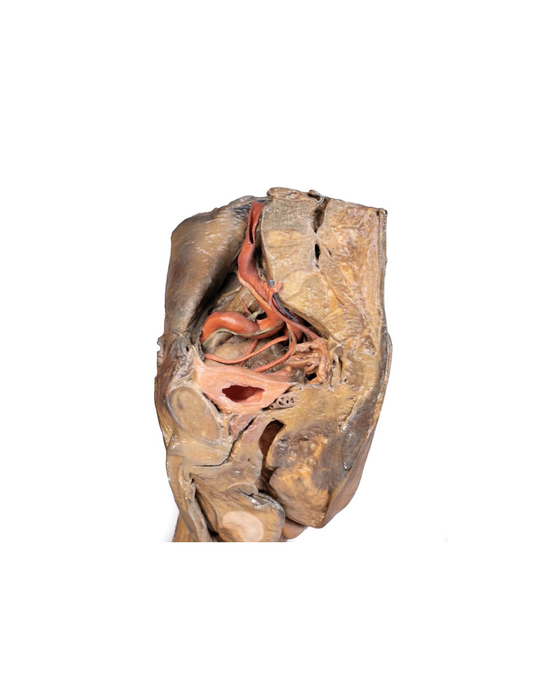
The common iliac artery is preserved with several key branches visible, particularly the distribution of the internal iliac within the true pelvis. Several major vessels, including the obturator artery and the partially obliterated umbilical artery, pass toward the anterior abdominal wall (to form the medial umbilical ligament) and give off the superior vesicular artery; while the roots of the ileolumbar, superior gluteal, inferior gluteal, and internal pudendal arteries are visible lateral to the urinary bladder. The ureter descends superficially to these vessels to approach the urinary bladder, which in this model is covered with peritoneum. The ductus deferens is exposed from entering the space through the deep inguinal ring and passes posteriorly (although dissected from its normal route of insertion and resting on the internal iliac artery). Adjacent to the ureter and on the superficial surface of the psoas major muscle is an enlarged iliac lymph node and part of the ascending lymphatic vascular system along the external iliac artery. Most of the pelvis was left unsectioned, allowing an appreciation of the rectovesicular pouch and the exposed superior rectal artery and vein approaching the preserved portion of the rectum. In cross section, the rectum, seminal vesicle, and prostate are visible (the plane of section preserves parts of both the prostatic urethra and ejaculatory duct). allowing an appreciation of the rectovesicular pocket and the exposed superior rectal artery and vein approaching the preserved portion of the rectum. In transverse section, the rectum, seminal vesicle, and prostate are visible (the plane of section preserves parts of both the prostatic urethra and the ejaculatory duct). allowing an appreciation of the rectovesicular pocket and the exposed superior rectal artery and vein approaching the preserved portion of the rectum. In cross section, the rectum, seminal vesicle, and prostate are visible (the plane of section preserves portions of both the prostatic urethra and ejaculatory duct).
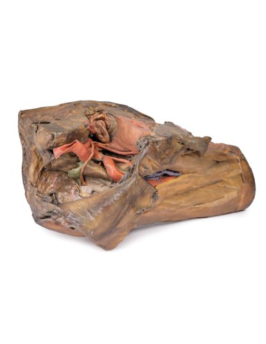
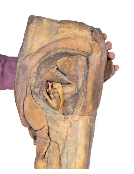
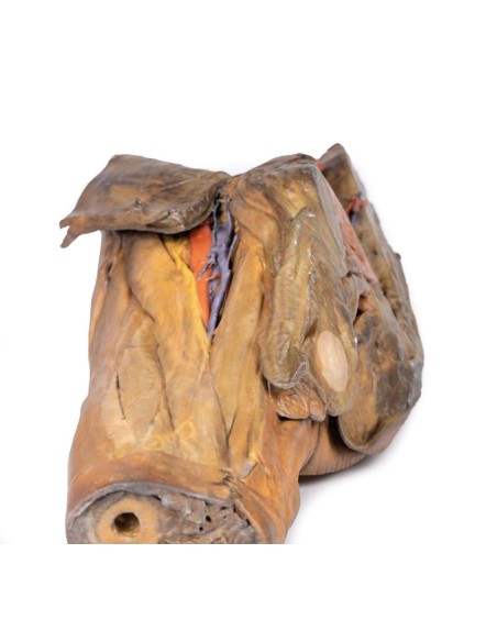
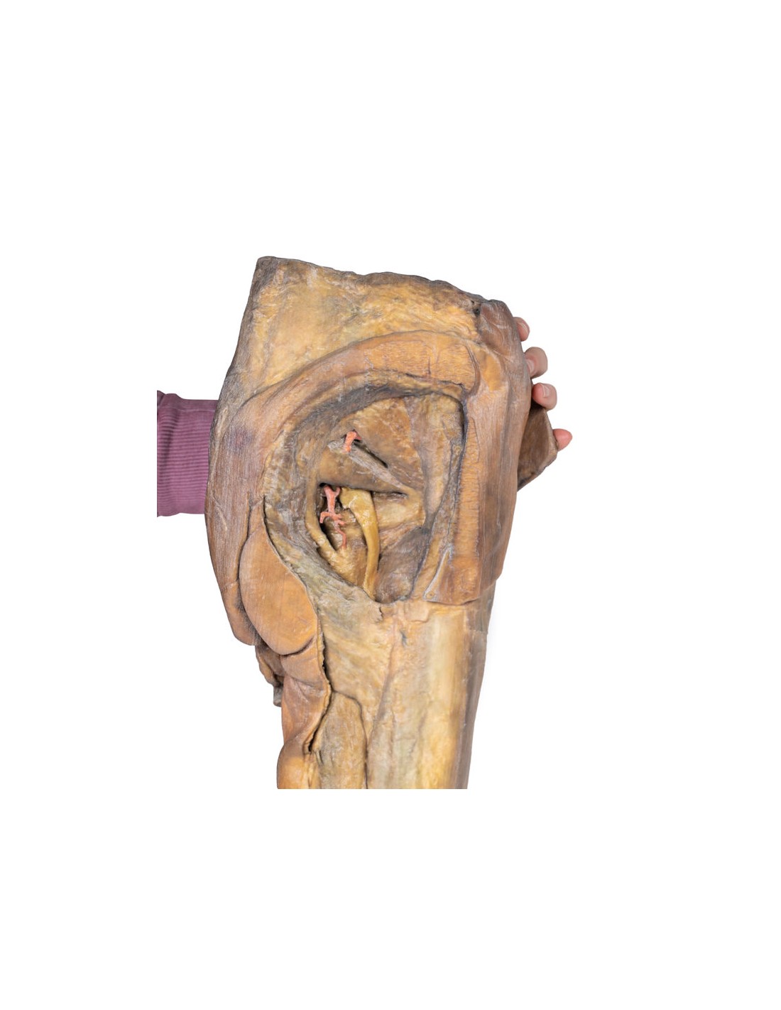
At the front of the thigh, the margins and contents of the femoral triangle are well preserved, with partial coverage by the anterior abdominal wall flap. Posteriorly, the skin over the gluteal region and the gluteus maximus muscle have been removed as sequential windows to expose the gluteus medius and minimus muscle, piriformis, internal obturator with twin muscles, and the quadratus femoris muscle. The superior and inferior gluteal arteries are retained superior and inferior to the piriformis, respectively; with the sciatic nerve exiting inferior to the piriformis before passing deep to the retained portion of the great gluteus.
What advantages does the Monash University anatomical dissection collection offer over plastic models or plastinated human specimens?
- Each body replica has been carefully created from selected patient X-ray data or human cadaver specimens selected by a highly trained team of anatomists at the Monash University Center for Human Anatomy Education to illustrate a range of clinically important areas of anatomy with a quality and fidelity that cannot be achieved with conventional anatomical models-this is real anatomy, not stylized anatomy.
- Each body replica has been rigorously checked by a team of highly trained anatomists at the Center for Human Anatomy Education, Monash University, to ensure the anatomical accuracy of the final product.
- The body replicas are not real human tissue and therefore not subject to any barriers of transportation, import, or use in educational facilities that do not hold an anatomy license. The Monash 3D Anatomy dissection series avoids these and other ethical issues that are raised when dealing with plastinated human remains.
No reviews
Tap to zoom