Your cart
There are no more items in your cart


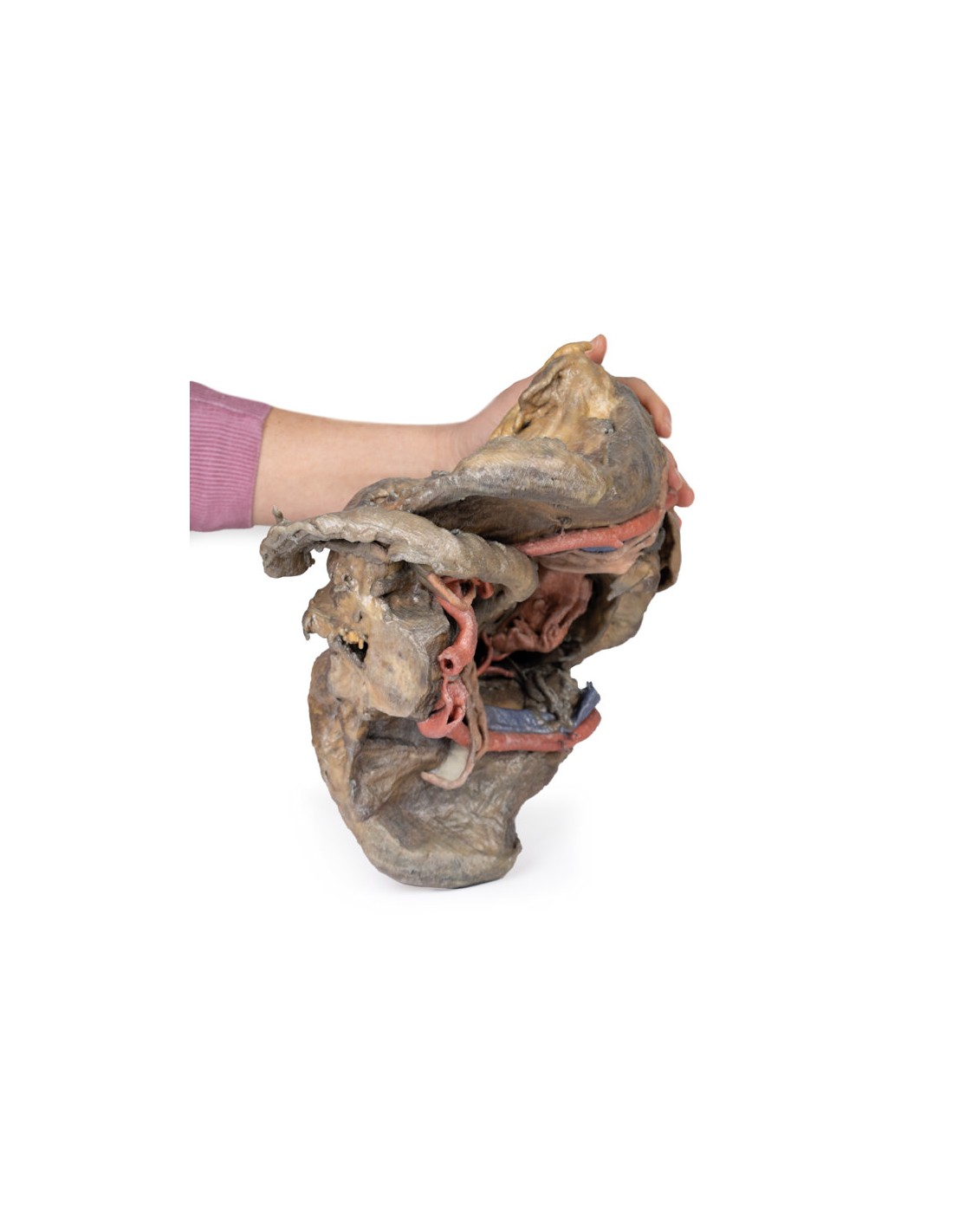
Deep dissection of the female pelvis - Erler Zimmer 3D anatomy Series MP1141
erler zimmer
EZ-MP1141
€6,171.86
Tax included
Made in ultra-high resolution 3D printing in full color.
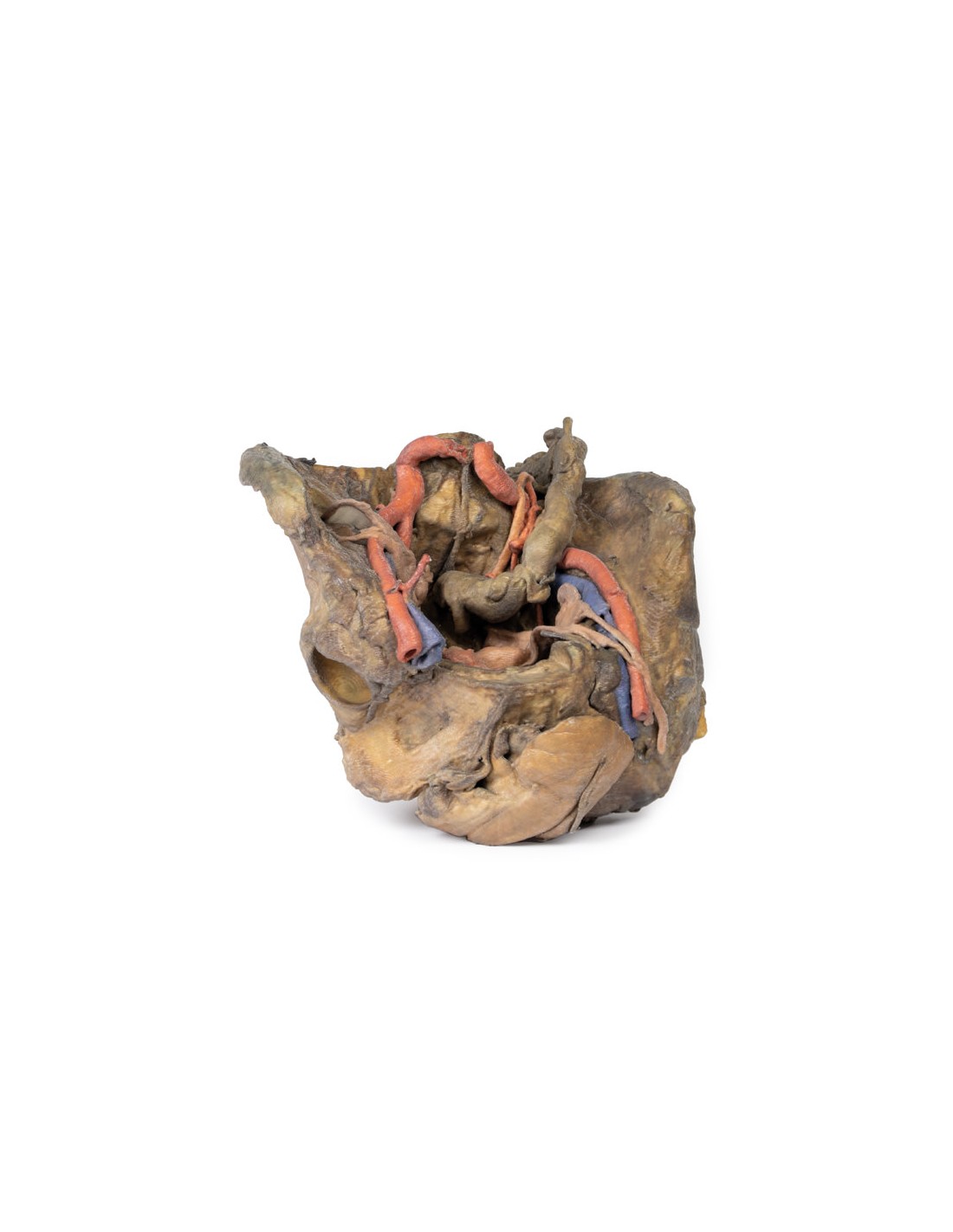
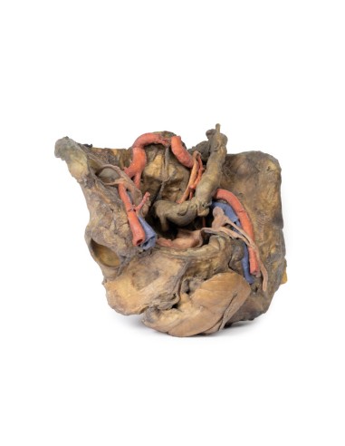
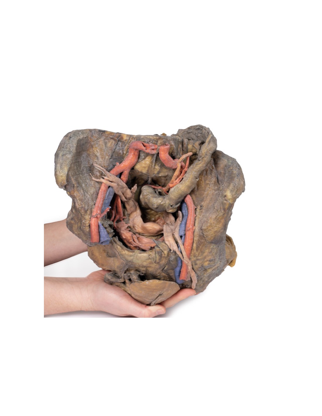
Deep dissection of the female pelvis - Erler Zimmer 3D anatomy Series MP1141
This model is part of the exclusive Monash 3D anatomy series, a comprehensive series of human dissections reproduced with ultra-high resolution color 3D printing.
This 3D model presents a deep dissection and isolation of the pelvis from surrounding regions, demonstrating in particular visceral and neurovascular structures related to deep ligaments and bony features.
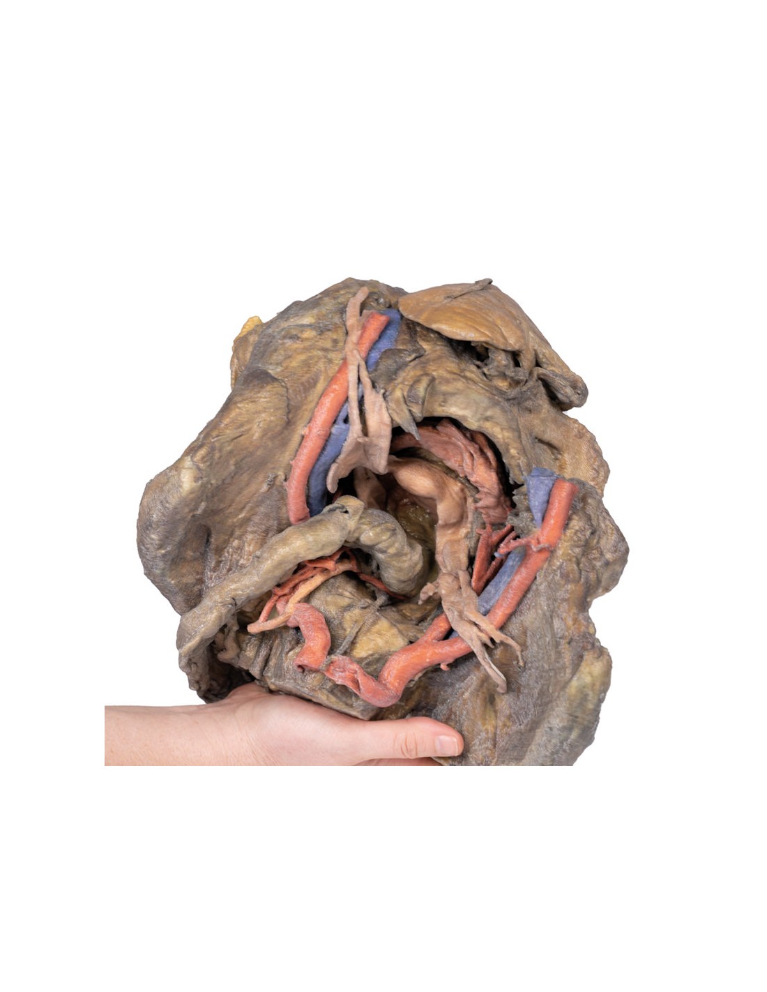
Within the false pelvis, the sigmoid colon descends on the left side of the specimen to the rectum, passing superficially through the pelvic rim and the passage of the common and external iliac artery and vein. Adjacent to the sigmoid colon are parts of the sigmoid arteries and the superior rectal artery, which rest on the surface relative to the common iliac vessels and near the descending ureter. Anteriorly in the true pelvis is the collapsed urinary bladder, and between the bladder and rectum rests the uterus. The organ is partially covered by the broad ligament, with both the suspensory ligament of the ovary and the round ligament having been separated and pulled away from the peritoneum on both sides to expose the surrounding blood vessels. While the ovarian ligaments, the round ligaments, uterine tubes and ovaries are trapped in the peritoneal fold of the broad ligament,
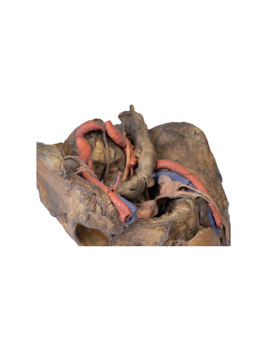
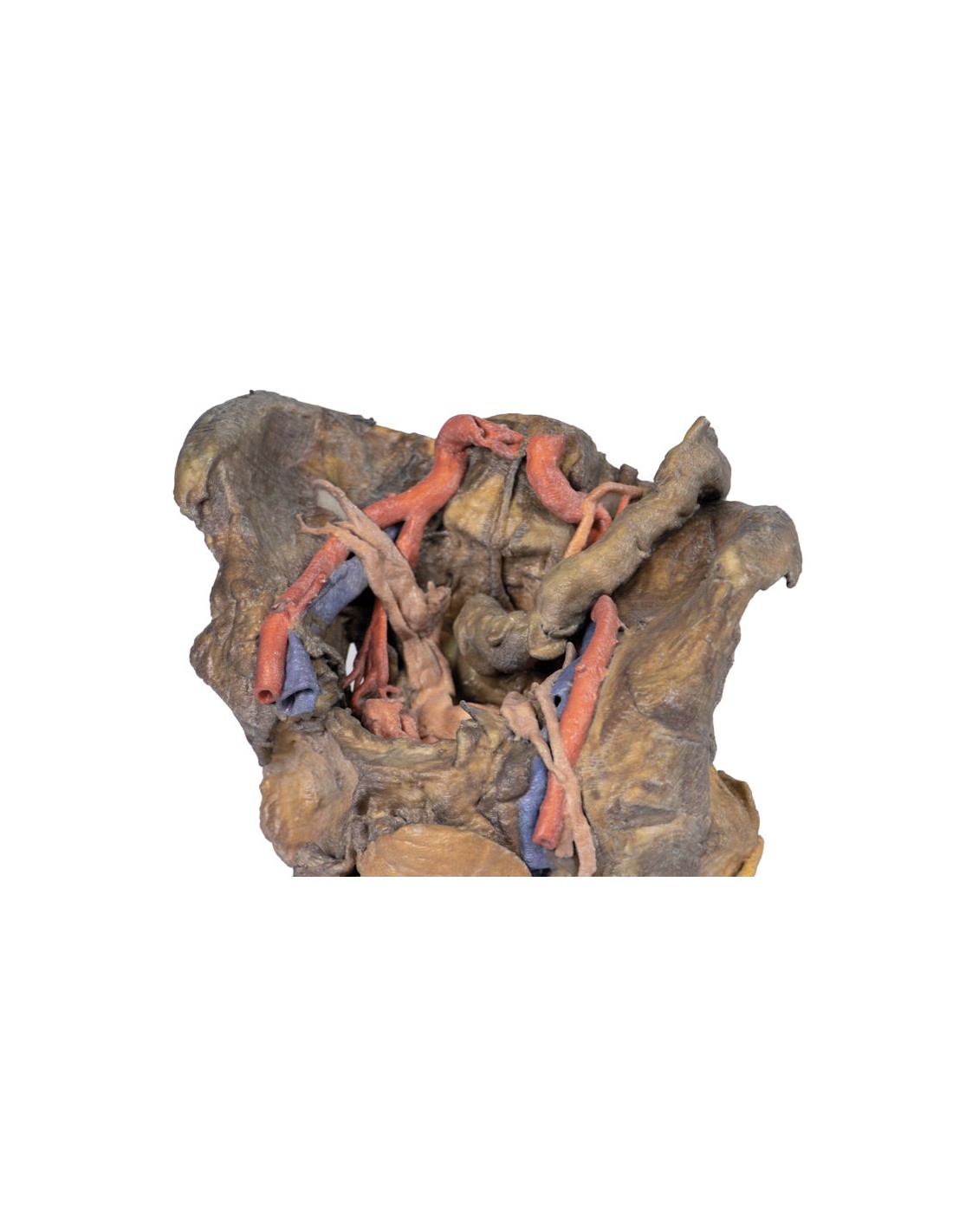
Lateral to these organs branches of the internal iliac artery can be identified, as well as a median sacral artery retained in the midline between the two common iliac arteries. On the left side only the uterine artery can be seen laterally. On the right side, the obturator, superior vesical, and uterine arteries can be seen. In addition, the origins of the inferior epigastric artery and vein can be seen arising from the external iliac vessels just before exiting the lower abdominal cavity.
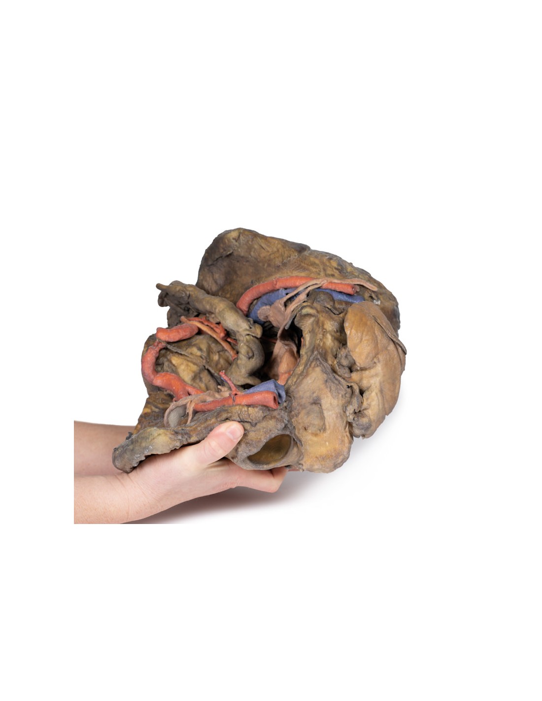
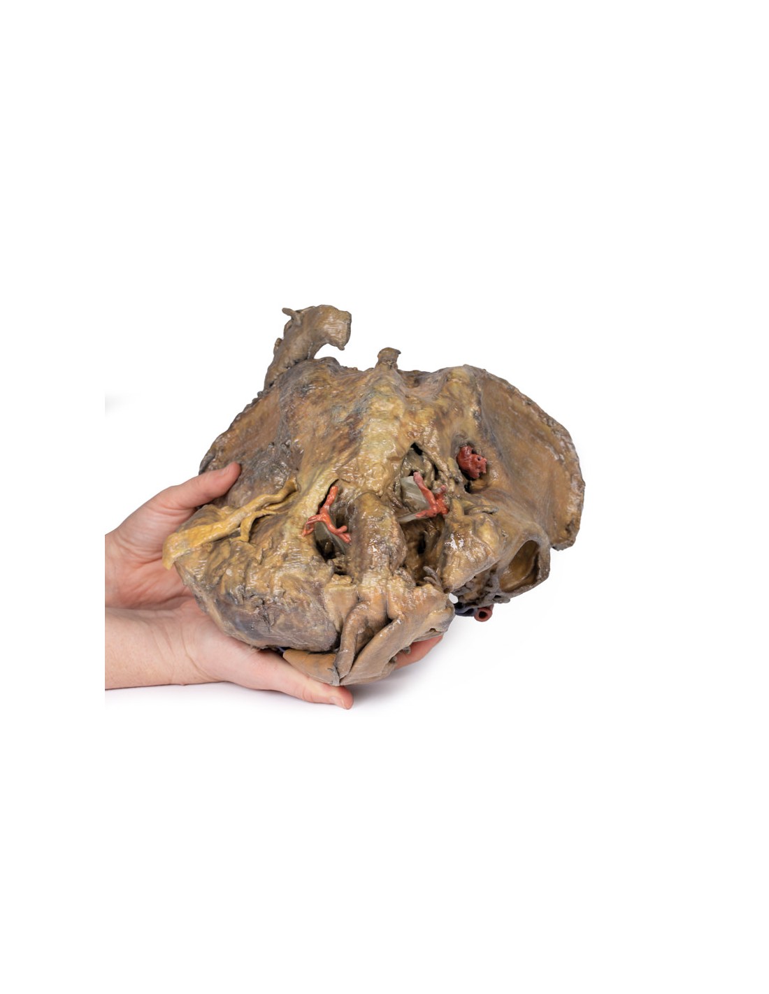
On the right side of the preserved pelvis, the entire musculature of the femur and thigh was removed to demonstrate the obturator membrane, the articular cartilage of the acetabulum, and the transverse ligament of the acetabulum. Posteriorly, the entire gluteal region was dissected to expose the superior gluteal foramen and the origin of the superior gluteal artery. The sacrotuberous ligament was removed to demonstrate the sacrospinous ligament, with some branches of the inferior rectus artery retained within the exposed iscoanal fossa.
On the left side of the preserved pelvis, the sciatic nerve was retained within the large sciatic foramen, as was the sacrotuberous ligament. The ischioanal fossa mirrors that of the right side, where the branches of the inferior rectal artery have been retained with respect to the fibers of the pelvic diaphragm, and integration of the external anal sphincter on the protruding external rectal surface.
What advantages does the Monash University anatomical dissection collection offer over plastic models or plastinated human specimens?
- Each body replica has been carefully created from selected patient X-ray data or human cadaver specimens selected by a highly trained team of anatomists at the Monash University Center for Human Anatomy Education to illustrate a range of clinically important areas of anatomy with a quality and fidelity that cannot be achieved with conventional anatomical models-this is real anatomy, not stylized anatomy.
- Each body replica has been rigorously checked by a team of highly trained anatomists at the Center for Human Anatomy Education, Monash University, to ensure the anatomical accuracy of the final product.
- The body replicas are not real human tissue and therefore not subject to any barriers of transportation, import, or use in educational facilities that do not hold an anatomy license. The Monash 3D Anatomy dissection series avoids these and other ethical issues that are raised when dealing with plastinated human remains.
No reviews
Tap to zoom