
Your cart
There are no more items in your cart


Dissection of hemipelvi and female thigh - Erler Zimmer 3D anatomy Series MP1140
erler zimmer
EZ-MP1140
€3,713.31
Tax included
Made in ultra-high resolution 3D printing in full color.
Dissection of hemipelvis and female thigh - Erler Zimmer 3D anatomy Series MP1140
This model of internal abdominal Perete is part of the exclusive Monash 3D anatomy series, a comprehensive series of human dissections reproduced with very high resolution color 3D printing.
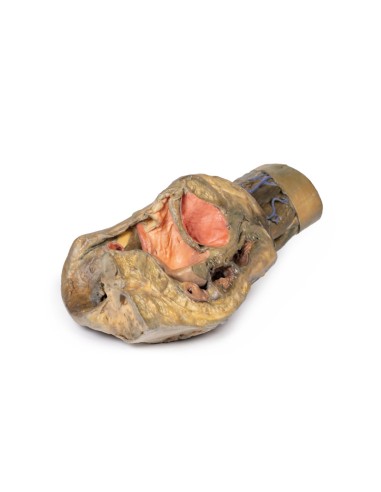
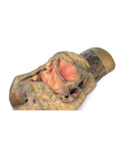
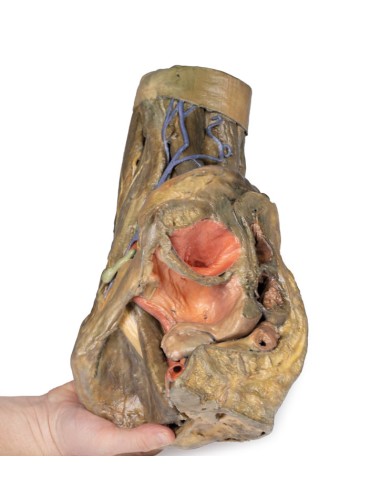
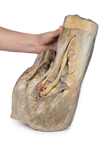
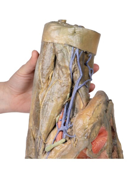
This 3D model preserves the left pelvis divided in the mediosagittal plane and the proximal thigh at approximately mid-thigh.
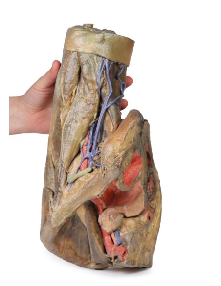
In the mediosagittal section, the urinary bladder, uterus, vagina, and rectum can be seen sequentially between the pubic symphysis (anteriorly) and sacrum (posteriorly). Retention of the peritoneum draped over the upper surface of these organs allows vesicouterine and rectouterine pockets to be seen. The reflection of the peritoneum from the uterus forms the broad ligament, with the uterine tube, fimbre, and left ovary closely associated in position near the pelvic brim. Lateral to the true pelvic contents can be seen the common and external iliac arteries that pass toward the subinguinal space between the common iliac vein and the greater psoas muscle. The descending course of the ureter can be traced through these vessels, and the femoral nerve is visible between the psoas major and the iliac muscle.
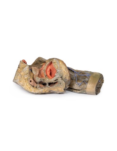
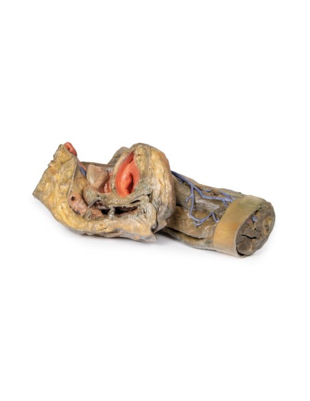
Superficial fascia has been removed across the thigh to the lateral margin of the perineum and near the lower section of the model itself. Anteriorly, the femoral triangle region was dissected to expose the contents as well as the horizontal group of inguinal lymph nodes immediately inferior to the inguinal ligament. Medially, the femoral vein receives drainage through the great saphenous vein and regional veins (including the superficial circumflex iliac, superficial external pudendal, and deep pudendal veins). The femoral artery can be seen immediately lateral to the vein, with parts of the femoral nerve descending just lateral to the artery and near the tendon of the iliopsoas muscle. Although somewhat disturbed by dissection,
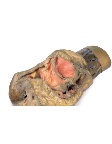
Posteriorly, the gluteal region was dissected with removal of the gluteus maximus to expose the underlying gluteal muscles, with the piriformis muscle reflex revealing neurovascular structures in the region. The sciatic nerve can be seen forming through contributions from the common tibial and peroneal nerves around the preserved portions of the superior and inferior gluteal arteries. Medially, the posterior cutaneous nerve of the thigh runs parallel with the sciatic nerve, with both muscles resting on the internal obturator tendon and twin muscles before descending into the thigh above the quadratus femoris and common hamstring origin, respectively. Deep down to the sacrotuberous ligament, the course of the internal pudendal artery and pudendal nerve to the ischioanal fossa can be followed,
What advantages does the Monash University anatomical dissection collection offer over plastic models or plastinated human specimens?
- Each body replica has been carefully created from selected patient X-ray data or human cadaver specimens selected by a highly trained team of anatomists at the Monash University Center for Human Anatomy Education to illustrate a range of clinically important areas of anatomy with a quality and fidelity that cannot be achieved with conventional anatomical models-this is real anatomy, not stylized anatomy.
- Each body replica has been rigorously checked by a team of highly trained anatomists at the Center for Human Anatomy Education, Monash University, to ensure the anatomical accuracy of the final product.
- The body replicas are not real human tissue and therefore not subject to any barriers of transportation, import, or use in educational facilities that do not hold an anatomy license. The Monash 3D Anatomy dissection series avoids these and other ethical issues that are raised when dealing with plastinated human remains.
No reviews
Tap to zoom
