Your cart
There are no more items in your cart


Endometrial Carcinoma - Erler Zimmer 3D anatomy Series MP2106
erler zimmer
EZ-MP2106
€1,109.83
Tax included
Made in ultra-high resolution 3D printing in full color.
Endometrial Carcinoma - Erler Zimmer 3D anatomy Series MP2106
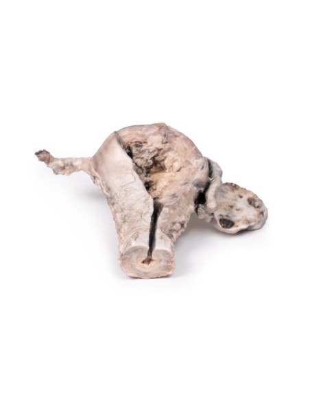
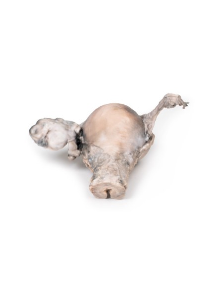
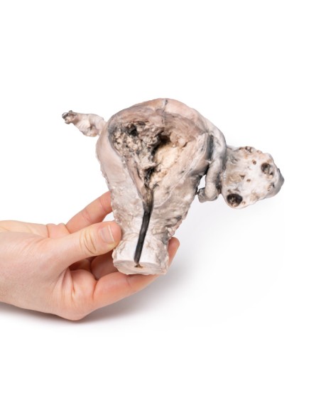
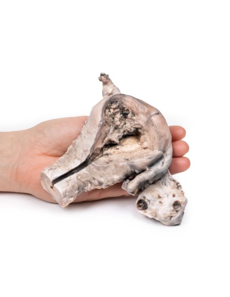
This dissection model highlighting Endometrial Carcinoma is part of the exclusive Monash 3D anatomy series, a comprehensive series of human dissections reproduced with ultra-high resolution color 3D printing.
Clinical History.
A 63-year-old woman presented with a history of dull lower abdominal pain for 2 months and heavy persistent vaginal bleeding for 1 week. Menopause had occurred 13 years earlier. Radical abdominal hysterectomy and bilateral salpingo-oophorectomy were commonly performed for the treatment of endometrial cancer after confirmation of endometrial cancer in biopsy.
Pathology
The specimen consists of the uterus, fallopian tubes, and ovaries. The endometrial cavity and endocervical canal were opened on the anterior side. The endometrial lining was severely abnormal especially on the right side, and a brown polypoid tumor invaded the myometrium and extended inferiorly into the cervical canal. Histologically, it was a well-differentiated adenocarcinoma of the endometrium. The left ovary, which was dissected in the coronal plane, was enlarged and had numerous large follicular cysts/cavities.
Additional Information.
Endometrial carcinoma is the most common gynecological malignancy in developed countries and the second most common in developing countries after cervical cancer. There are two main types of endometrial carcinoma. Endometrial carcinoma accounts for nearly 80% of endometrial carcinoma. They usually present early and therefore have a more favorable outcome. These tumors may result from atypical endometrial hyperplasia. Common genetic abnormalities observed in endometrial cancers are mutations in the PTEN, PIK3Ca, and ARID1A genes. Serous carcinoma is a less common form of endometrial carcinoma. These tumors are associated with mutations in the TP53 gene and have a worse prognosis. Endometrioid cancers tend to affect women between the ages of 55 and 65. Risk factors for developing endometrioid cancer of the endometrium include obesity, impaired glucose tolerance, infertility, uncontested estrogen therapy (e.g., early menarche, late menopause, or exogenous sources). Serous neoplasms affect older women aged 65 to 75 years with other risk factors for development include lower BMI and an atrophic uterus. Women with hereditary nonpolyposis colorectal cancer (Lynch syndrome) have a significantly higher risk of developing endometrial cancer.
The most common symptom of endometrial cancer is abnormal vaginal bleeding. Most frequently it presents as postmenopausal bleeding, which often allows for early presentation. Others may be asymptomatic or an incidental finding of an abnormal endometrium on abdominopelvic imaging. The main radiologic sign of endometrial cancer is abnormally thickened endometrium on pelvic ultrasound or CT scan. Diagnosis is made on endometrial biopsy, endometrial curettage, or hysterectomy. Treatment depends on the stage of the tumor and includes local radiotherapy, systemic chemotherapy, and surgical hysterectomy +/- salpingo-oophorectomy.
What advantages does the Monash University anatomical dissection collection offer over plastic models or plastinated human specimens?
- Each body replica has been carefully created from selected patient X-ray data or human cadaver specimens selected by a highly trained team of anatomists at the Monash University Center for Human Anatomy Education to illustrate a range of clinically important areas of anatomy with a quality and fidelity that cannot be achieved with conventional anatomical models-this is real anatomy, not stylized anatomy.
- Each body replica has been rigorously checked by a team of highly trained anatomists at the Center for Human Anatomy Education, Monash University, to ensure the anatomical accuracy of the final product.
- The body replicas are not real human tissue and therefore not subject to any barriers of transportation, import, or use in educational facilities that do not hold an anatomy license. The Monash 3D Anatomy dissection series avoids these and other ethical issues that are raised when dealing with plastinated human remains.
No reviews
Tap to zoom
