
Your cart
There are no more items in your cart


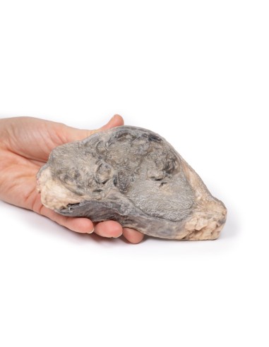
Breast Carcinoma - Erler Zimmer 3D anatomy Series MP2105
erler zimmer
EZ-MP2105
€507.28
Tax included
Made in ultra-high resolution 3D printing in full color.
Breast Carcinoma - Erler Zimmer 3D anatomy Series MP2105
This dissection model highlighting Breast Carcinoma is part of the exclusive Monash 3D anatomy series, a comprehensive series of human dissections reproduced with ultra-high resolution color 3D printing.
Clinical history
A 76-year-old woman presented to the emergency department with a sudden loss of consciousness. She had signs of a left-sided cerebrovascular accident. She was intubated and her stroke was treated. During her admission to the intensive care unit, a fixed mass was noted in the left breast with palpable lymphadenopathy in the left axilla. She died of respirator-associated pneumonia.
Pathology
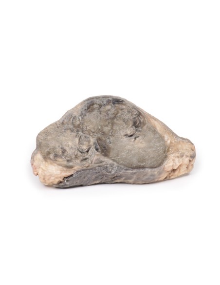
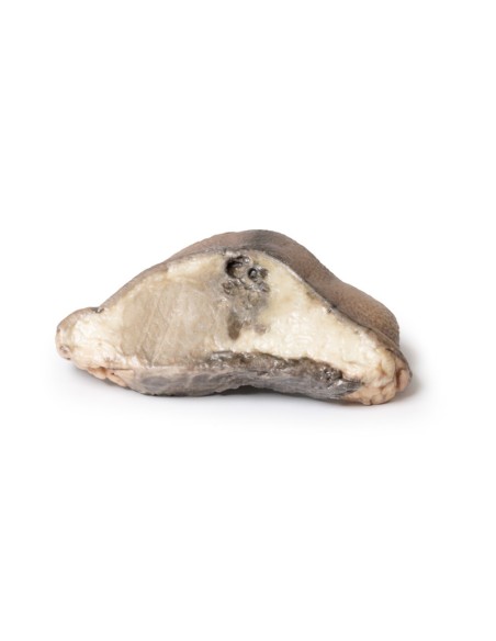
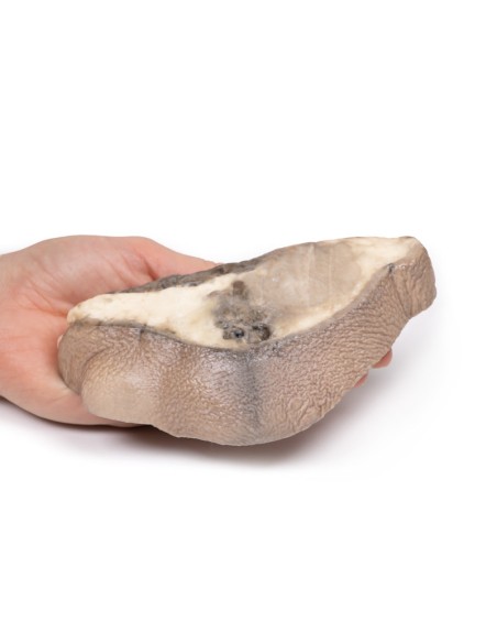
The specimen is the patient's left breast mounted to visualize the cut surface. Immediately below and attached to the skin is a large oval tumor mass 11 cm in maximum diameter. The tumor is adherent to the underlying muscle. The tumor is not encapsulated and has a variegated cut surface with areas of necrosis, hemorrhage, and cyst formation. This is a breast adenocarcinoma, which has involved regional lymph nodes.
Further information
Breast carcinoma is the second most commonly diagnosed cancer in women worldwide. It is rare in women younger than 30 years of age, but the incidence increases significantly after age 30 with the peak occurring between the ages of 70 and 80. The incidence has decreased since the introduction of breast cancer screening programs, which offer mammography to women at risk and public awareness and education on breast self-examination. However, breast cancer remains a leading cause of cancer death in women.
Major risk factors for the development of breast cancer include female sex (men account for 1 percent of breast cancer diagnoses), estrogen exposure (early menarche, late menopause, exogenous estrogen), family history of breast cancer, nulliparous, non-breastfeeding, radiation exposure, and obesity. Germline mutations in oncosuppressor genes, such as BRCA1, BRCA2, TP53, ATM, CDH1 and CHEK2, are linked to some hereditary cases of breast cancer.
Most breast neoplasms are adenocarcinomas that start in the duct/lobular system as carcinoma in situ (DCIS). These neoplasms are further subdivided according to their expression of estrogen receptors (ER) and human epidermal growth factor 2 (HER2), which guides treatment. The most frequent sites for distant metastasis are bone, liver, lung and brain.
In developed countries with screening programs, most patients present after an abnormal mammogram. Symptomatic patients present with a breast mass that is classically hard, irregular, and immobile. Other clinical symptoms are axillary lymphadenopathy, overlying skin changes (erythematous or thickened skin, rippled skin (peau d'orange), and nipple retraction. Symptoms of distant spread of the disease may also cause the patient to present.
Treatment depends on the stage of the disease and the ER and HER2 status of the tumor. Surgical treatments include single- or bilateral mastectomy or conservative breast lumpectomy. Surgical clearance of the axillary lymph node is performed in cases with positive lymph node disease. Radiation therapy is given to patients at high risk of local recurrence. Patients with HER2-positive tumors are treated with targeted drugs, such as trastuzumab (Herceptin). Patients with ER-positive tumors can be treated with antiestrogen therapy, such as tamoxifen. Systemic chemotherapy is also used to treat some patients with breast cancer.
What advantages does the Monash University anatomical dissection collection offer over plastic models or plastinated human specimens?
- Each body replica has been carefully created from selected patient X-ray data or human cadaver specimens selected by a highly trained team of anatomists at the Monash University Center for Human Anatomy Education to illustrate a range of clinically important areas of anatomy with a quality and fidelity that cannot be achieved with conventional anatomical models-this is real anatomy, not stylized anatomy.
- Each body replica has been rigorously checked by a team of highly trained anatomists at the Center for Human Anatomy Education, Monash University, to ensure the anatomical accuracy of the final product.
- The body replicas are not real human tissue and therefore not subject to any barriers of transportation, import, or use in educational facilities that do not hold an anatomy license. The Monash 3D Anatomy dissection series avoids these and other ethical issues that are raised when dealing with plastinated human remains.
No reviews
Tap to zoom
