Your cart
There are no more items in your cart


Uterine Leiomyoma - Erler Zimmer 3D anatomy Series MP2107
erler zimmer
EZ-MP2107
€348.92
Tax included
Made in ultra-high resolution 3D printing in full color.
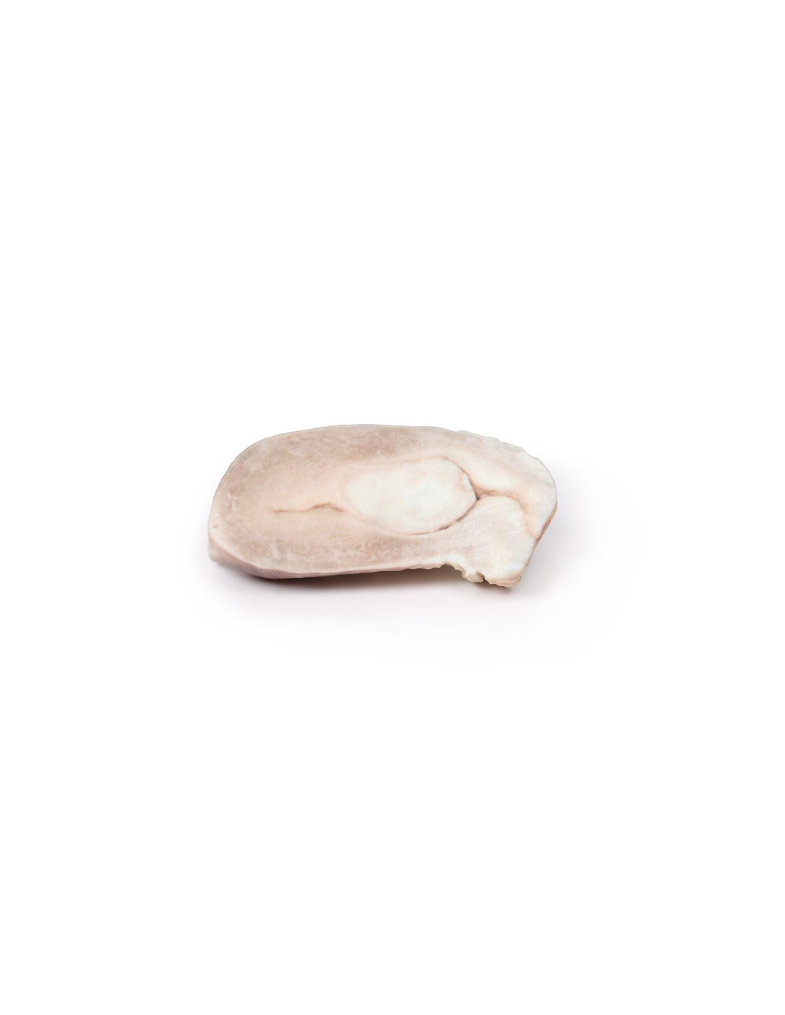
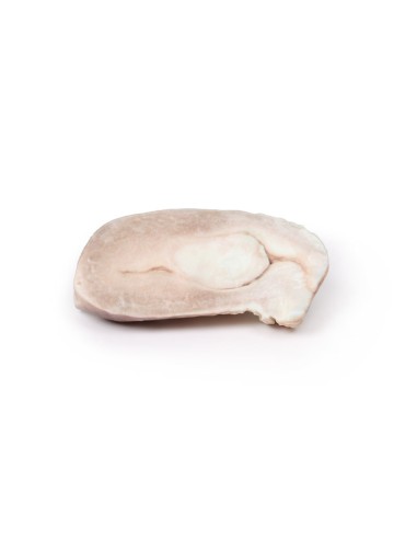
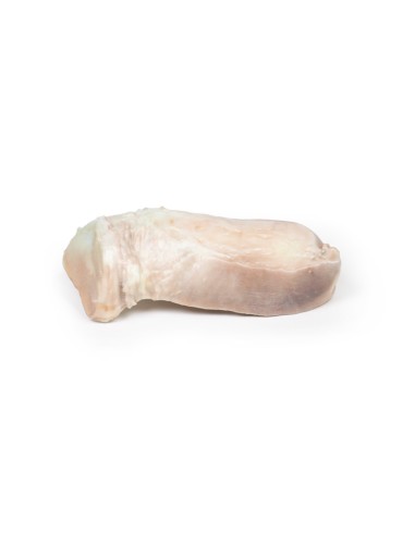
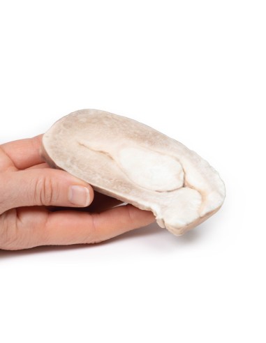
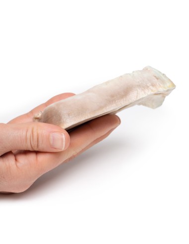
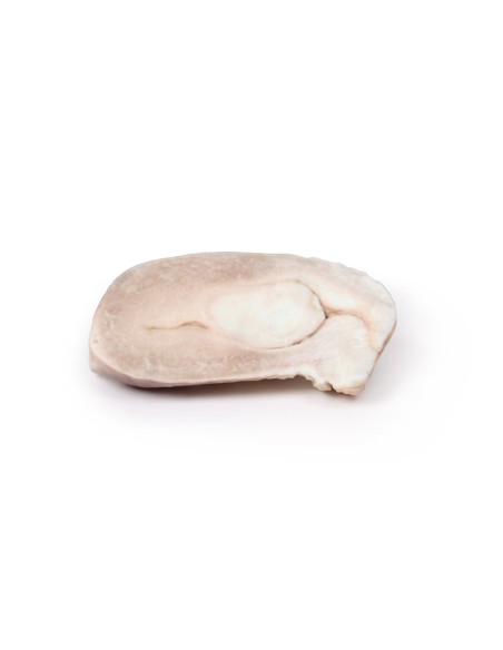
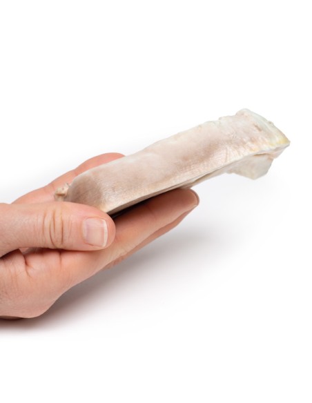
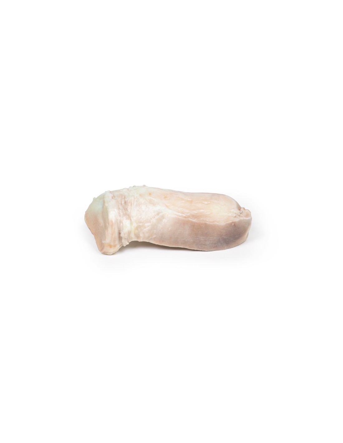
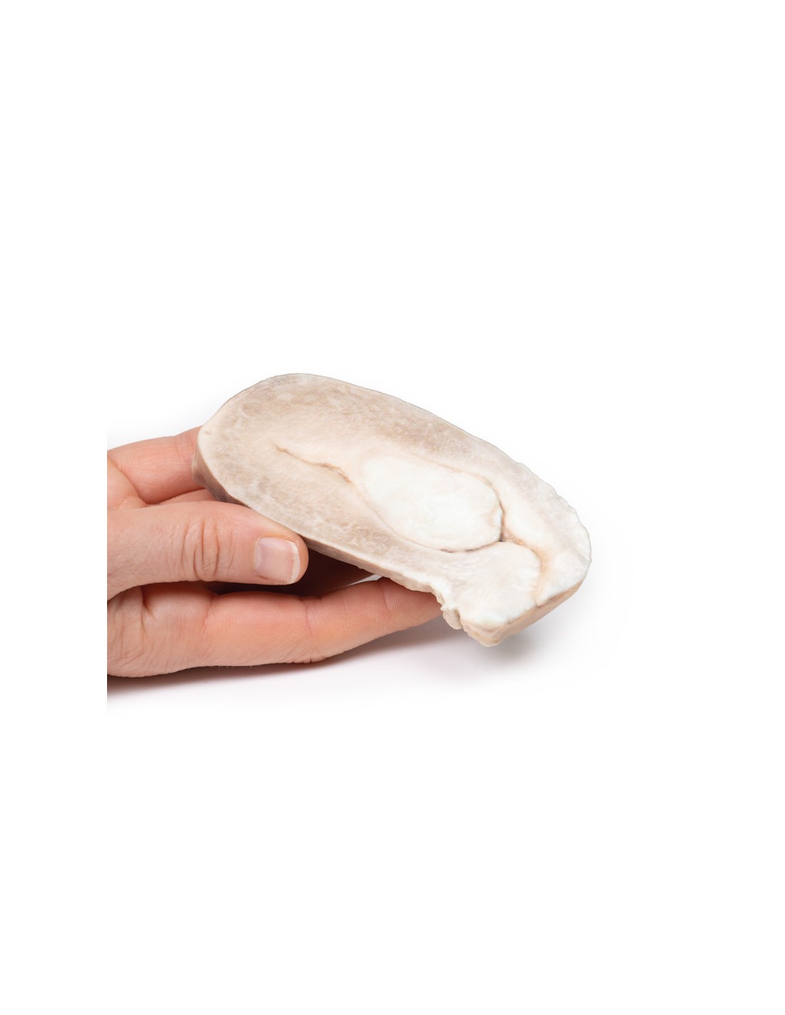
Uterine Leiomyoma - Erler Zimmer 3D anatomy Series MP2107
This dissection model highlighting a uterine leiomyoma (or uterine myoma) is part of the exclusive Monash 3D anatomy series, a comprehensive series of human dissections reproduced with ultra-high resolution color 3D printing.
Clinical history
A 30-year-old woman presents with inability to conceive. She also reports a history of intermittent pelvic discomfort, menorrhagia, and painful periods. On examination, a pelvic mass was palpable. All her blood tests were within normal range. A pelvic ultrasound showed a hypoechogenic mass within the myometrium of her uterus. She had a hysteroscopic myomectomy, but unfortunately complications meant that her surgery was converted to an emergency hysterectomy. She made a complete postoperative recovery.
Pathology
The specimen includes the cervix, body, and fundus of the uterus. The normal-sized uterus was cut in the sagittal plane. A large ovoid mass approximately 4 cm x 2 cm protrudes into the uterine cavity and extends inferiorly to the opening of the cervix. It originates from the posterior part of the uterus. The cervical canal is clearly visible.
Further information
Uterine leiomyomas, also called fibroids, are the most common pelvic tumors in females. They are present in nearly 25% of reproductive females. They are benign tumors arising from the smooth muscle and fibroblasts of the myometrium. They usually involve the myometrium of the uterine body. Rarely they may involve the lower uterus or cervix. Leiomyomas can occur as single or multiple lesions and can become very large. There are rare variants, which can extend and spread distally, but are still considered benign: e.g., benign metastasizing leiomyoma, which commonly spreads to the lining; or disseminated peritoneal leiomyomatosis, which appears on the peritoneum covering the uterus
Risk factors for fibroid development include reproductive age, being a black woman, and early menarche. Greater parity has been found to be protective. Most leiomyomas have normal karyotypes, but there are some that show mutations in HMG genes. Transformation to malignant leiomyosarcoma is very rare.
Common symptoms of uterine fibroids include abnormal vaginal bleeding, pelvic pain, dyspareunia, dysmenorrhea, and pelvic structure compression symptoms such as urinary symptoms or venous compression symptoms. Leiomyomas can decrease fertility and in pregnant women increase the rate of early pregnancy loss, fetal malpresentation, and postpartum hemorrhage. Pelvic ultrasound is usually used to diagnose leiomyomas. CT and MRI scans are rarely used to diagnose.
Leiomyomas can grow but can also regress. Treatment is reserved for persistent or severely symptomatic fibroids. Hormonal treatment may be used to regulate symptoms of irregular menstrual bleeding. Surgical treatments include myomectomy (removal of fibroids by myomectomy), hysterectomy, myolysis (thermal ablation of leiomyoma), and uterine artery ablation/embolization.
What advantages does the Monash University anatomical dissection collection offer over plastic models or plastinated human specimens?
- Each body replica has been carefully created from selected patient X-ray data or human cadaver specimens selected by a highly trained team of anatomists at the Monash University Center for Human Anatomy Education to illustrate a range of clinically important areas of anatomy with a quality and fidelity that cannot be achieved with conventional anatomical models-this is real anatomy, not stylized anatomy.
- Each body replica has been rigorously checked by a team of highly trained anatomists at the Center for Human Anatomy Education, Monash University, to ensure the anatomical accuracy of the final product.
- The body replicas are not real human tissue and therefore not subject to any barriers of transportation, import, or use in educational facilities that do not hold an anatomy license. The Monash 3D Anatomy dissection series avoids these and other ethical issues that are raised when dealing with plastinated human remains.
No reviews
Tap to zoom