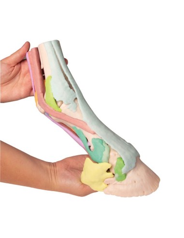
Horse foot reproduction Model 1 Erler Zimmer VET4400
erler zimmerMade of PVC
Horse foot reproduction Model 1 Erler Zimmer VET4400:
Anatomical model for veterinary study
This model of a stallion's foot was derived from CT and MRI data and is anatomically accurate and life-size. Produced using color 3D printing, each anatomical structure is depicted in a different color. The horn capsule is available separately and can be attached to the standing limb. Four models are available: from full anatomy, consisting of 25 colored structures, to progressively reduced models in which deeper structures are visible.
Model 1:
Structures: Pastoral-navicular ligaments; common extensor of fingers; collateral ligaments of the coffin joint; collateral ligaments of the pastern joint; collateral ligaments of the coronal joint and ligaments of the palmar coronal joint; crossed bands on equal legs; deep flexor tendon; triangular bone; navicular bone; navicular bone-triangular bone ligament (Lig. impar); horn capsule; cannon bone; coronary bone; oblique ligaments of the equal leg; Axial palmar ligaments; metacarpal; proximal scutum; Palmar ligament of equal legs; equal legs; superficial flexor tendon; short ligaments of the same leg; suspensory ligament (M. interosseus) and its supporting branches; straight band for equal legs.
Why buy it
Erler Zimmer Anatomical Models are the most widely used teaching aids for the study of veterinary anatomy in schools and universities. High quality, faithfulness to detail, excellent value for money.
Ideal for teaching, providing clarification to patients, and for scientific medical training.
They can also be used as furnishing accessories to personalize one's medical office.
- Weight
- 1


