Your cart
There are no more items in your cart

Craniopharyngioma - Erler Zimmer 3D anatomy Series MP2017
erler zimmer
EZ-MP2017
€1,040.05
Tax included
Made in ultra-high resolution 3D printing in full color.
Craniopharyngioma - Erler Zimmer 3D anatomy Series MP2017
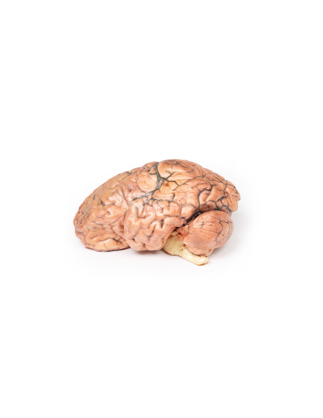
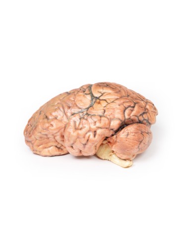
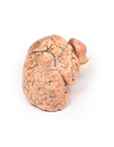
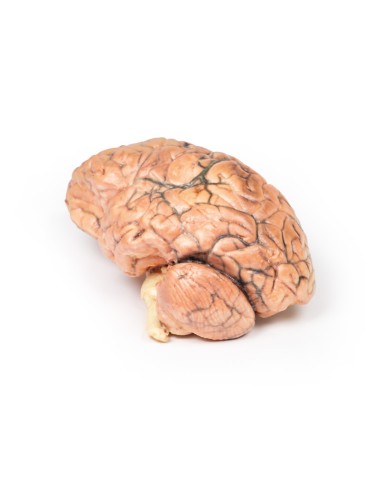
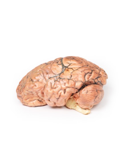
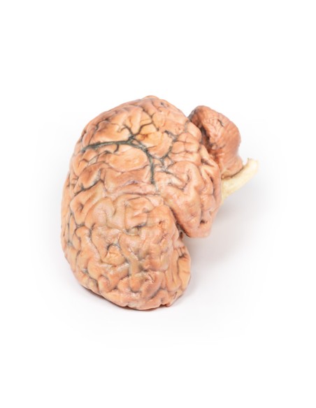
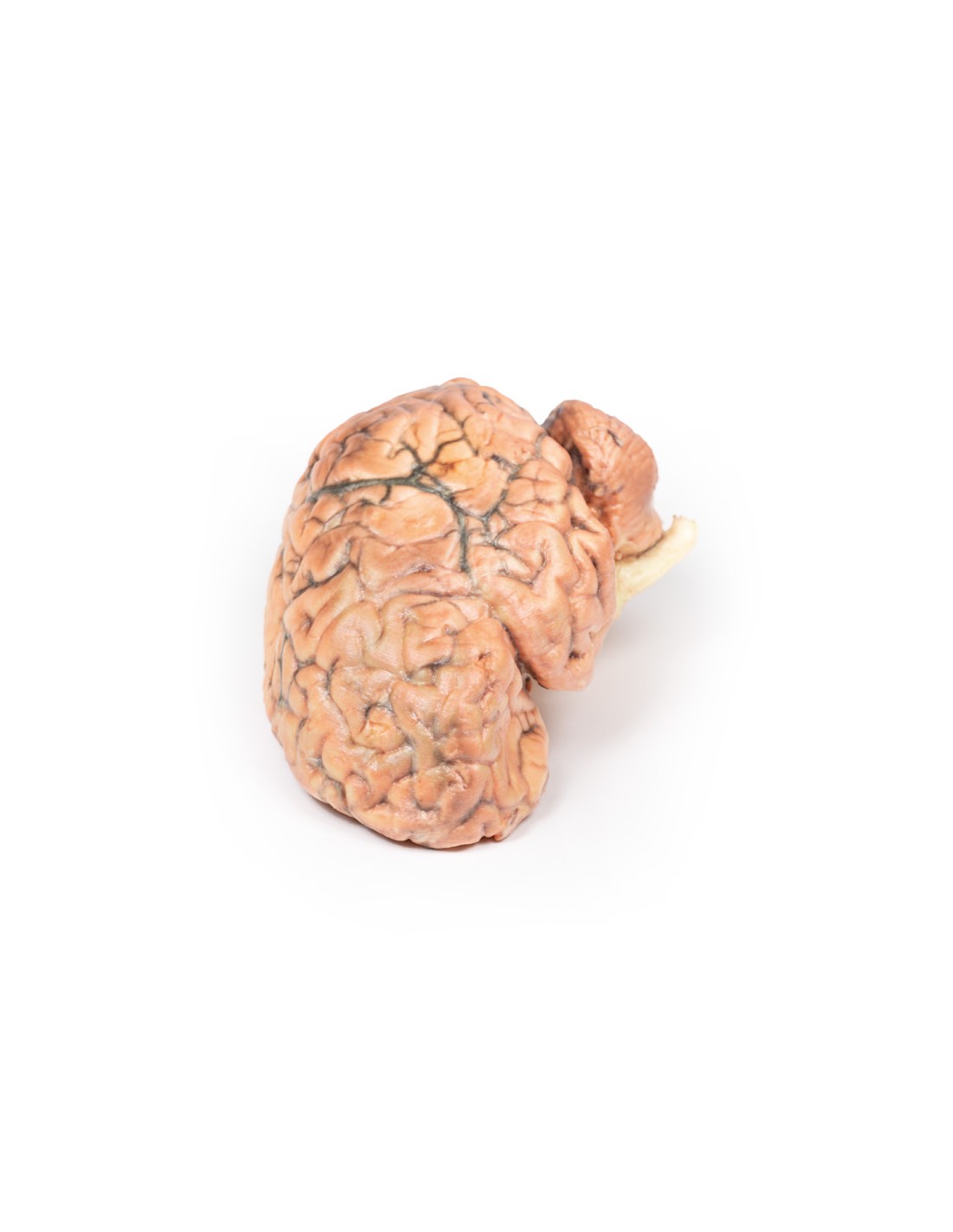
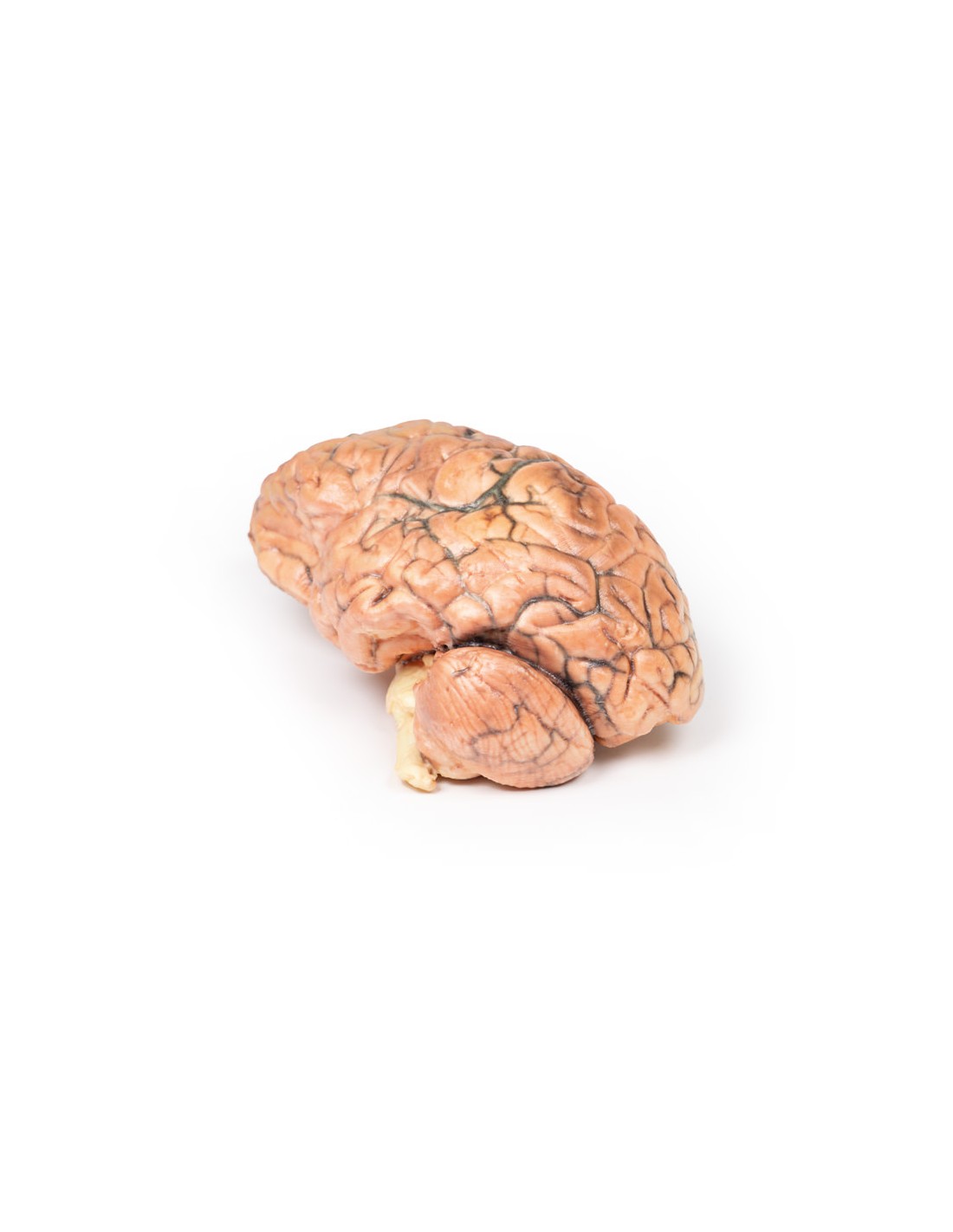
This dissection model highlighting a Craniopharyngioma is part of the exclusive Monash 3D anatomy series, a comprehensive series of human dissections reproduced with ultra-high resolution color 3D printing.
Clinical History.
A 62-year-old woman presented with disorientation with respect to time, place, and person. Objective examination revealed no localized neurological signs. Radiological investigations revealed a space-occupying lesion in the floor of the 3rd ventricle. The tissue was removed during surgery, but the lesion could not be completely excised. Histology confirmed the diagnosis of craniopharyngioma. After surgery, the patient developed complex metabolic disorders, probably of hypothalamic origin. She gradually deteriorated, and 10 weeks after admission she died following an episode of gastric aspiration.
Pathology
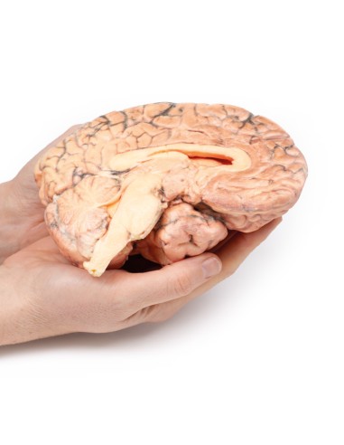
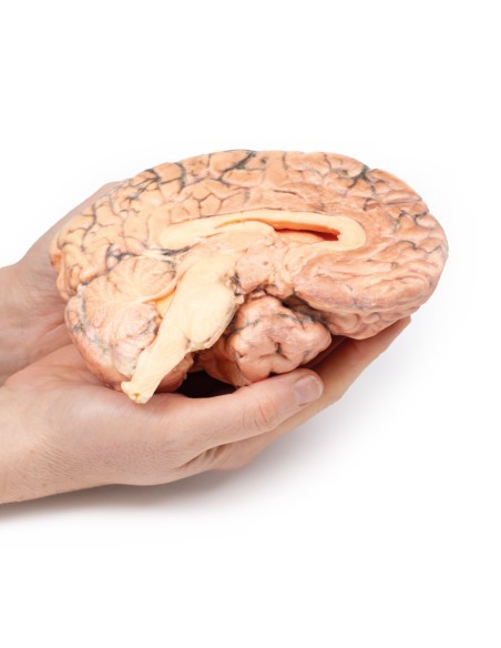
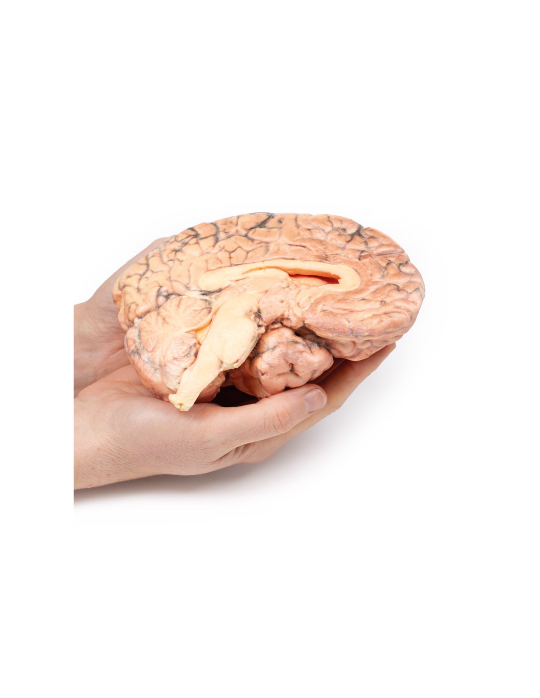
The brain was dissected in the sagittal plane, showing the medial surface. A pink-gray ovoid tumor measuring 2.5 x 1.5 cm on the cut surface is centered in the hypothalamus region. It is encapsulated except at its ventral pole where tissue was removed in a previous surgery, and the cut surface reveals a microcystic or spongy appearance. The tumor distorts the 3rd ventricle and extends to obliterate Munro's foramen. The optic chiasm is displaced caudally (arrow). The previous ventriculoatrial shunt prevented dilatation of the lateral ventricles despite this obstruction.
Additional Information.
Craniopharyngiomas constitute 1-3% of all brain tumors and 5-10% in children, with a bimodal distribution favoring age 5-14 years, and a second peak between 50-75 years. There is a higher incidence in Japan and parts of Africa. Craniopharyngiomas are epithelial tumors generally originating in the pituitary pedicle. Other sites of origin include the sella turcica, optic system and third ventricle. Solid and cystic components are often present, the latter containing cholesterol crystals. Craniopharyngiomas can be divided into two categories: adamantinomatous and papillary types, each with distinct histology and genetic alterations, although the prognostic significance of these types remains unclear.
Treatment includes surgical resection and radiation therapy (RT) for treatment and post-surgical residual disease. The prognosis depends largely on tumor resection, control, and treatment-related complications from endocrine and local sequelae and local sequelae.
What advantages does the Monash University anatomical dissection collection offer over plastic models or plastinated human specimens?
- Each body replica has been carefully created from selected patient X-ray data or human cadaver specimens selected by a highly trained team of anatomists at the Monash University Center for Human Anatomy Education to illustrate a range of clinically important areas of anatomy with a quality and fidelity that cannot be achieved with conventional anatomical models-this is real anatomy, not stylized anatomy.
- Each body replica has been rigorously checked by a team of highly trained anatomists at the Center for Human Anatomy Education, Monash University, to ensure the anatomical accuracy of the final product.
- The body replicas are not real human tissue and therefore not subject to any barriers of transportation, import, or use in educational facilities that do not hold an anatomy license. The Monash 3D Anatomy dissection series avoids these and other ethical issues that are raised when dealing with plastinated human remains.
No reviews
Tap to zoom