
Your cart
There are no more items in your cart


Left cerebral infarction - Erler Zimmer 3D anatomy Series MP2005
erler zimmer
EZ-MP2005
€307.32
Tax included
Made in ultra-high resolution 3D printing in full color.
Left Cerebral Infarction - Erler Zimmer 3D anatomy Series MP2005
This dissection model highlighting a left cerebral infarction is part of the exclusive Monash 3D anatomy series, a comprehensive series of human dissections reproduced with ultra-high resolution color 3D printing.
Clinical history.
The patient was a 51-year-old woman who had had a cerebrovascular accident resulting in left hemiplegia 2 years before her death. At autopsy, she had severe generalized atherosclerosis and an old left ventricular myocardial infarction with overlying mural thrombus.
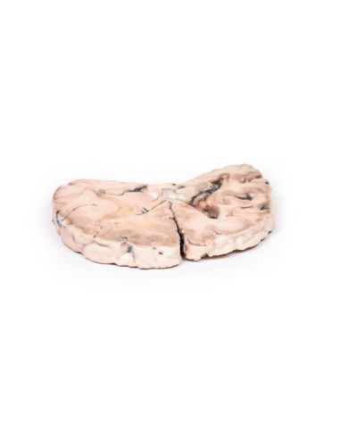
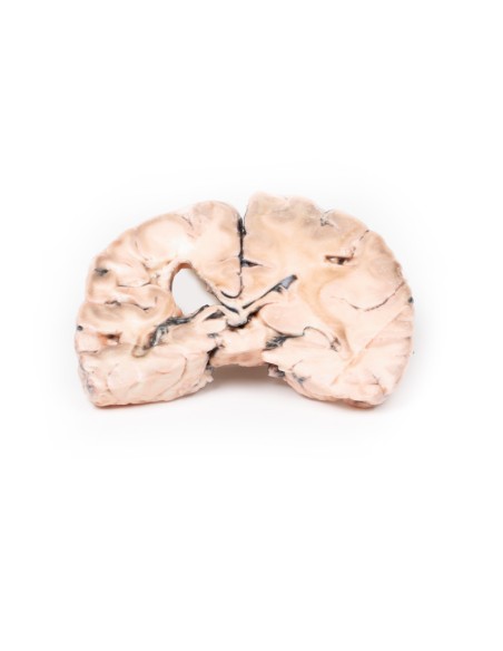
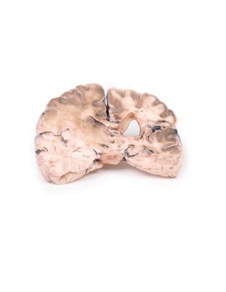
Pathology
A coronal section of the cerebral hemispheres shows irregular cystic cavities in the distribution territory of the right middle cerebral artery. The infarct cavities have irregular, yellow walls and show partial collapse. There is compensatory dilatation of the left lateral ventricle. On the posterior side, the arteries under the mammillary bodies were moderately atheromatous, although this is difficult to visualize macroscopically.
Additional Information.
Because of the underlying history of myocardial disease with the presence of mural thrombus, it is assumed that her cerebral infarction was probably caused by thromboembolism.
What advantages does the Monash University anatomical dissection collection offer over plastic models or plastinated human specimens?
- Each body replica has been carefully created from selected patient X-ray data or human cadaver specimens selected by a highly trained team of anatomists at the Monash University Center for Human Anatomy Education to illustrate a range of clinically important areas of anatomy with a quality and fidelity that cannot be achieved with conventional anatomical models-this is real anatomy, not stylized anatomy.
- Each body replica has been rigorously checked by a team of highly trained anatomists at the Center for Human Anatomy Education, Monash University, to ensure the anatomical accuracy of the final product.
- The body replicas are not real human tissue and therefore not subject to any barriers of transportation, import, or use in educational facilities that do not hold an anatomy license. The Monash 3D Anatomy dissection series avoids these and other ethical issues that are raised when dealing with plastinated human remains.
No reviews
Tap to zoom
