Your cart
There are no more items in your cart


Made in ultra-high resolution 3D printing in full color.
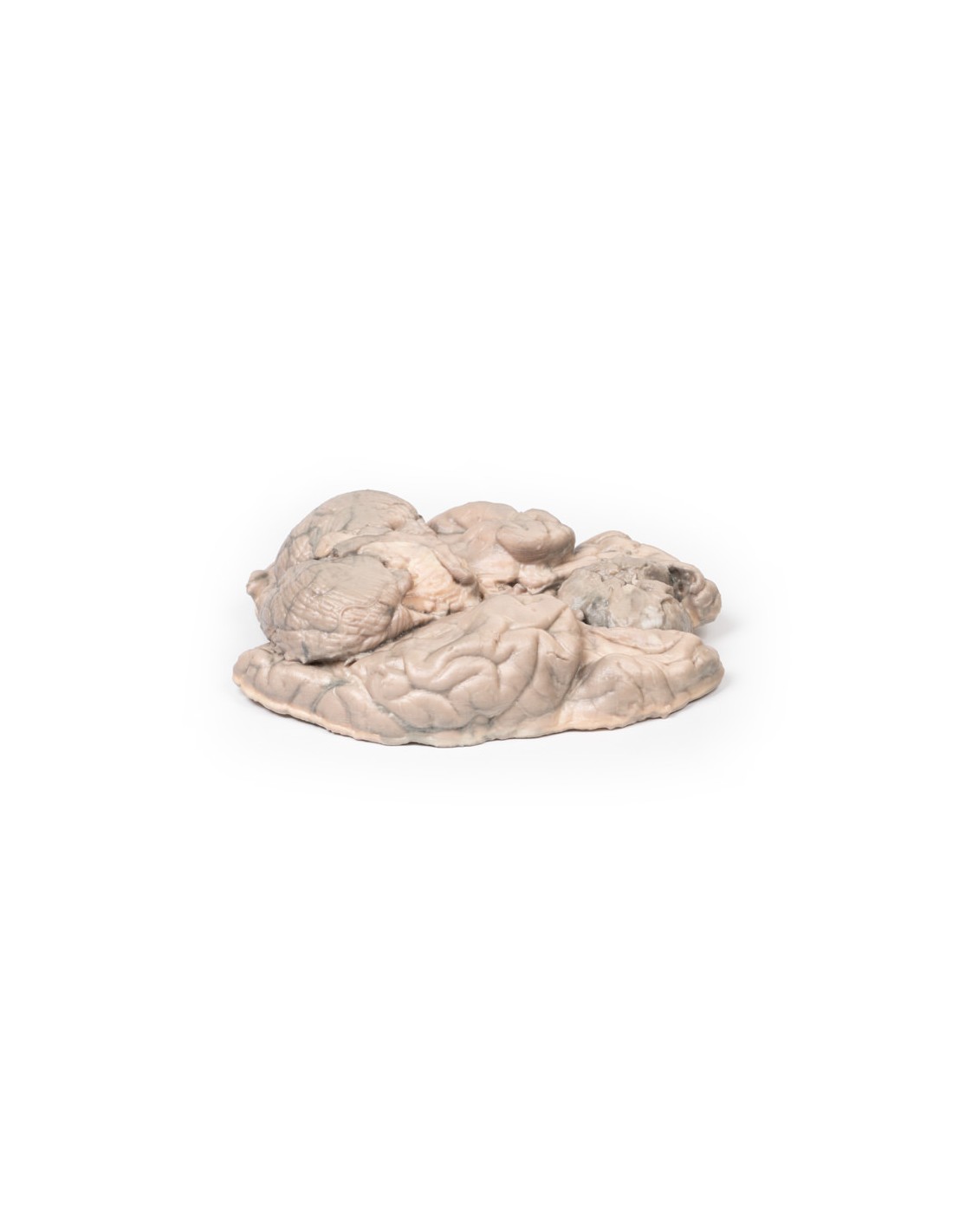
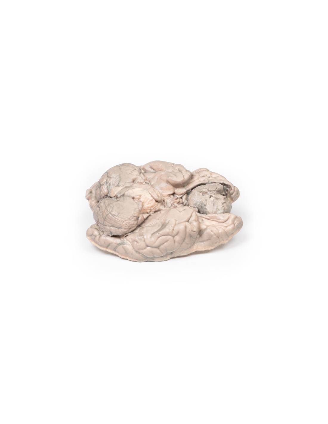
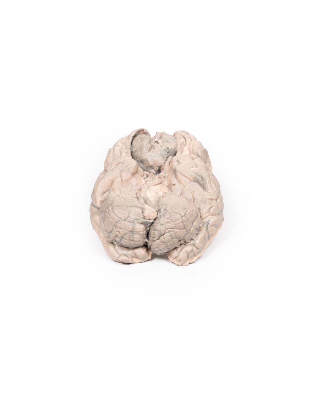
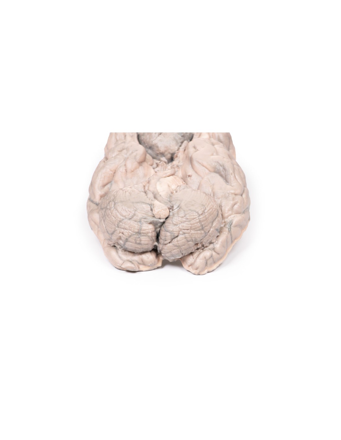
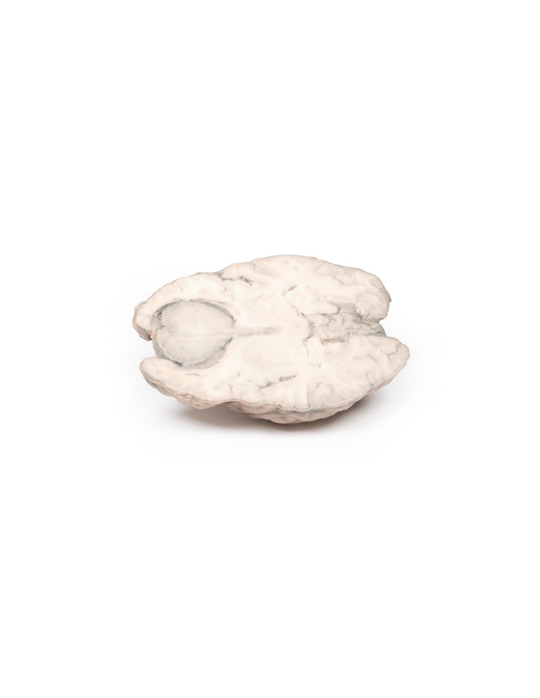
Meningioma - Erler Zimmer 3D anatomy Series MP2004
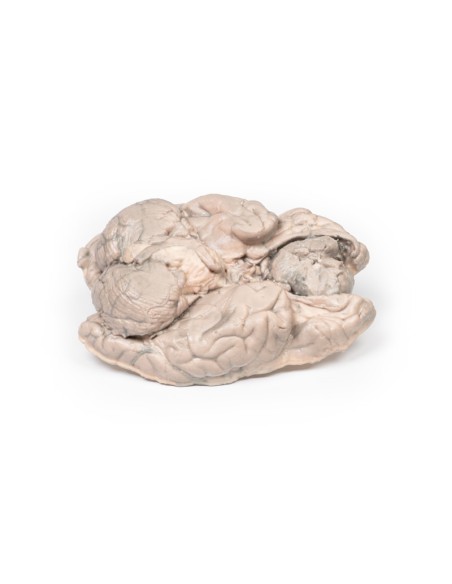
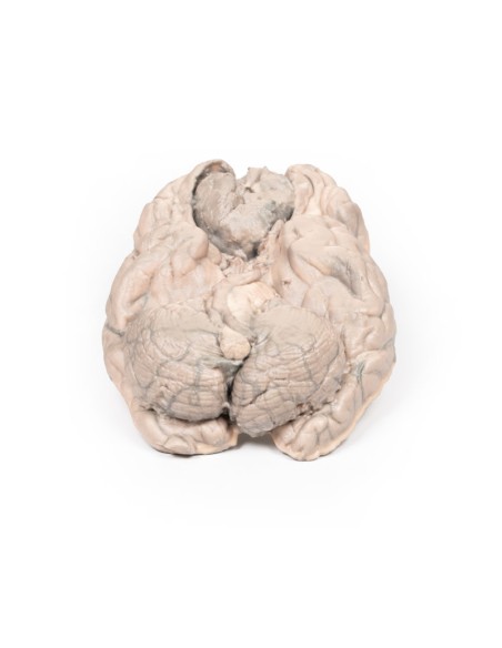
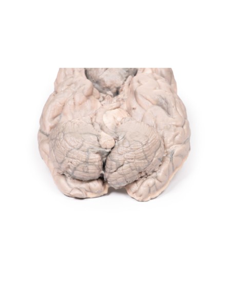
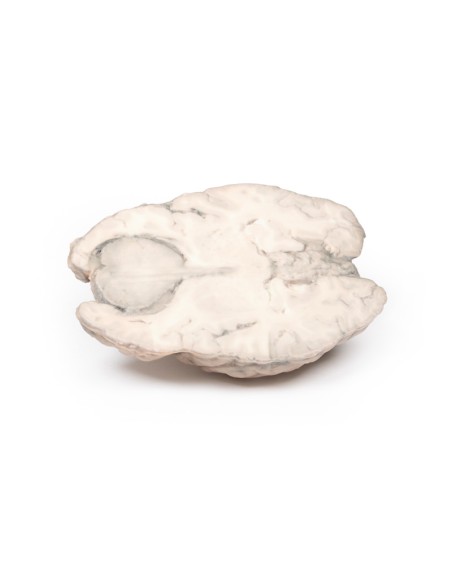
This dissection model highlighting a meningioma is part of the exclusive Monash 3D anatomy series, a comprehensive series of human dissections reproduced with very high resolution color 3D printing.
Clinical History.
A 68-year-old woman presented with a recent onset of seizures and was diagnosed with epilepsy. The collateral history revealed a gradual change in the patient's personality. She subsequently died several months later from a myocardial infarction.
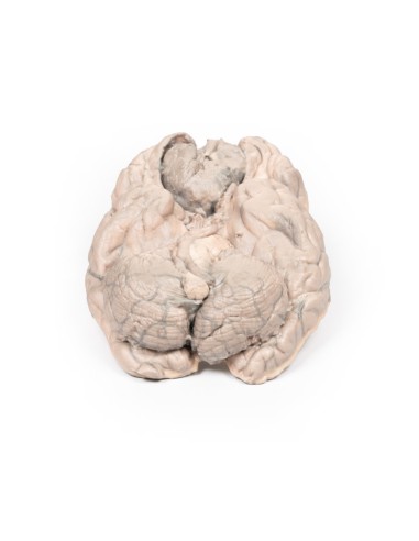
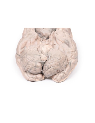
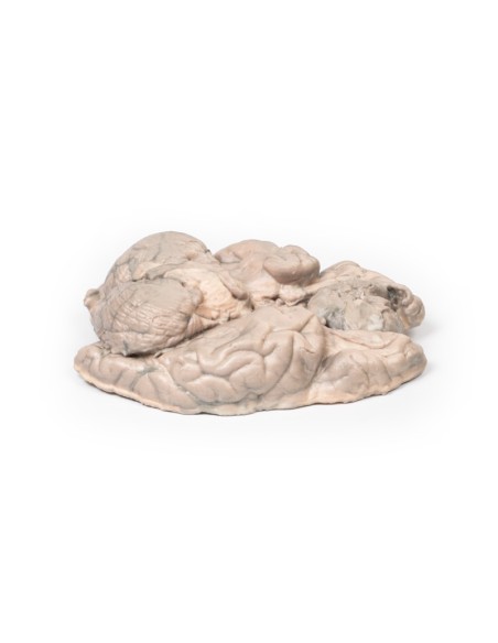
Pathology
This brain specimen was sliced horizontally. A well-circumscribed 6-cm tumor is evident between the two frontal lobes. The tumor is compressing the frontal lobes. It has a pinkish cut surface with some yellow areas indicating necrosis. It was attached to the dura anteriorly. This is an example of a meningioma.
Further information
Meningiomas are often said to be the most common tumors of the central nervous system (CNS); however, they actually arise in the meninges (dura, arachnoid and pia), which strictly speaking are not part of the CNS per se. They arise from arachnoid cells closely associated with the dura; therefore, these tumors may be associated with the dura or dural folds (falx cerebri and tentorium cerebelli). Meningiomas are predominantly slow-growing benign tumors. Symptoms are determined by the location of the tumor and the rate of growth. Symptoms include seizures, change in mental status, changes in vision, hearing or smell, and symptoms of increased intracranial pressure. Meningiomas are often asymptomatic. Treatment includes observation, surgery, or radiation therapy, depending on the clinical setting and morphology of the tumor.
Meningiomas are rare in children with a median age of 65 years at diagnosis. There is a female predominance of 3:2. Exposure to ionizing radiation, including cranial radiotherapy, increases the risk of meningioma development. The greatest genetic predisposition to development is observed in patients with neurofibromatosis type 2 (NF2). NF2 is an autosomal dominant disease caused by mutations in the NF2 gene on chromosome 22 that leads to multiple tumors associated with the nervous system.
What advantages does the Monash University anatomical dissection collection offer over plastic models or plastinated human specimens?
- Each body replica has been carefully created from selected patient X-ray data or human cadaver specimens selected by a highly trained team of anatomists at the Monash University Center for Human Anatomy Education to illustrate a range of clinically important areas of anatomy with a quality and fidelity that cannot be achieved with conventional anatomical models-this is real anatomy, not stylized anatomy.
- Each body replica has been rigorously checked by a team of highly trained anatomists at the Center for Human Anatomy Education, Monash University, to ensure the anatomical accuracy of the final product.
- The body replicas are not real human tissue and therefore not subject to any barriers of transportation, import, or use in educational facilities that do not hold an anatomy license. The Monash 3D Anatomy dissection series avoids these and other ethical issues that are raised when dealing with plastinated human remains.
No reviews
Tap to zoom