Your cart
There are no more items in your cart

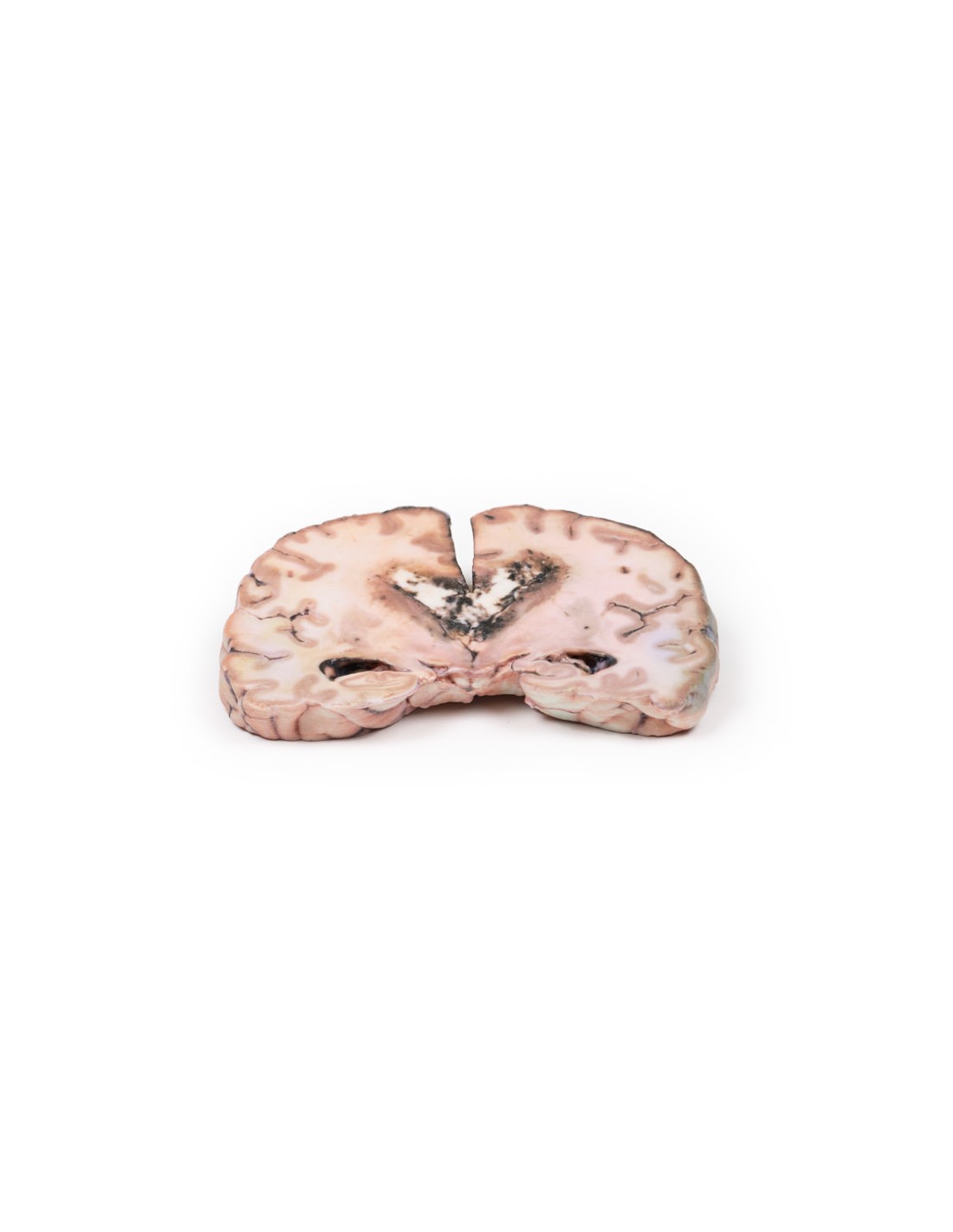
Grade 3-4 glioma that caused papilledema - Erler Zimmer 3D anatomy Series MP2003
erler zimmer
EZ-MP2003
€500.57
Tax included
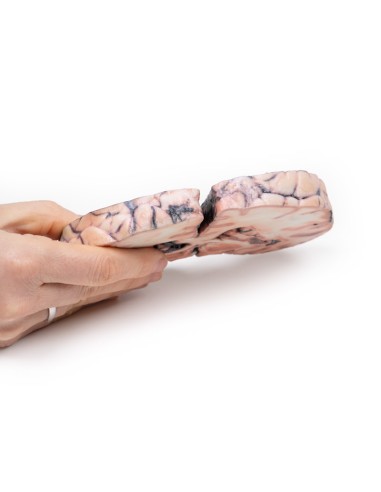
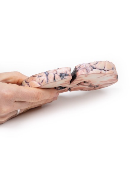
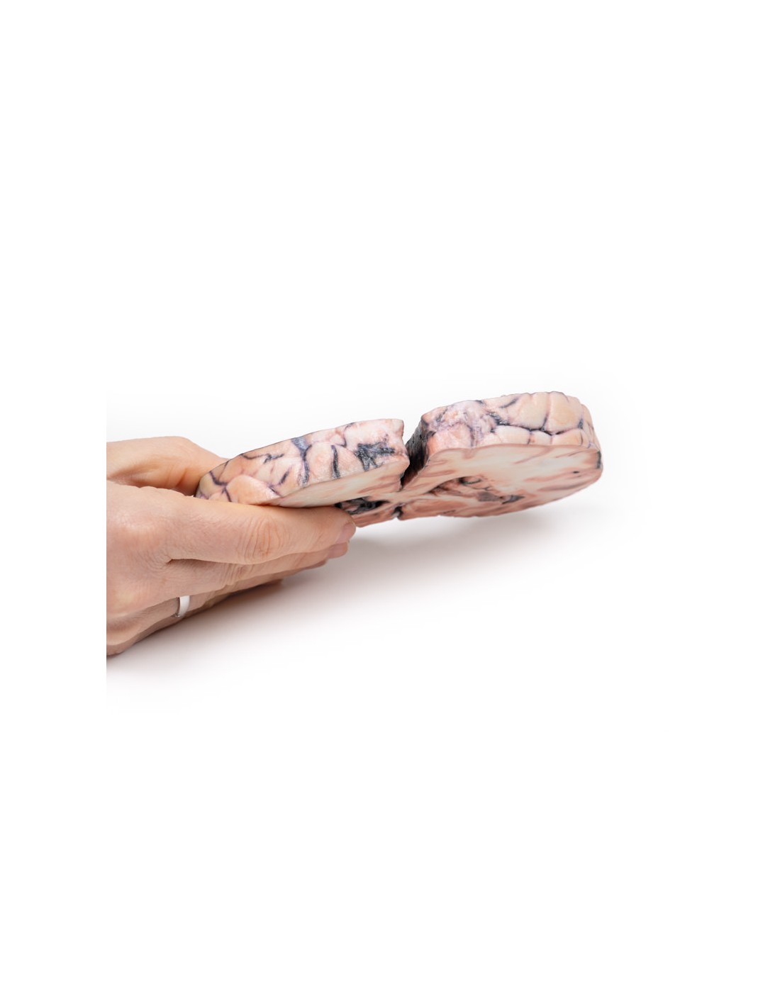
Made in ultra-high resolution 3D printing in full color.
Grade 3-4 glioma that caused papilledema - Erler Zimmer 3D anatomy Series MP2003
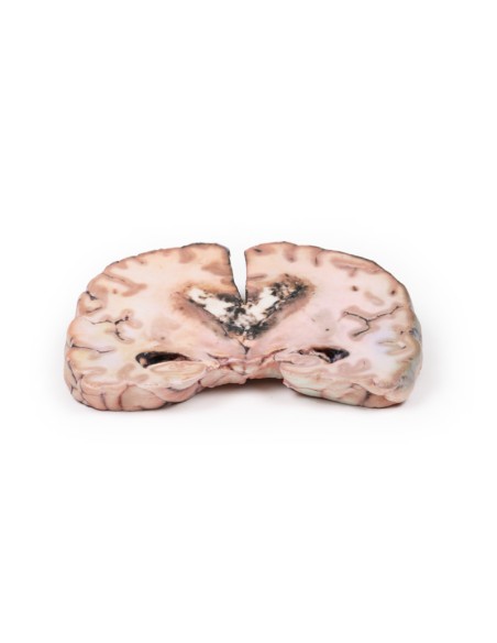
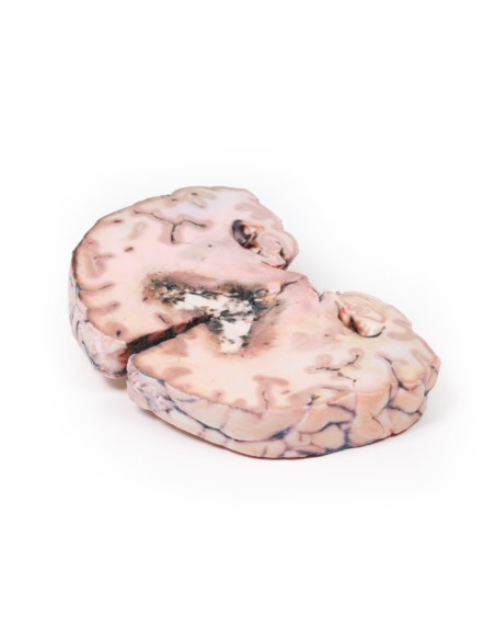
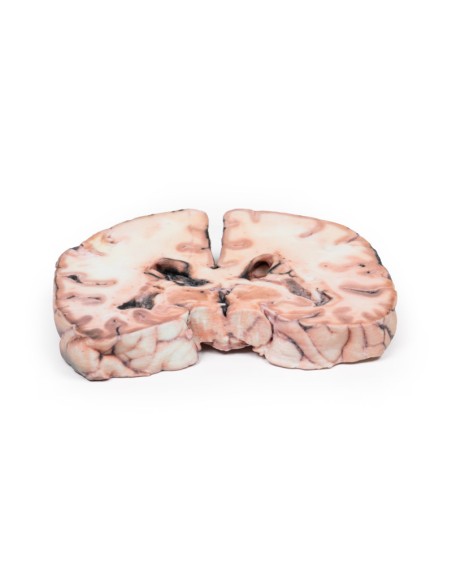
This glioma brain dissection model is part of the exclusive Monash 3D anatomy series, a comprehensive series of human dissections reproduced with ultra-high resolution color 3D printing.
Clinical History.
The patient was a 24-year-old woman who presented 18 months before death with an abnormal electroencephalogram (EEG) after a single seizure. Six months later she complained of blurred vision and headache. Bilateral papilledema was observed at ophthalmoscopy; however, there were no signs of localization to explain this. Further investigation showed a space-occupying mass, which was biopsied and diagnosed histologically as a grade III-IV glioma. The patient was treated with radiotherapy. One month after the start of treatment, the patient experienced weakness in her left arm and leg.
Shortly thereafter, she was hospitalized with drowsiness and vomiting; from then on, she deteriorated rapidly and died.
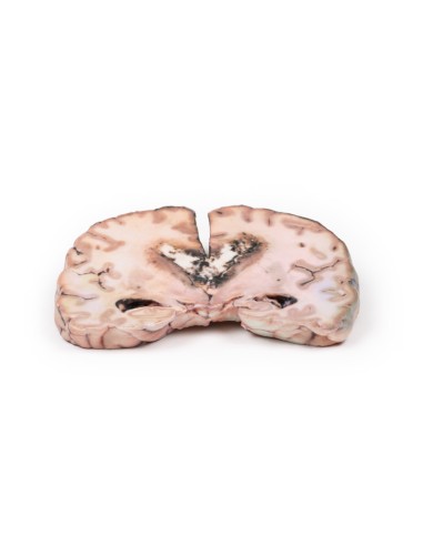
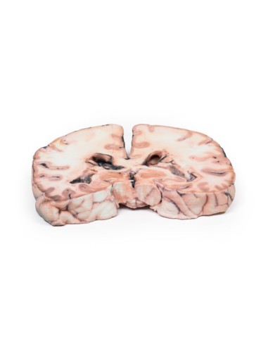
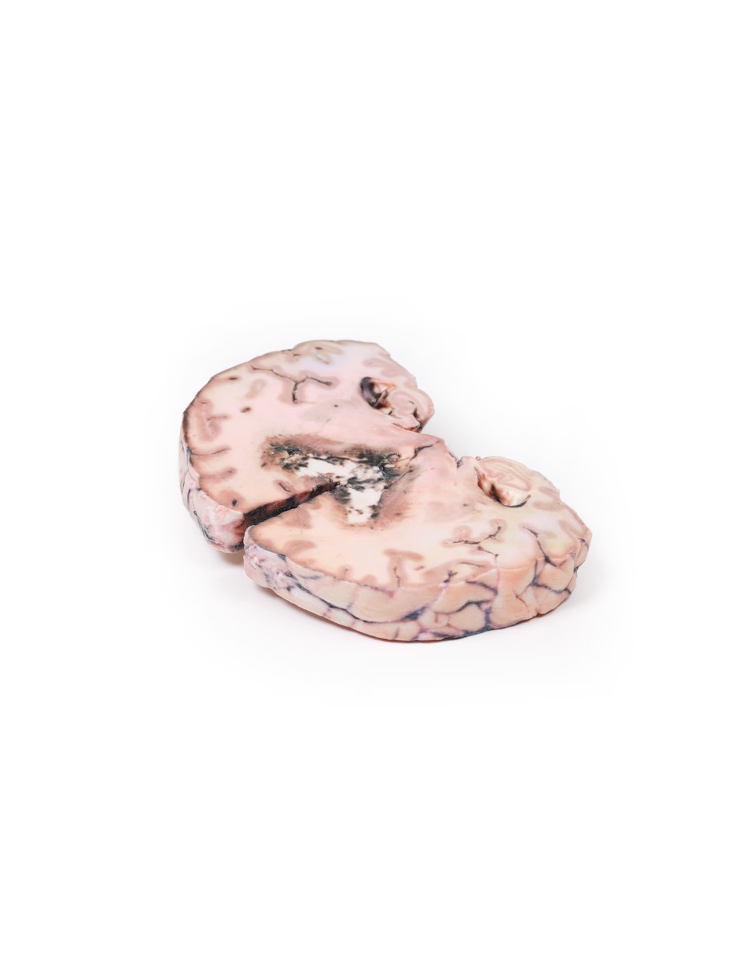
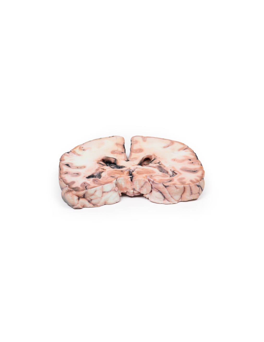
Pathology
The specimen shows a large intracerebral lesion, which has obliterated the lateral ventricles and the inner 2/3 of the internal capsule and basal ganglia on the right side. It is infiltrating through the corpus callosum and distorting the mesencephalic duct. The tumor is fairly well demarcated and vascular with numerous areas of hemorrhage and necrosis, resulting in its mottled and variegated appearance.
What advantages does the Monash University anatomical dissection collection offer over plastic models or plastinated human specimens?
- Each body replica has been carefully created from selected patient X-ray data or human cadaver specimens selected by a highly trained team of anatomists at the Monash University Center for Human Anatomy Education to illustrate a range of clinically important areas of anatomy with a quality and fidelity that cannot be achieved with conventional anatomical models-this is real anatomy, not stylized anatomy.
- Each body replica has been rigorously checked by a team of highly trained anatomists at the Center for Human Anatomy Education, Monash University, to ensure the anatomical accuracy of the final product.
- The body replicas are not real human tissue and therefore not subject to any barriers of transportation, import, or use in educational facilities that do not hold an anatomy license. The Monash 3D Anatomy dissection series avoids these and other ethical issues that are raised when dealing with plastinated human remains.
No reviews
Tap to zoom