Your cart
There are no more items in your cart


Metastatic melanoma - Erler Zimmer 3D anatomy Series MP2018
erler zimmer
EZ-MP2018
€857.54
Tax included
Made in ultra-high resolution 3D printing in full color.
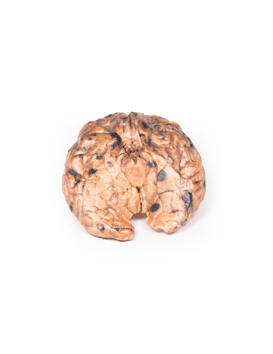
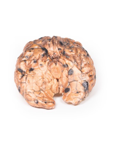
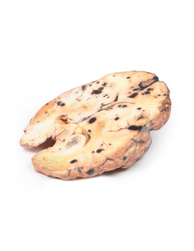
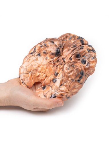
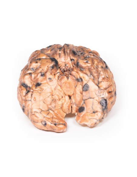
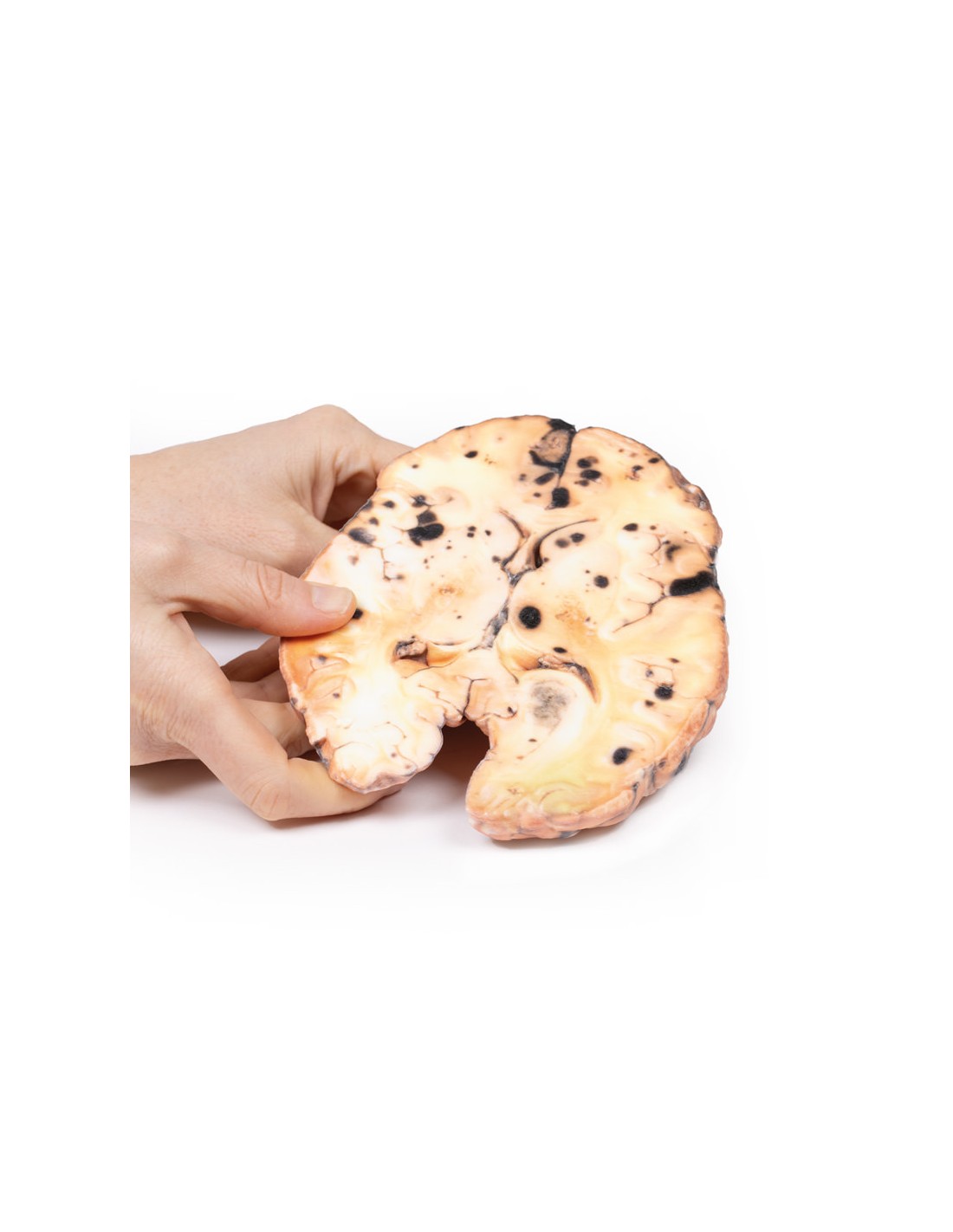
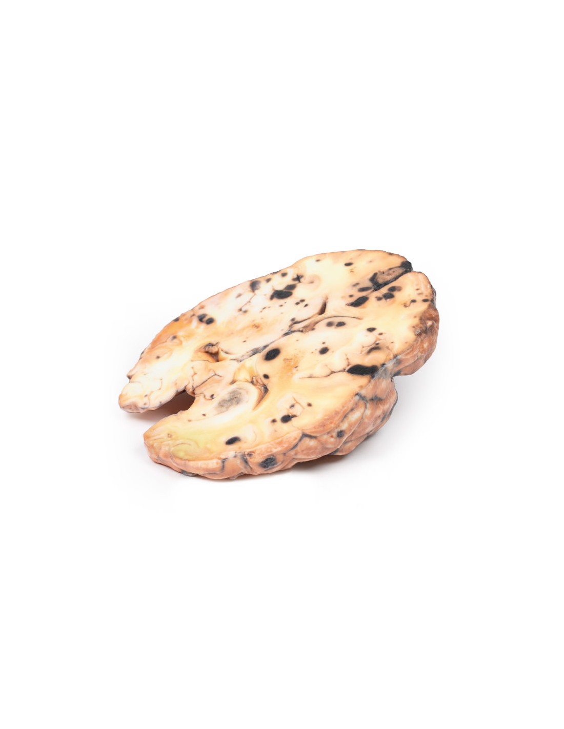
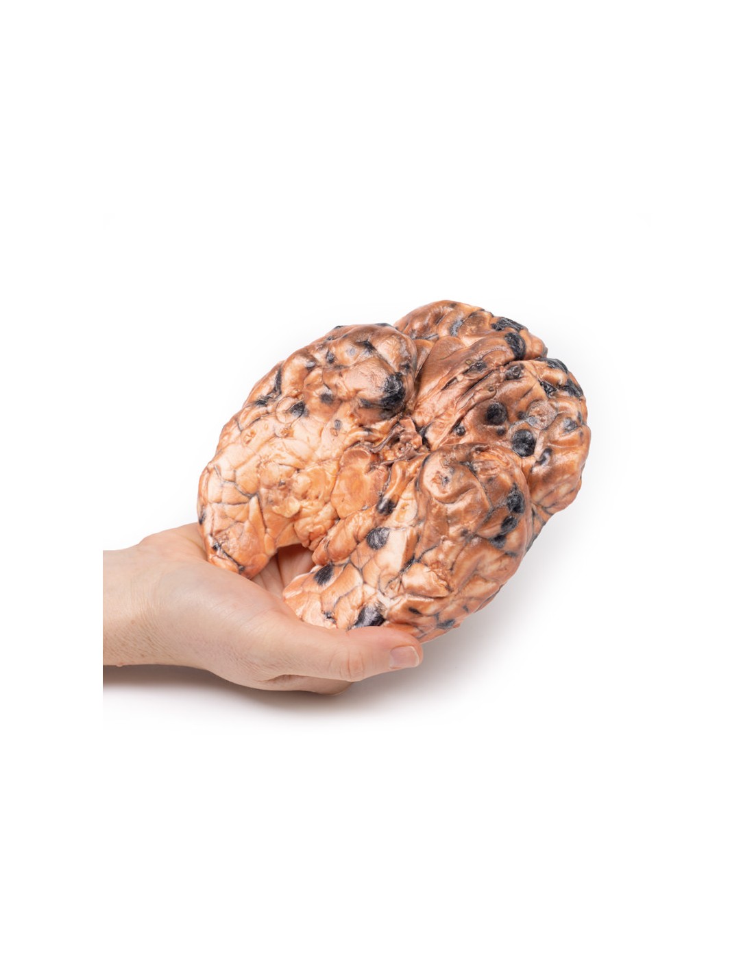
Metastatic Melanoma - Erler Zimmer 3D anatomy Series MP2018
This dissection model highlighting a metastatic Melanoma is part of the exclusive Monash 3D anatomy series, a comprehensive series of human dissections reproduced with ultra-high resolution color 3D printing.
Clinical history
In the 1970s, a 31-year-old woman presented with severe headache and diplopia against the background of a pigmented skin lesion (diagnosed as invasive cutaneous melanoma) removed from her neck 8 months earlier. Clinical examination revealed no abnormality, and after discharge the patient was subsequently admitted with persistent vomiting. Her condition worsened and she died.
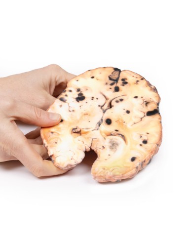
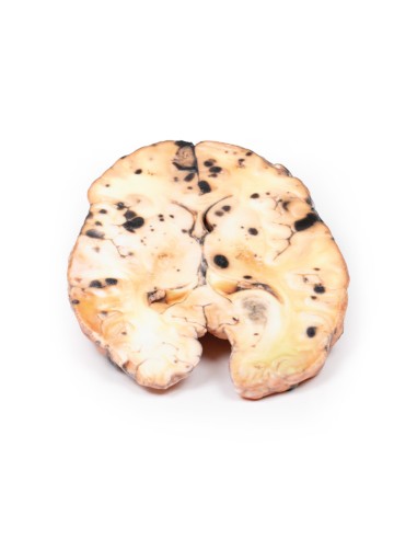
Pathology
This specimen shows diffuse metastases of intracerebral melanoma. The lower surface is characterized by numerous elevated dark nodules up to 1.5 cm in diameter. Similar lesions are present on the upper surface of the cut where it is seen that these secondary melanotic deposits are confined exclusively to the gray matter. The tumor deposits are not encapsulated and are invading the cortex. Some necrosis and hemorrhage are present.
Further information
Of all patients who have metastatic disease to the brain, 10% are from cutaneous melanoma. Risk increases with age over 60 years, male sex, duration of disease, and more advanced tumor/metastatic stage. BRAF and NRAS mutations, expression of CCR4 receptors on tumor cells, and activation of the PI3K pathway are all risk factors for the development of brain metastases. Eighty percent of brain metastases from melanoma are supratentorial. The presentation is often with headache, neurological deficits, and/or seizures. In addition, these lesions are at risk for spontaneous hemorrhage. Modern diagnosis is based on neuroimaging and often histology from a stereotactic brain biopsy, if a previous diagnosis has not been made. Treatment includes stereotactic radiosurgery (SRS), radiation therapy, and/or systemic therapy with "checkpoint inhibitor immunotherapy" or targeted treatments.
What advantages does the Monash University anatomical dissection collection offer over plastic models or plastinated human specimens?
- Each body replica has been carefully created from selected patient X-ray data or human cadaver specimens selected by a highly trained team of anatomists at the Monash University Center for Human Anatomy Education to illustrate a range of clinically important areas of anatomy with a quality and fidelity that cannot be achieved with conventional anatomical models-this is real anatomy, not stylized anatomy.
- Each body replica has been rigorously checked by a team of highly trained anatomists at the Center for Human Anatomy Education, Monash University, to ensure the anatomical accuracy of the final product.
- The body replicas are not real human tissue and therefore not subject to any barriers of transportation, import, or use in educational facilities that do not hold an anatomy license. The Monash 3D Anatomy dissection series avoids these and other ethical issues that are raised when dealing with plastinated human remains.
No reviews
Tap to zoom