Your cart
There are no more items in your cart


Intracranial injury - Erler Zimmer 3D anatomy Series MP2011
erler zimmer
EZ-MP2011
€516.67
Tax included
Made in ultra-high resolution 3D printing in full color.
Intracranial Lesion - Erler Zimmer 3D anatomy Series MP2011
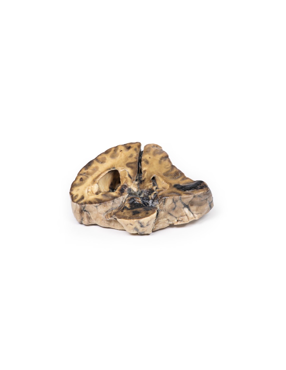
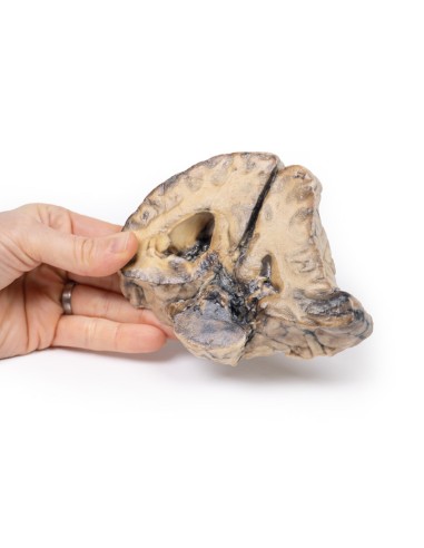
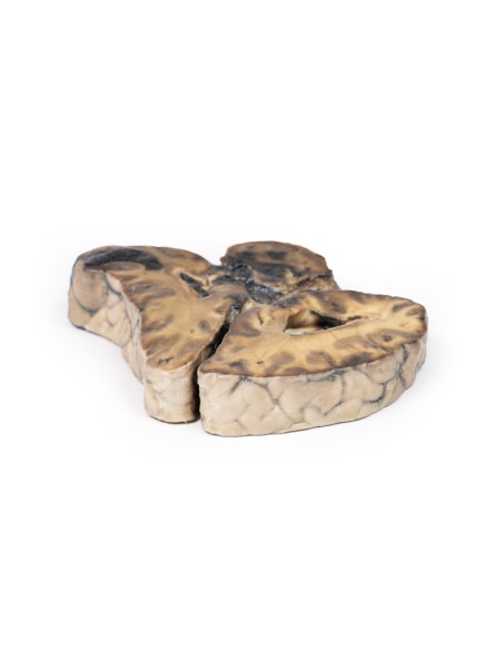
This dissection model highlighting an Intracranial Lesion is part of the exclusive Monash 3D anatomy series, a comprehensive series of human dissections reproduced with ultra-high resolution color 3D printing.
Clinical History.
A 56-year-old woman with 6 months of intermittent headaches and vomiting was admitted to the hospital in a coma after a grand mal seizure and did not regain consciousness.
Pathology
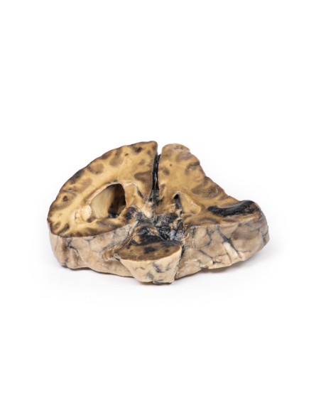
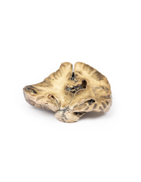
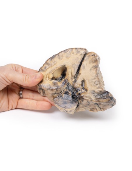
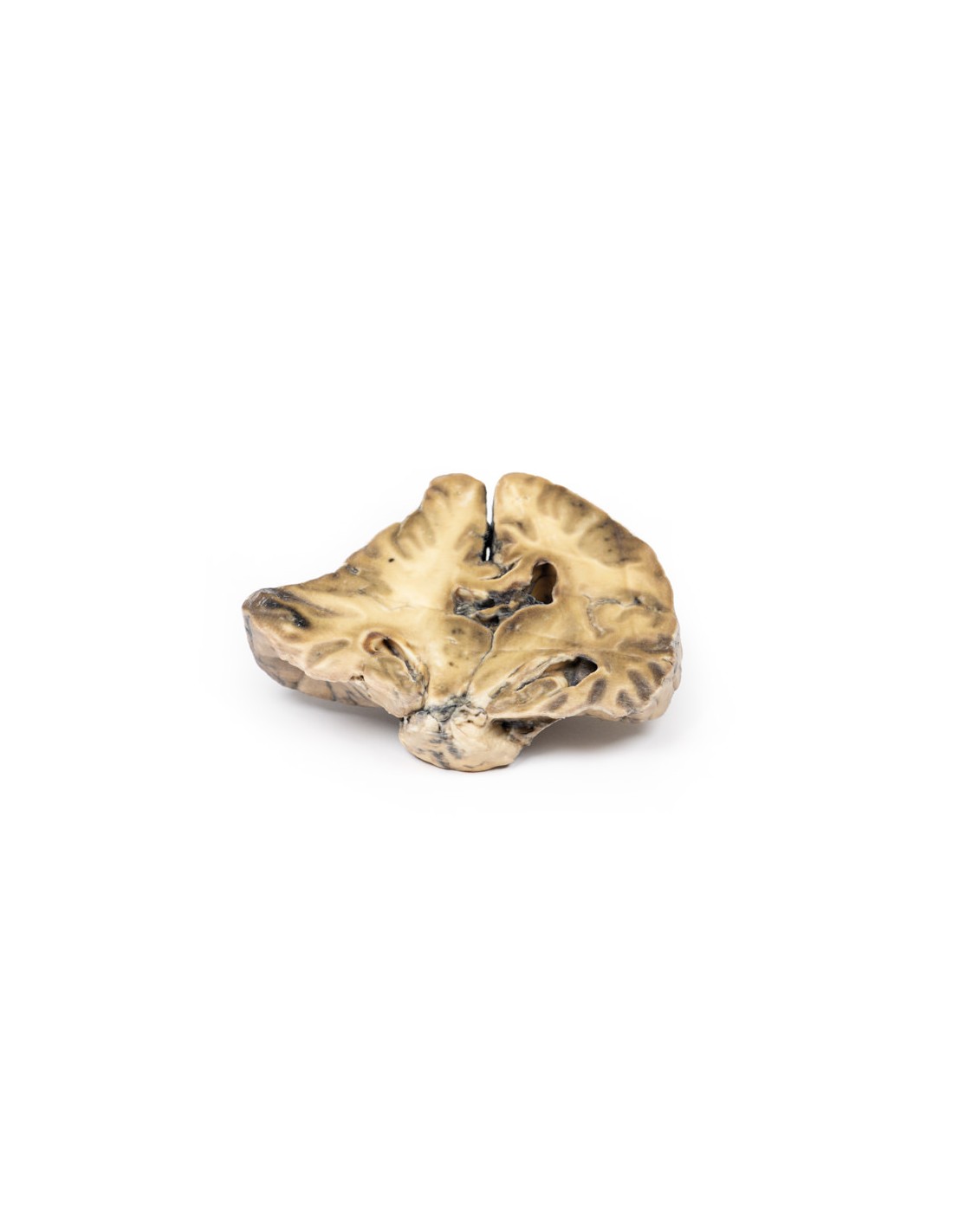
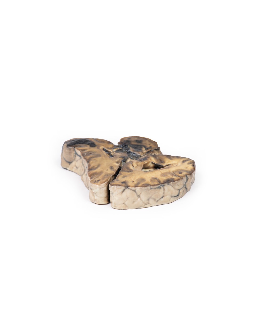
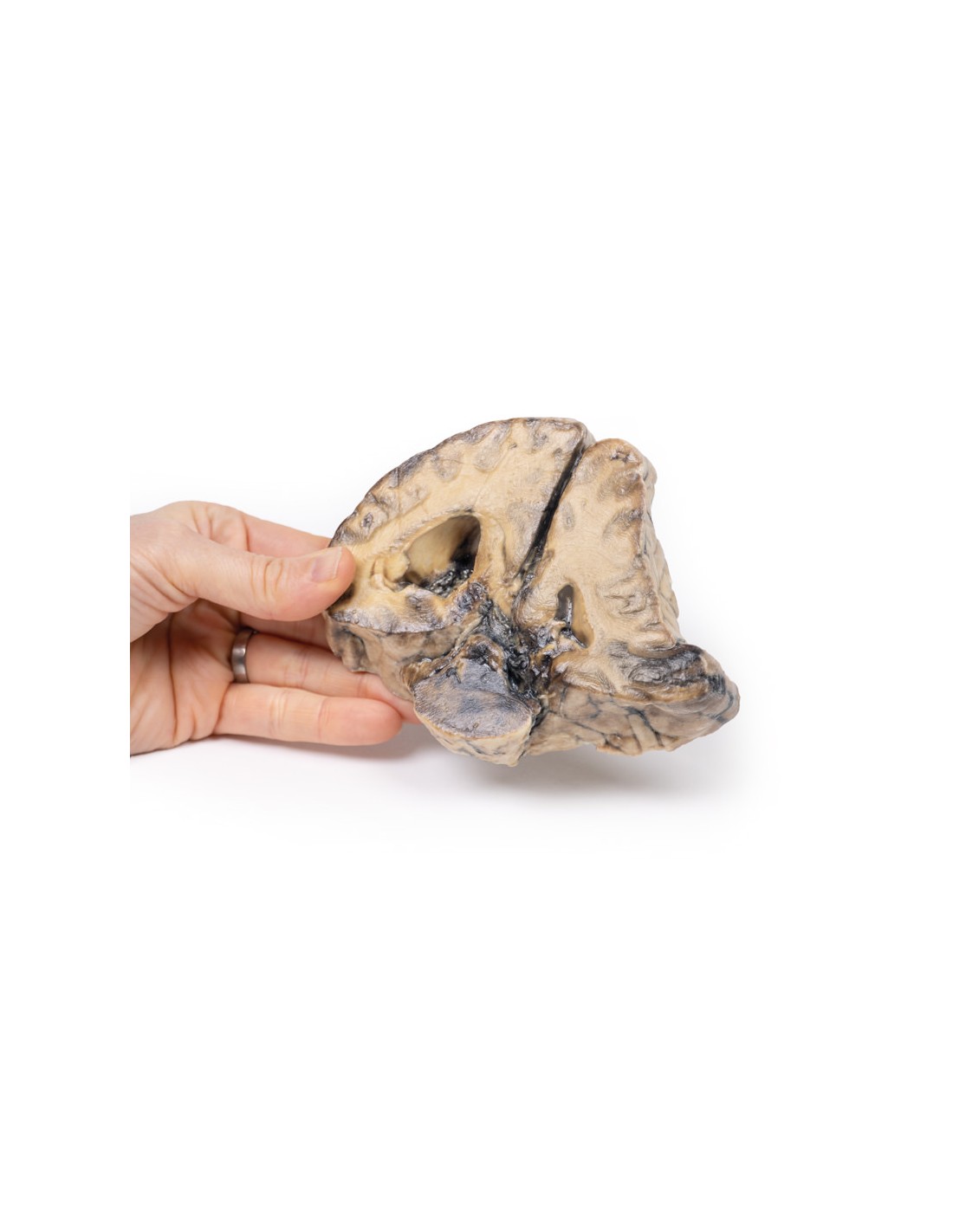
The specimen is a coronal section of a brain. It is evident that the brain has been compressed laterally and downward by an expanding intracranial mass on the right side, probably a meningioma. The original mass is not present. The anterior face shows displacement of midline structures with subfalcine* herniation of the cingulate gyrus. The posterior face (see photo) shows hemorrhages of various ages within the temporal lobe and the pons, typical of massive supratentorial lesions. There is also ventricular asymmetry.
*In subfalcine (or cingulate) herniation, the most common type of brain herniation, the innermost part of the frontal lobe is pushed under part of the cerebral sickle, between the two hemispheres of the brain.
More information
Symptoms of a meningioma occupying space in the cranial cavity can be caused by the tumor mass pressing on the brain, which can lead to atrophy and displacement of the brain parenchyma, leading to symptoms resulting from disruptions in cranial nerve function, blood flow, and normal brain function. General symptoms may include:
Muscle convulsions: e.g., myoclonic (single or multiple muscle contractions, jerks and/or spasms) or tonic-clonic (great evil: loss of consciousness and body tone, followed by muscle contractions and relaxing muscle contractions, loss of control of bodily functions, period of shortness of breath and the person may take on a blue tinge, a person may be sleepy and experience headaches, confusion, weakness, numbness and muscle aches)
Sensory changes: changes in vision, smell and/or hearing without loss of consciousness .
Symptoms and signs may vary with tumor location.
What advantages does the Monash University anatomical dissection collection offer over plastic models or plastinated human specimens?
- Each body replica has been carefully created from selected patient X-ray data or human cadaver specimens selected by a highly trained team of anatomists at the Monash University Center for Human Anatomy Education to illustrate a range of clinically important areas of anatomy with a quality and fidelity that cannot be achieved with conventional anatomical models-this is real anatomy, not stylized anatomy.
- Each body replica has been rigorously checked by a team of highly trained anatomists at the Center for Human Anatomy Education, Monash University, to ensure the anatomical accuracy of the final product.
- The body replicas are not real human tissue and therefore not subject to any barriers of transportation, import, or use in educational facilities that do not hold an anatomy license. The Monash 3D Anatomy dissection series avoids these and other ethical issues that are raised when dealing with plastinated human remains.
No reviews
Tap to zoom