Your cart
There are no more items in your cart


Berry's aneurysm of the basilar artery - Erler Zimmer 3D anatomy Series MP2001
erler zimmer
EZ-MP2001
€334.16
Tax included
Made in ultra-high resolution 3D printing in full color.
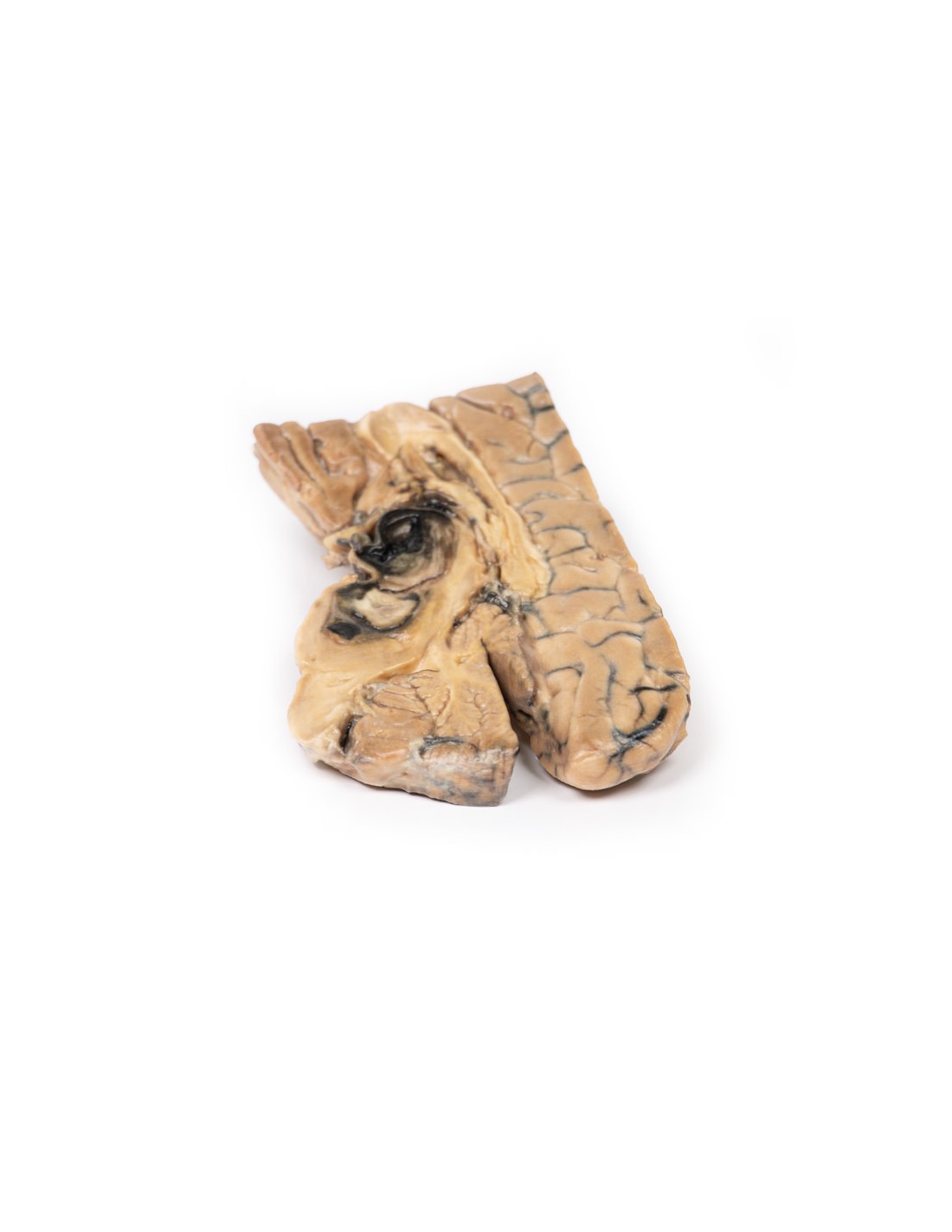
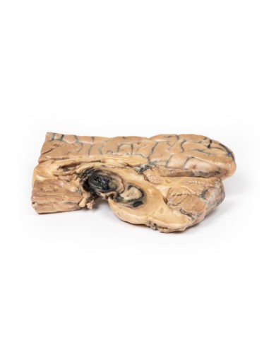
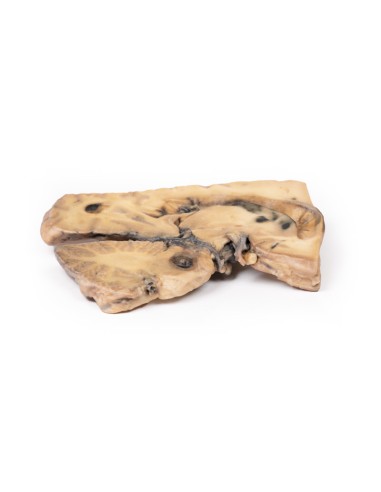
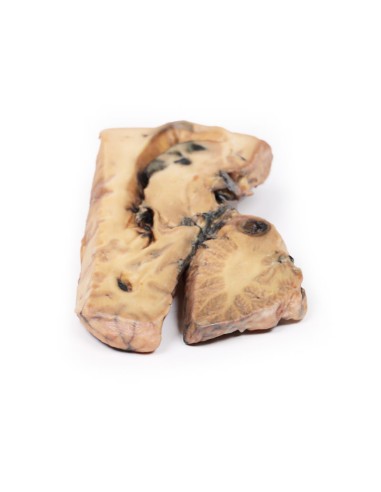
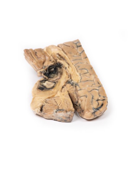
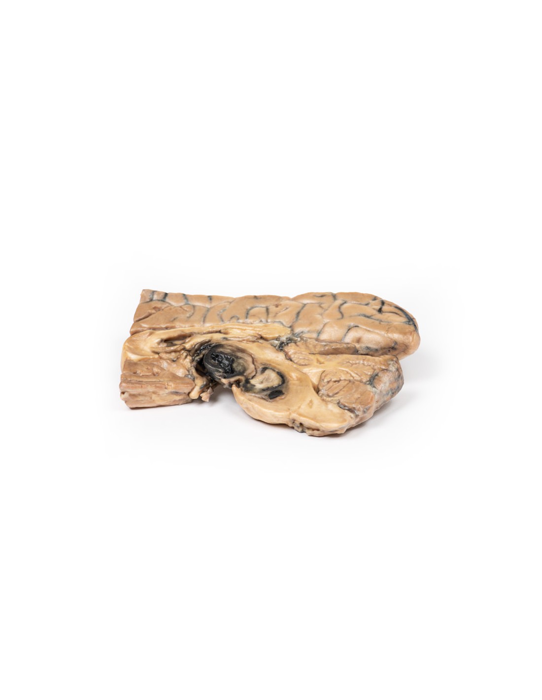
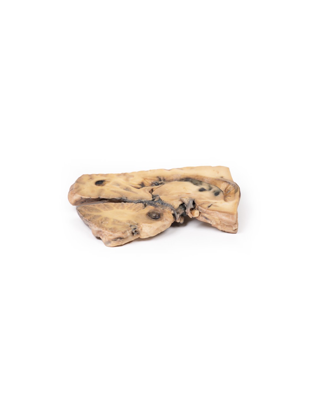
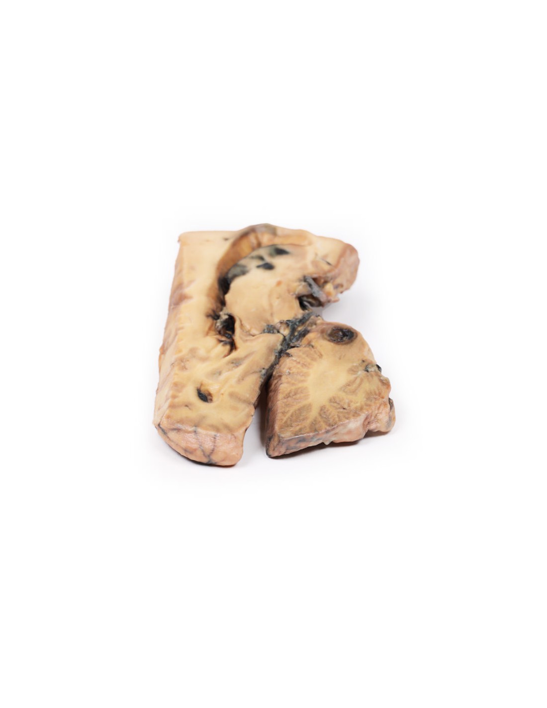
Berry aneurysm of the basilar artery - Erler Zimmer 3D anatomy Series MP2001
This brain dissection model with aneurysm is part of the exclusive Monash 3D anatomy series, a comprehensive series of human dissections reproduced with ultra-high resolution color 3D printing.
Clinical History.
A 37-year-old patient presented to the hospital after falling and hitting his head, resulting in symptoms of headache, vomiting and disorientation. CT showed dilatation of the lateral ventricles associated with a large mass projecting posteriorly into the third ventricle. A shunt for hydrocephalus was performed 1 week later. An angiography revealed a partially thrombosed aneurysm, measuring 1 x 1 cm, originating from the basilar artery. At 3 months after surgery, the shunt was revised due to obstruction, with repeated cerebral angiographies revealing enlargement of the aneurysm interval. Attempted ligation of the aneurysm was unsuccessful. The patient remained unconscious despite several attempts to revise the shunt and died.
Pathology
This brain was sliced in the mid-sagittal plane. It includes an entire half-section of the brain about 1 cm thick. A large dark-colored ovoid berry aneurysm 5 x 2 cm in diameter, originating from the basilar artery, is clearly visible on the medial surface. It has eroded into the midbrain, compressing the third ventricle from below and inferiorly into the substance of the pons. The wall of the aneurysm appears intact although a blood clot is seen in the third ventricle that appears to leak from the lateral wall of that ventricle. The aneurysm is filled with a laminated thrombus. A small area of mucoid degeneration 0.4 cm in diameter is seen posterior to the aneurysm within the bridge.
Examination of the lateral aspect of the sagittal section shows dilatation of the lateral ventricle, hematic staining of the ventricular wall, and patchy hemorrhagic infarction of the caudate nucleus. There was some discoloration of the meninges over the tip of the left temporal lobe and cerebellum (not included in the 3D print), consistent with subarachnoid hemorrhage.
More information on cerebral aneurysm.
The prevalence of aneurysms is about 3.2 percent in the population, while rupture is much less common, occurring in only 7.9 per 100,000 person-years. A minority of intracranial aneurysms originate from the posterior circulation and are mostly located at junctional sites around the basilar, vertebral, and cerebellar arteries. Symptoms are secondary to subarachnoid hemorrhage or a mass effect with associated compression of adjacent brain parenchyma and cranial nerves. Rupture results in complications due to bleeding and increased intracranial pressure. Hydrocephalus, rebleeding, and vasospasm may also occur. Management is by surgical means; In recent years, new therapies include endovascular coil surgery and subsequent monitoring.
What advantages does the Monash University anatomical dissection collection offer over plastic models or plastinated human specimens?
- Each body replica has been carefully created from selected patient X-ray data or human cadaver specimens selected by a highly trained team of anatomists at the Monash University Center for Human Anatomy Education to illustrate a range of clinically important areas of anatomy with a quality and fidelity that cannot be achieved with conventional anatomical models-this is real anatomy, not stylized anatomy.
- Each body replica has been rigorously checked by a team of highly trained anatomists at the Center for Human Anatomy Education, Monash University, to ensure the anatomical accuracy of the final product.
- The body replicas are not real human tissue and therefore not subject to any barriers of transportation, import, or use in educational facilities that do not hold an anatomy license. The Monash 3D Anatomy dissection series avoids these and other ethical issues that are raised when dealing with plastinated human remains.
No reviews
Tap to zoom