Your cart
There are no more items in your cart

Pituitary adenoma - Erler Zimmer 3D anatomy Series MP2006
erler zimmer
EZ-MP2006
€1,053.47
Tax included
Made in ultra-high resolution 3D printing in full color.
Pituitary Adenoma - Erler Zimmer 3D anatomy Series MP2006
This dissection model highlighting a pituitary adenoma is part of the exclusive Monash 3D anatomy series, a comprehensive series of human dissections reproduced with ultra-high resolution color 3D printing.
Clinical History.
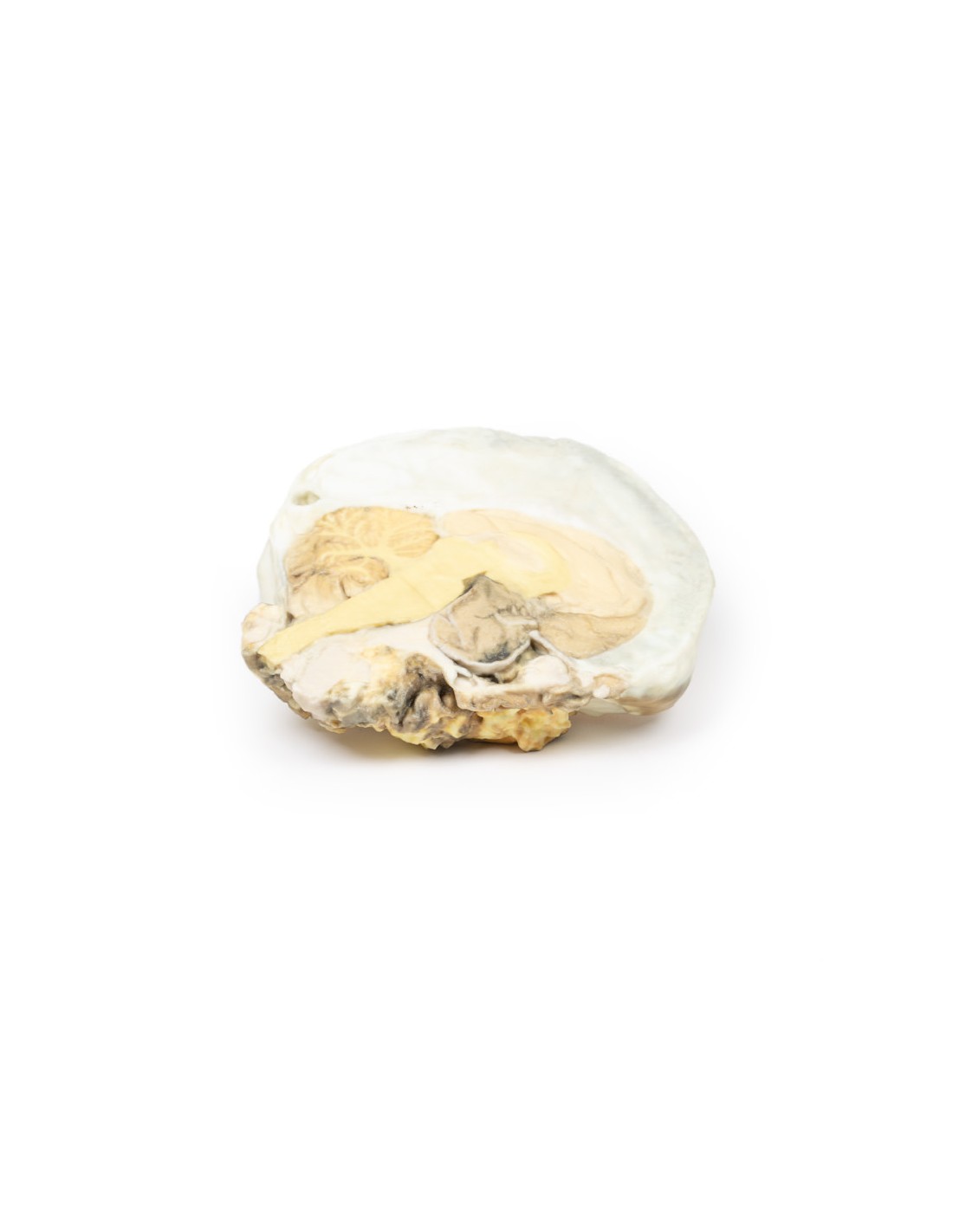
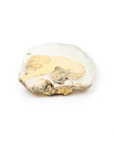
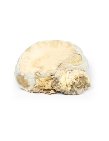
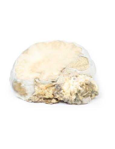
A 29-year-old man presented with a 22-month history of headache and blurred vision. Examination revealed bi-temporal hemianopsia and left VI nerve palsy. Skull radiography showed erosion of most of the sphenoid body with some dorso sellae and anterior clinoid process intact. Carotid angiography showed upward and lateral displacement of the anterior and middle cerebral arteries. Pneumoencephalography (a common imaging procedure used until the 1970s in which cerebrospinal fluid was drained and replaced by air, oxygen, or helium that served as a contrast agent in X-ray examinations) showed upward displacement of the lateral and third ventricles from below. A craniotomy was performed but the patient died soon after.
Pathology
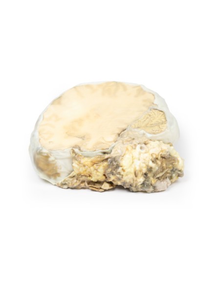
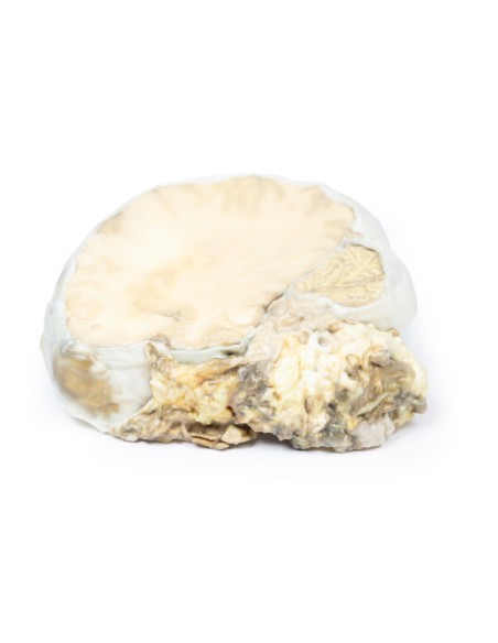
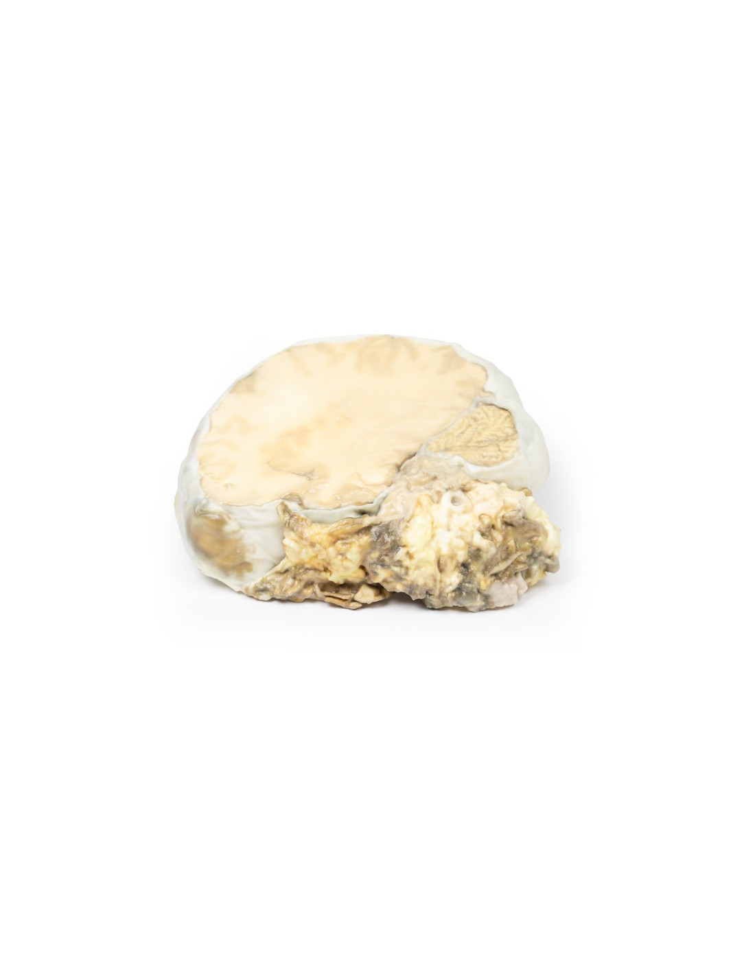
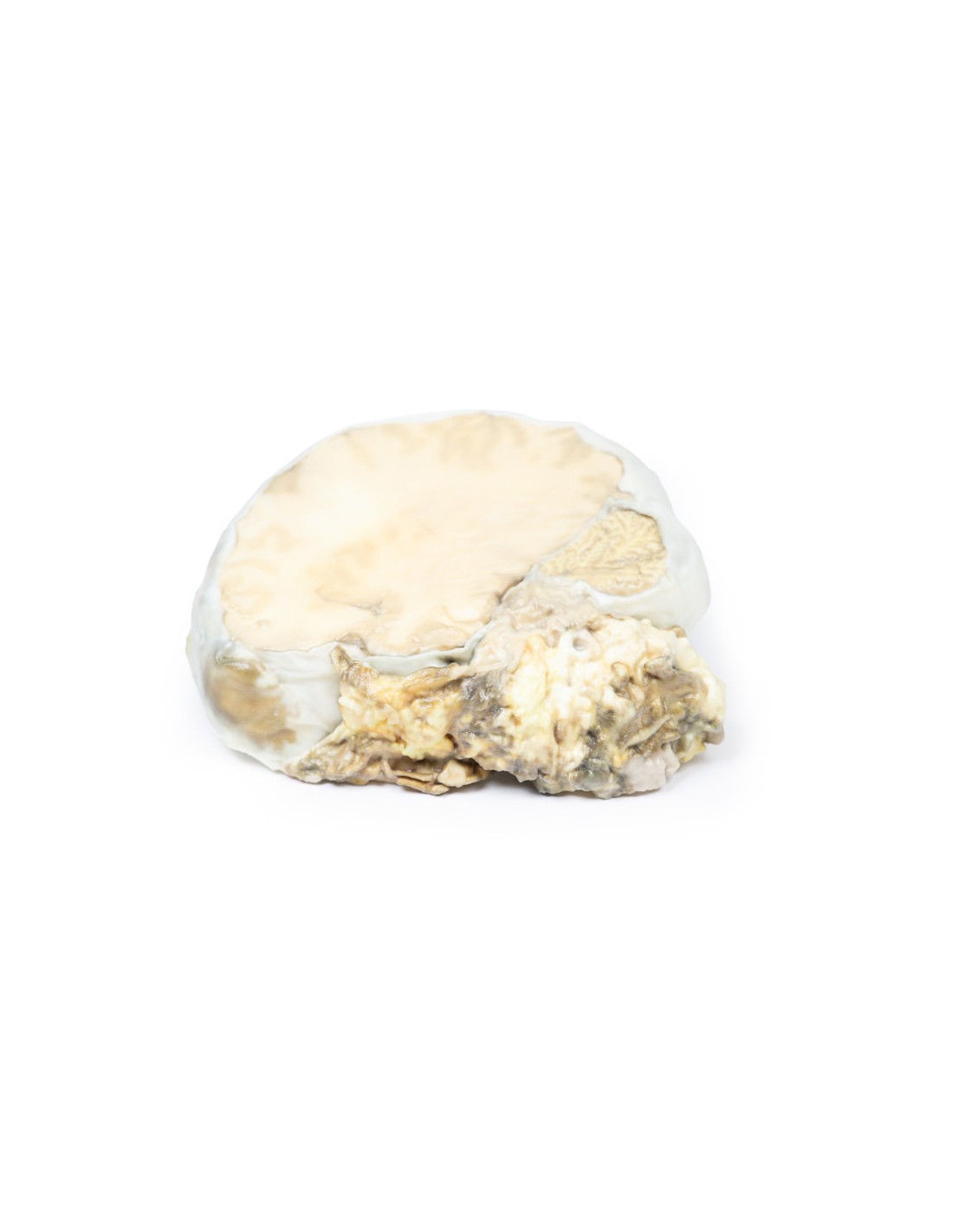
The brain specimen is sliced in the sagittal plane to the right of the sickle cerebri, which remains in situ. The pituitary gland has been completely replaced by a round tumor with a maximum diameter of 4 cm. The cut surface of the tumor is light brown and homogeneous (except for an area of hemorrhage superiorly, probably caused by surgical trauma). The tumor caused upward displacement of the midbrain. Erosion of the tumor has destroyed the sphenoid bone; thus the sella turca is enlarged (arrow). The optic chiasm is compressed by the tumor. Histologically, this tumor was a chromophobe adenoma arising from the anterior pituitary gland.
Additional Information.
This specimen is from an old case, and the investigations used would now be considered antiquated. Modern investigation would include an initial brain CT scan followed by MRI of the brain to further visualize the pituitary lesion prior to any surgical intervention.
Pituitary adenomas are the most common pituitary tumor and are most commonly found in adults with a peak incidence between the ages of 35 and 60. Primary pituitary carcinoma is very rare, and the pituitary gland is an uncommon site for metastasis. Clinical manifestations of pituitary adenomas are related to local mass effect and tumor function. Local effects include increased intracranial pressure (headache, nausea, and vomiting), saddle expansion, bone erosion, and compression of decussating nerve fibers in the optic chiasm, causing bitemporal hemianopsia.
Pituitary adenomas can be functioning (i.e., associated with hormone excess) or nonfunctioning (i.e., without clinical symptoms of hormone excess). About 75% of adenomas are functional: they usually secrete prolactin, growth hormone, or ACTH. Secretion of TSH, LH, and FSH from pituitary adenomas is very rare. Some adenomas may secrete two hormones, with growth hormone and prolactin being the most common combination. Nonfunctional pituitary adenomas come to clinical attention later than those associated with endocrine abnormalities and can lead to hypopituitarism due to compression atrophy of the surrounding normal gland.
What advantages does the Monash University anatomical dissection collection offer over plastic models or plastinated human specimens?
- Each body replica has been carefully created from selected patient X-ray data or human cadaver specimens selected by a highly trained team of anatomists at the Monash University Center for Human Anatomy Education to illustrate a range of clinically important areas of anatomy with a quality and fidelity that cannot be achieved with conventional anatomical models-this is real anatomy, not stylized anatomy.
- Each body replica has been rigorously checked by a team of highly trained anatomists at the Center for Human Anatomy Education, Monash University, to ensure the anatomical accuracy of the final product.
- The body replicas are not real human tissue and therefore not subject to any barriers of transportation, import, or use in educational facilities that do not hold an anatomy license. The Monash 3D Anatomy dissection series avoids these and other ethical issues that are raised when dealing with plastinated human remains.
No reviews
Tap to zoom