Your cart
There are no more items in your cart


Metastatic adenocarcinoma in the brain - Erler Zimmer 3D anatomy Series MP2007
erler zimmer
EZ-MP2007
€307.32
Tax included
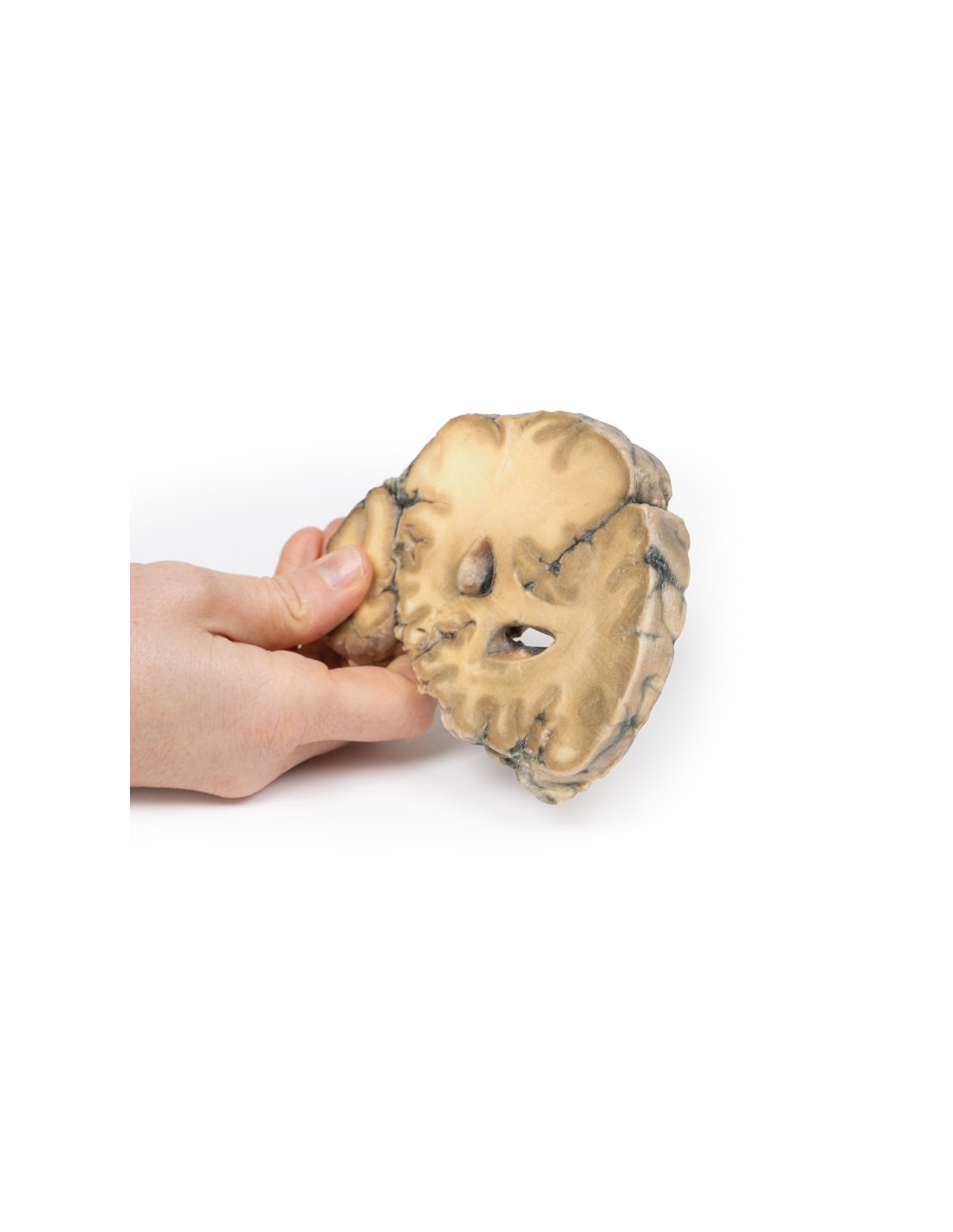
Made in ultra-high resolution 3D printing in full color.
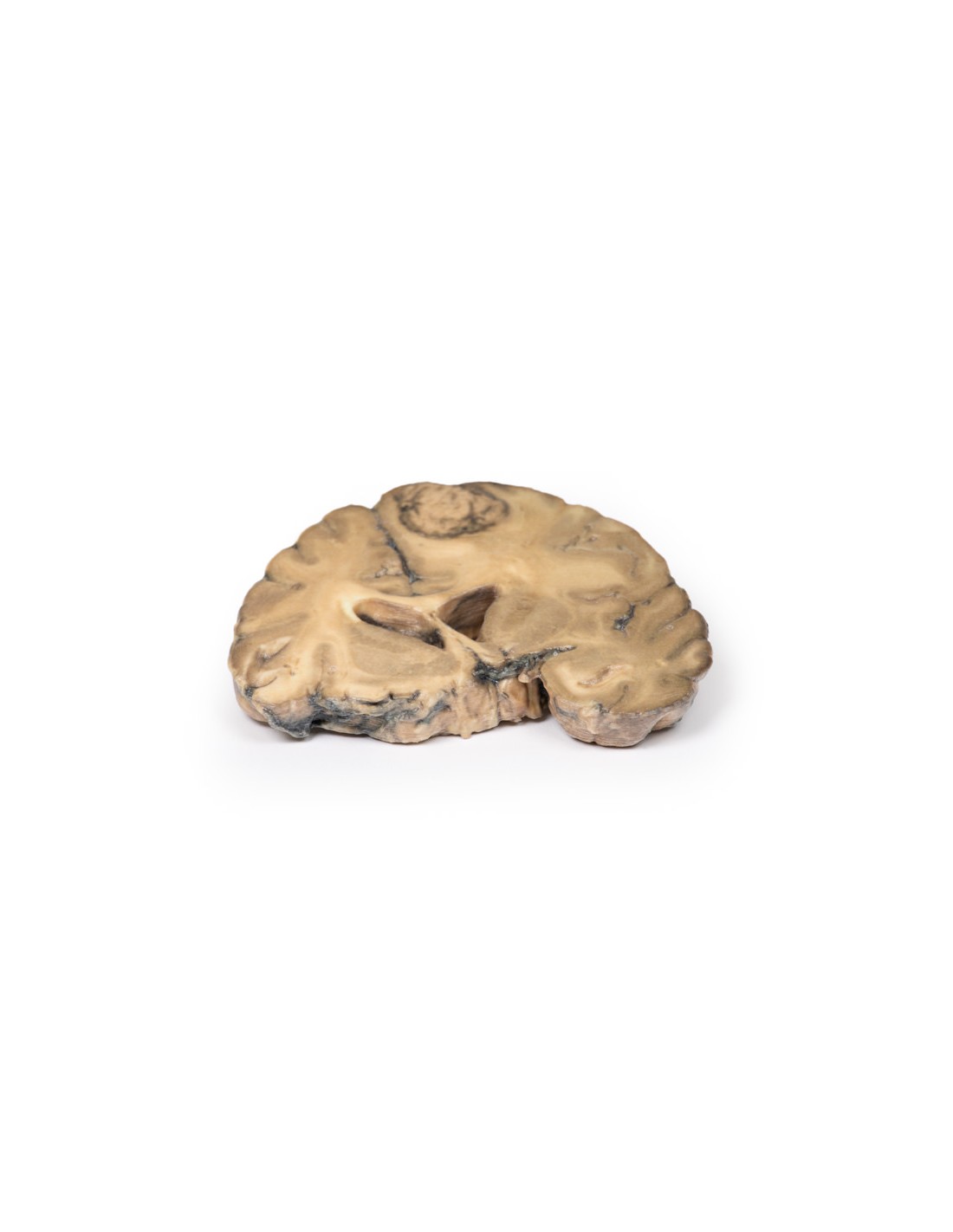
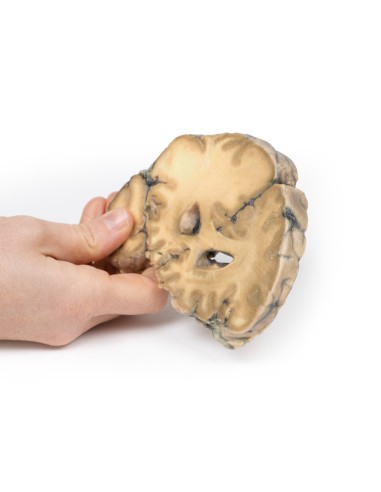
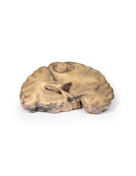
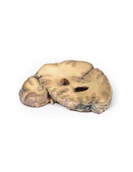
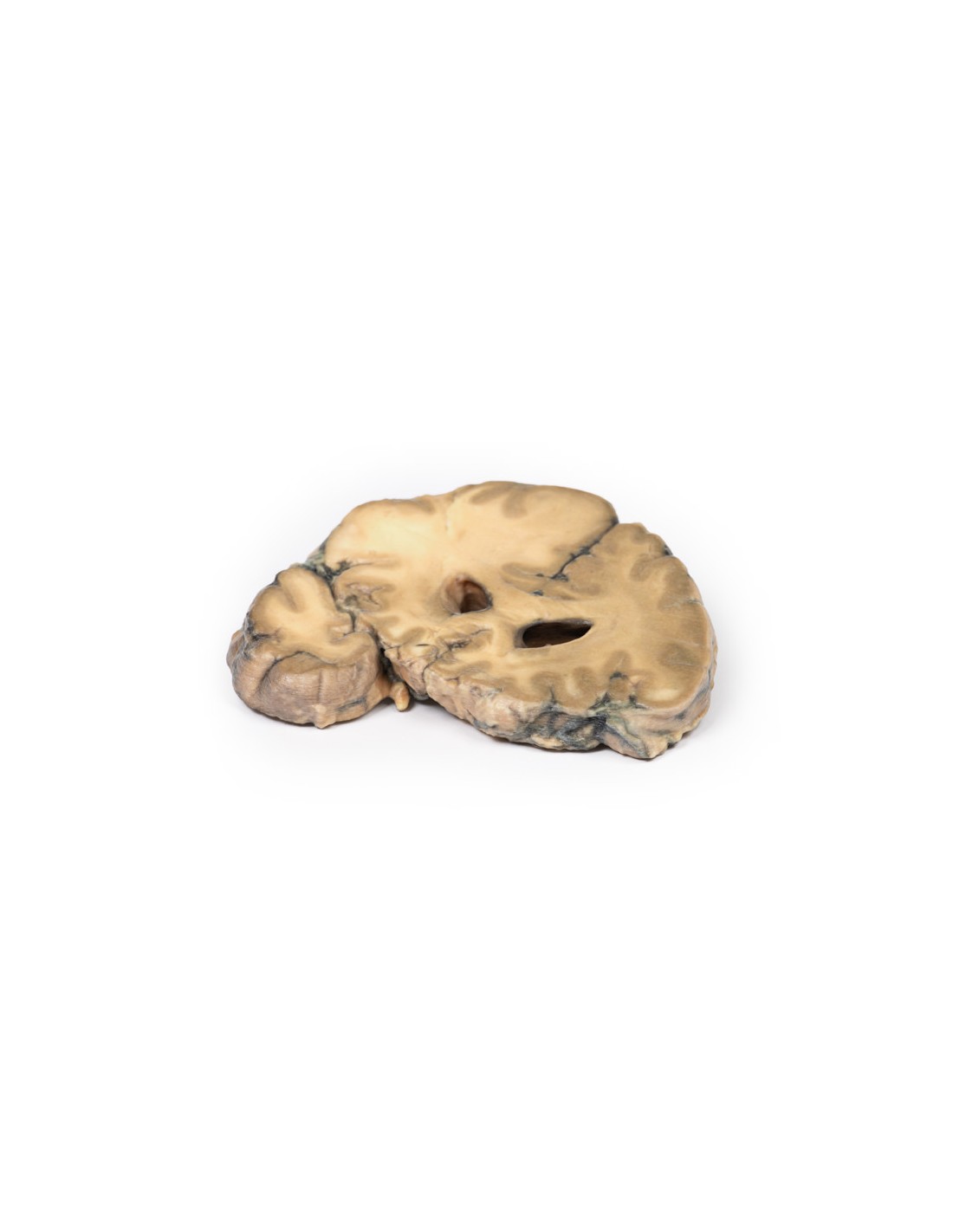
Metastatic Adenocarcinoma in the Brain - Erler Zimmer 3D anatomy Series MP2007
This dissection model highlighting a metastatic Adenocarcinoma in the brain is part of the exclusive Monash 3D anatomy series, a comprehensive series of human dissections reproduced with ultra-high resolution color 3D printing.
Clinical History.
A 56-year-old man underwent total gastrectomy and splenectomy for gastric adenocarcinoma. Over a two-month period, he developed a progressively unsteady gait, increasing left hand weakness, and frontal headaches associated with nausea and vomiting. Imaging revealed a lesion in the right frontal lobe. He underwent craniotomy with resection of the lesion, which was confirmed to be metastatic gastric adenocarcinoma. He manifested gradually increasing symptoms as well as jaundice, deterioration of consciousness, and papilledema due to increased intracranial pressure. Repeat imaging revealed recurrence of the right frontal metastatic lesion and liver metastases. The patient died 9 months after the initial gastrectomy surgery.
Pathology
This brain specimen is cut in the coronal plane. A circumscribed, variegated, pinkish-gray tumor is evident in the right frontal lobe. The tumor involves gray and white matter. Compression of the right lateral ventricle by the lesion is evident with displacement of the midline structures as well.
Further information
Stomach cancer is one of the most common causes of cancer death worldwide. Risk factors include male sex, diet, smoking, and chronic Helicobacter pylori infection. The most common sites for gastric adenocarcinoma metastasis are the liver, peritoneum, lungs and bones. Brain metastases are rare and occur in <1% of cases. Isolated brain metastases are very rare, being more commonly seen in disseminated disease and associated with poor prognosis. Palliative treatment may include surgery, radiotherapy, steroids, chemotherapy, or a combination thereof.
What advantages does the Monash University anatomical dissection collection offer over plastic models or plastinated human specimens?
- Each body replica has been carefully created from selected patient X-ray data or human cadaver specimens selected by a highly trained team of anatomists at the Monash University Center for Human Anatomy Education to illustrate a range of clinically important areas of anatomy with a quality and fidelity that cannot be achieved with conventional anatomical models-this is real anatomy, not stylized anatomy.
- Each body replica has been rigorously checked by a team of highly trained anatomists at the Center for Human Anatomy Education, Monash University, to ensure the anatomical accuracy of the final product.
- The body replicas are not real human tissue and therefore not subject to any barriers of transportation, import, or use in educational facilities that do not hold an anatomy license. The Monash 3D Anatomy dissection series avoids these and other ethical issues that are raised when dealing with plastinated human remains.
No reviews
Tap to zoom