Your cart
There are no more items in your cart


Right ventricular hypertrophy - Erler Zimmer 3D anatomy Series MP2031
erler zimmer
EZ-MP2031
€1,054.81
Tax included
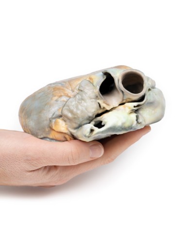
Made in ultra-high resolution 3D printing in full color.
Right Ventricular Hypertrophy - Erler Zimmer 3D anatomy Series MP2031
This dissection model highlighting a Right Ventricular Hypertrophy is part of the exclusive Monash 3D anatomy series, a comprehensive series of human dissections reproduced with ultra-high resolution color 3D printing.
Medical History.
This 56-year-old woman suffered from emphysema and for 2 years had been presenting with increasing shortness of breath on exertion associated with recurrent attacks of bronchitis. On examination, she had a blood pressure of 160/90 mm Hg, a heart rate of 96 beats/min, and 6 cm of jugular venous congestion. The apical beat was impalpable, bilateral crepitations were heard, and peripherally pitting edema was present. Special investigations: the ECG showed a right heart strain pattern. Arterial blood examination showed respiratory acidosis. Despite treatment there was constant deterioration and death.
Pathology
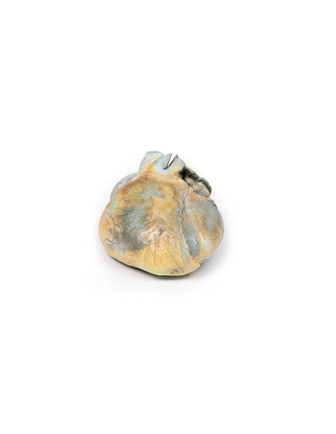
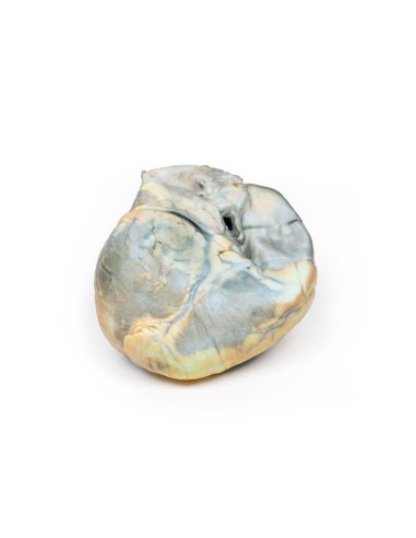
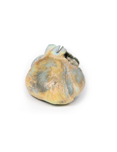
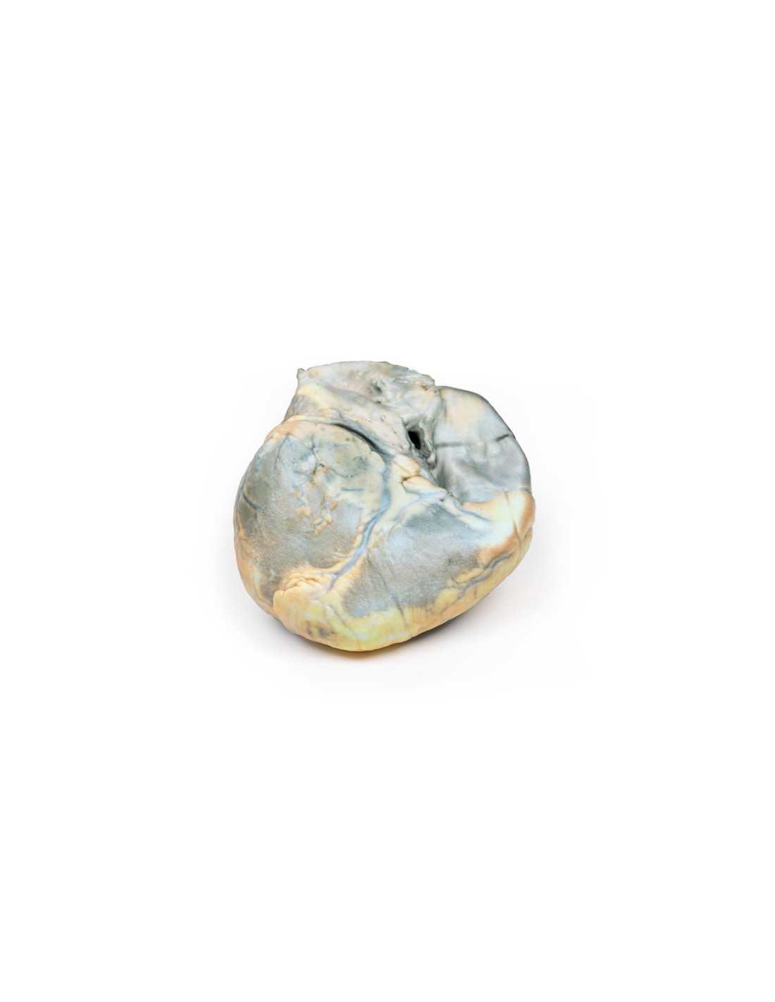
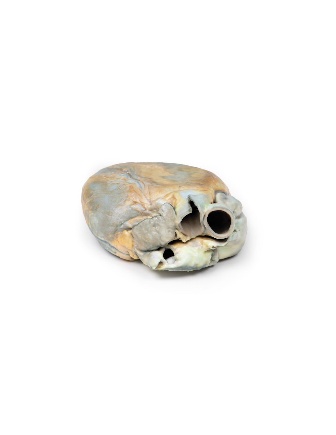
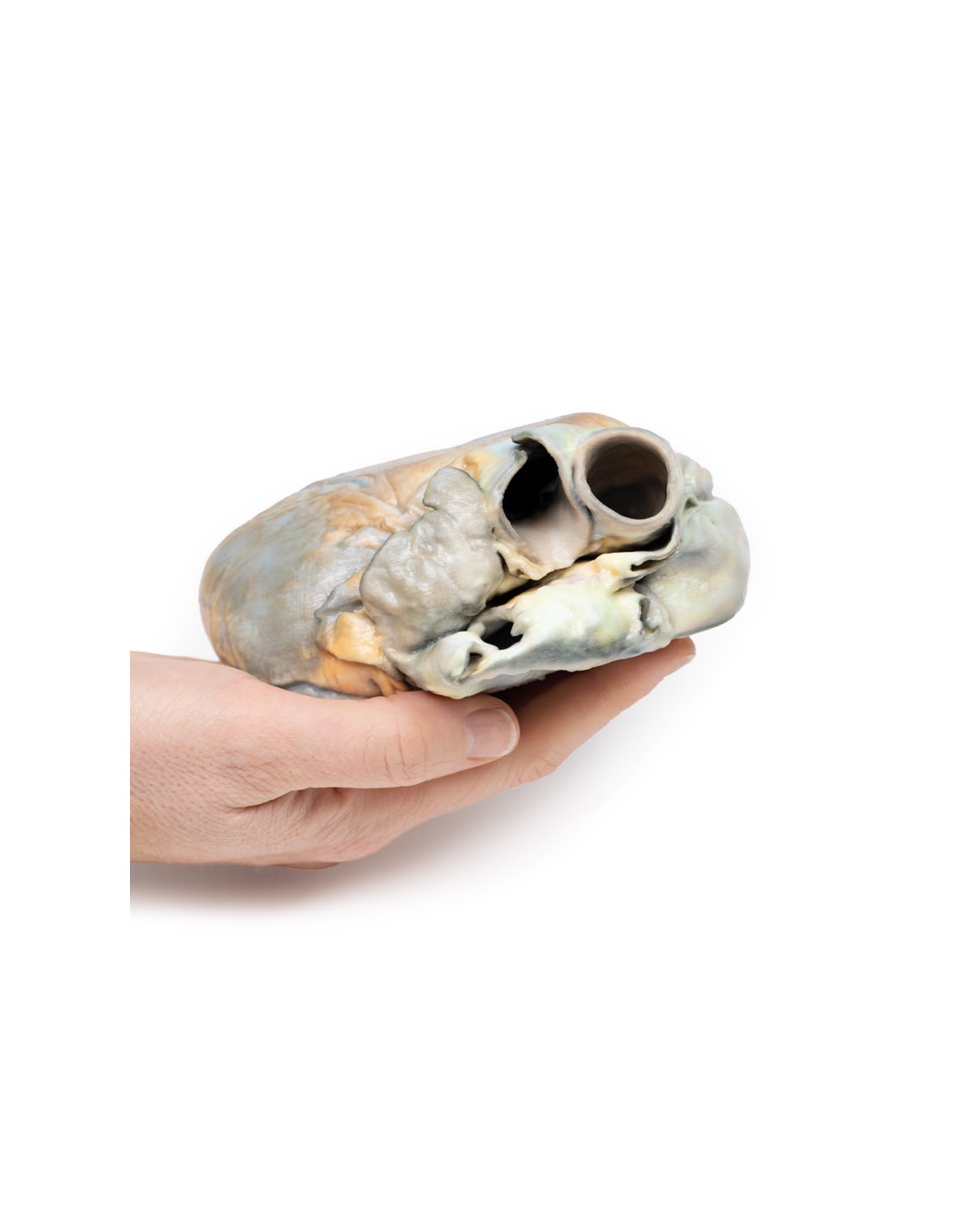
The specimen is of the outer surface of the heart seen from the anterior aspect. The right ventricle is greatly enlarged and hypertrophied. Everything appears to be normal otherwise. This is an example of right ventricular hypertrophy (RVH) in a patient with emphysema.
Additional Information.
RVH usually occurs due to chronic lung disease or structural defects of the heart. One of the most common causes of RVH is pulmonary hypertension (PH), which leads to increased pulmonary arterial pressure. When the right ventricle tries to compensate for this increased pressure, it changes shape and size causing hypertrophy and wall thickness of the right ventricle. The global incidence of PH is 4 per 1 million people: RVH occurs in about 30 percent of these cases. Common causes of PH include chronic obstructive pulmonary disease (COPD), pulmonary embolism, and other restrictive lung diseases. RVH also occurs in response to structural defects in the heart, such as tricuspid insufficiency, which allows backward flow of blood into the ventricle. Other structural defects that lead to RVH include tetralogy of Fallot, ventricular septal defects, pulmonary valve stenosis, and atrial septal defects. RVH is also associated with abdominal obesity and elevated systolic blood pressure.
What advantages does the Monash University anatomical dissection collection offer over plastic models or plastinated human specimens?
- Each body replica has been carefully created from selected patient X-ray data or human cadaver specimens selected by a highly trained team of anatomists at the Monash University Center for Human Anatomy Education to illustrate a range of clinically important areas of anatomy with a quality and fidelity that cannot be achieved with conventional anatomical models-this is real anatomy, not stylized anatomy.
- Each body replica has been rigorously checked by a team of highly trained anatomists at the Center for Human Anatomy Education, Monash University, to ensure the anatomical accuracy of the final product.
- The body replicas are not real human tissue and therefore not subject to any barriers of transportation, import, or use in educational facilities that do not hold an anatomy license. The Monash 3D Anatomy dissection series avoids these and other ethical issues that are raised when dealing with plastinated human remains.
No reviews
Tap to zoom