
Your cart
There are no more items in your cart


Rheumatic endocarditis - Erler Zimmer 3D anatomy Series MP2039
erler zimmer
EZ-MP2039
€838.75
Tax included
Made in ultra-high resolution 3D printing in full color.
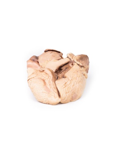
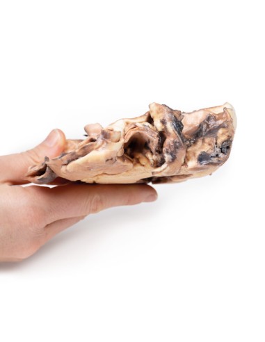
Rheumatic Endocarditis - Erler Zimmer 3D anatomy Series MP2039
This dissection model highlighting Rheumatic Endocarditis is part of the exclusive Monash 3D anatomy series, a comprehensive series of human dissections reproduced with ultra-high resolution color 3D printing.
Clinical History.
The patient was a 52-year-old woman with increasing dyspnea. She related a past history of fever with fluctuating joint pain during childhood following a sore throat. On examination: cyanotic, pulse showed atrial fibrillation, elevated jugular venous pulse, pansystolic murmur at apex, hepatomegaly and edema dependent. She was being treated with digoxin, lasix (furosemide) and penicillin but died after cardiac arrest.
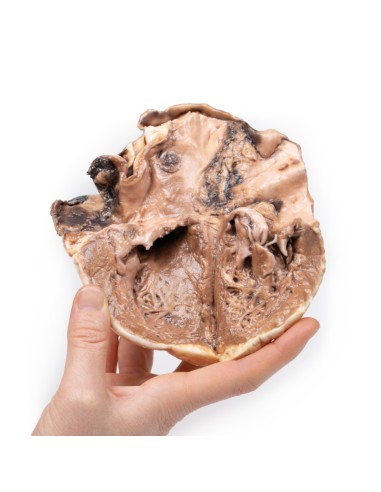
Pathology
The specimen is that of an open heart to show the left atrium and left ventricle. The mitral valve has been cut, but those visible parts show considerable thickening. The left atrial wall shows deposition of blood and fibrin. The left auricular appendage is filled with blood clots, caused by atrial fibrillation. The mural thrombus on the atrial wall is found at the typical site:- the deep layers of the endocardium forming irregular thickenings, called MacCallum plaques (arrows).
Further information
In this patient, the history of fever and joint pain following sore throat is very indicative of a history of rheumatic fever. Rheumatic fever is an inflammatory disease that can involve the heart, joints, skin, and brain. Typical symptoms include fever, multiple painful joints, involuntary muscle movements (chorea), and occasionally a characteristic nonpruritic rash known as "erythema marginalis."
Rheumatic fever may occur 2-4 weeks after an infection of the throat by the bacterium Streptococcus pyogenes. If the infection is not treated (with pencillin), rheumatic fever occurs in up to three percent of people. The underlying mechanism is believed to involve the production of antibodies against a person's own tissues (autoimmune disease). Because of their genetics, some people are more likely to get the disease when exposed to bacteria than others. Other risk factors include malnutrition and poverty, which occur more commonly in low- and middle-income countries and particularly in indigenous communities.
The heart is involved in about half of the cases. Damage to the heart valves, known as rheumatic heart disease (RHD), usually occurs after repeated attacks (carditis), but can sometimes occur after one. Carditis can evolve into chronic rheumatic heart disease, which usually affects the heart valves. The mitral valve is the most commonly affected valve, with fibrosis leading to mitral valve stenosis, and this specimen shows mitral valve thickening. Stenosis is thought to occur due to Aschoff's nodules, which are granulomatous lesions with a central area of fibrinoid necrosis and surrounded by an infiltration of autoreactive T cells. Aschoff nodules also contain "giant cells," which are thought to be a type of degenerative connective or endothelial tissue.
The stenosis may progress over the years, and as it worsens, the left atrium will become increasingly dilated. As a result, atrial fibrillation may develop and mural thrombi may form. In addition, narrow mitral stenosis can cause severe heart failure.
.
What advantages does the Monash University anatomical dissection collection offer over plastic models or plastinated human specimens?
- Each body replica has been carefully created from selected patient X-ray data or human cadaver specimens selected by a highly trained team of anatomists at the Monash University Center for Human Anatomy Education to illustrate a range of clinically important areas of anatomy with a quality and fidelity that cannot be achieved with conventional anatomical models-this is real anatomy, not stylized anatomy.
- Each body replica has been rigorously checked by a team of highly trained anatomists at the Center for Human Anatomy Education, Monash University, to ensure the anatomical accuracy of the final product.
- The body replicas are not real human tissue and therefore not subject to any barriers of transportation, import, or use in educational facilities that do not hold an anatomy license. The Monash 3D Anatomy dissection series avoids these and other ethical issues that are raised when dealing with plastinated human remains.
No reviews
Tap to zoom
