Your cart
There are no more items in your cart


Traumatic esophageal-aortic fistula - Erler Zimmer 3D anatomy Series MP2040
erler zimmer
EZ-MP2040
€307.32
Tax included
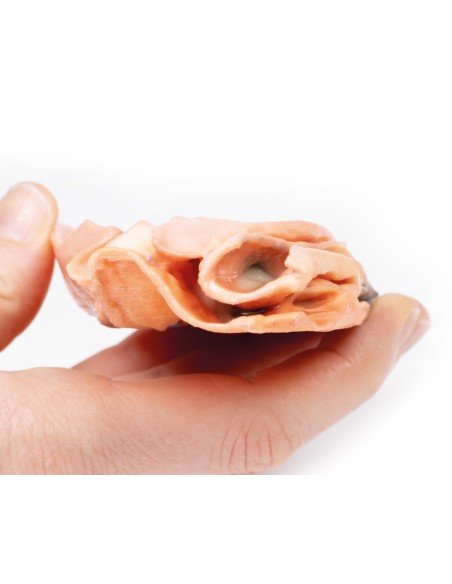
Made in ultra-high resolution 3D printing in full color.
Traumatic esophageal-aortic fistula - Erler Zimmer 3D anatomy Series MP2040
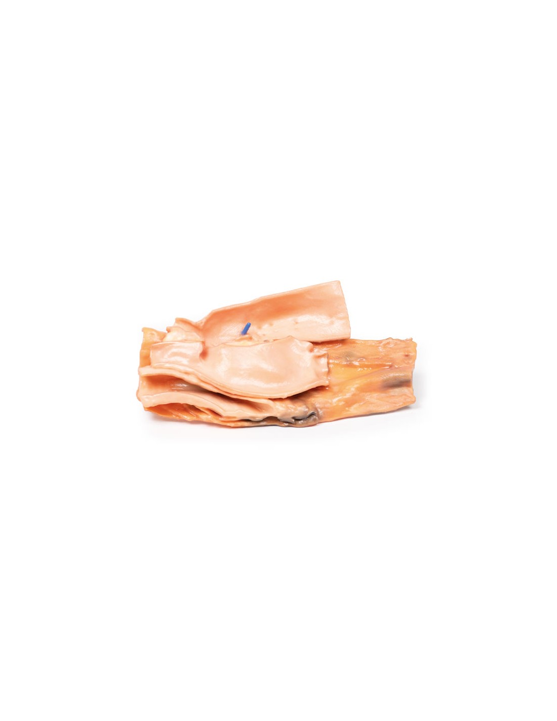
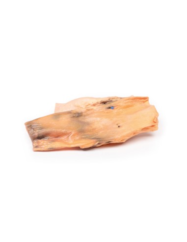
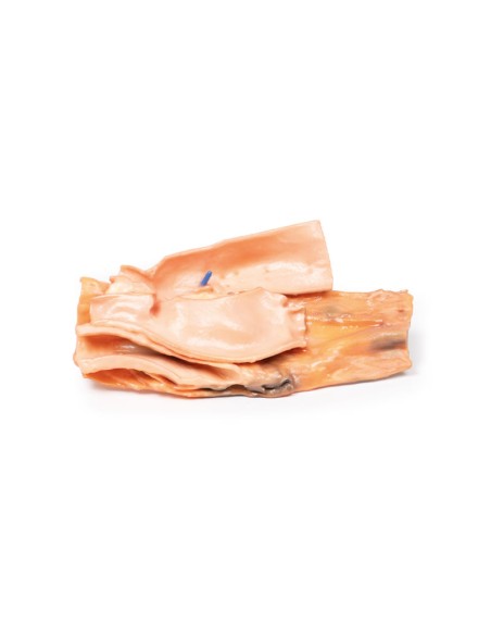
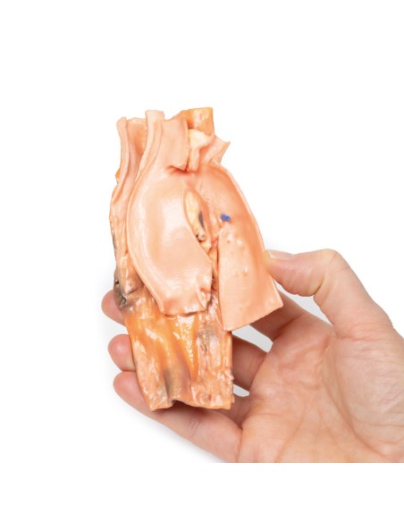
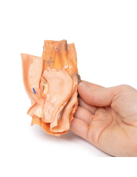
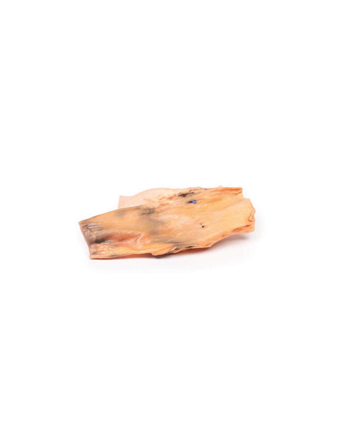
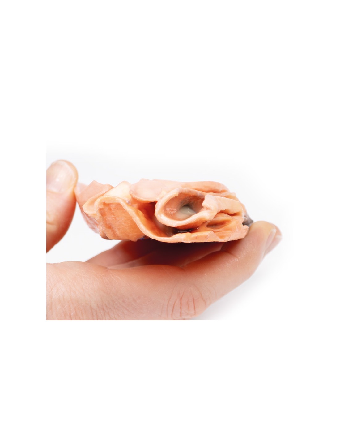
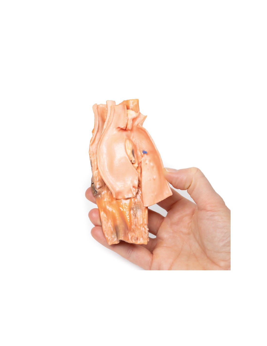
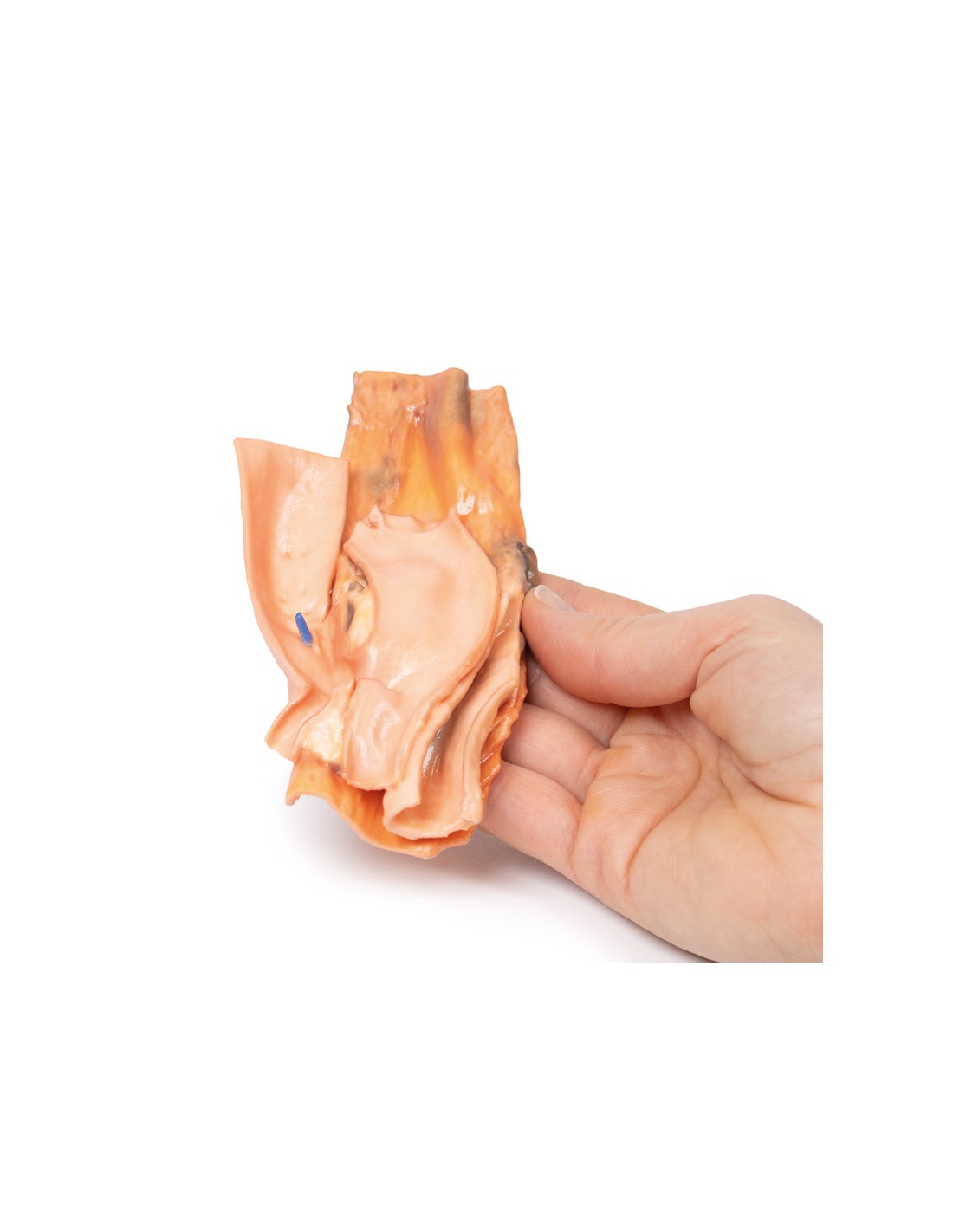
This dissection model highlighting an esophageal-aortic Traumatic Fistula is part of the exclusive Monash 3D anatomy series, a comprehensive series of human dissections reproduced with ultra-high resolution color 3D printing.
Clinical History.
A woman who ingested a bone chop during lunch collapsed later in the afternoon and suffered massive hematemesis. At laparotomy, her stomach was filled with fresh blood but the cause was not identified. She died the next day and the autopsy revealed a communication between the aorta and esophagus. The stomach was swollen with blood and contained some bone fragments.
Pathology
The specimen is a block dissection of the distal trachea (posterolateral on the right margin), aortic arch (open in the coronal plane and seen from the anterior aspect) and esophagus (posterior and open longitudinally). The esophageal mucosa is ulcerated and hemorrhagic. A small blue probe identifies a fistula between the esophagus and the posterior wall of the descending thoracic aorta.
Additional note.
Although this scenario has been a traumatic cause of esophageal-aortic fistula, it should be noted that there are nontraumatic causes of the same. Indeed, these fistulas can be caused by compression of the aorta by an aneurysm, advanced gastrointestinal neoplasms, or erosion of an aortic graft into the adjacent gastrointestinal tract and can occur anywhere along the length of the aorta.
Aorto-enteric fistulas are life-threatening. The most common presentation is gastrointestinal hemorrhage and may present as minor bleeding or as a large, life-threatening hemorrhage that results in hemodynamic compromise. Patients may present with melena (dark sticky stools containing partially digested blood) or frank hemorrhage in the stool. In smaller fistulas with slow, minor bleeding, patients may present with malaise or lower extremity ischemia due to decreased blood flow from aortic bleeding. Other presentations include hematemesis as occurred in this case.
Diagnosis of these fistulas can be difficult, depending on the cause, size, and location of the fistula. In a stable patient, endoscopic exploration or angio-CT may be the first-line options for diagnosis. However, diagnosis in hemodynamically unstable patients is more time-critical and may require laparotomy and stabilization with blood transfusions.
.
What advantages does the Monash University anatomical dissection collection offer over plastic models or plastinated human specimens?
- Each body replica has been carefully created from selected patient X-ray data or human cadaver specimens selected by a highly trained team of anatomists at the Monash University Center for Human Anatomy Education to illustrate a range of clinically important areas of anatomy with a quality and fidelity that cannot be achieved with conventional anatomical models-this is real anatomy, not stylized anatomy.
- Each body replica has been rigorously checked by a team of highly trained anatomists at the Center for Human Anatomy Education, Monash University, to ensure the anatomical accuracy of the final product.
- The body replicas are not real human tissue and therefore not subject to any barriers of transportation, import, or use in educational facilities that do not hold an anatomy license. The Monash 3D Anatomy dissection series avoids these and other ethical issues that are raised when dealing with plastinated human remains.
No reviews
Tap to zoom