
Your cart
There are no more items in your cart


Hypertrophic subaortic stenosis - Erler Zimmer 3D anatomy Series MP2036
erler zimmer
EZ-MP2036
€909.88
Tax included
Made in ultra-high resolution 3D printing in full color.
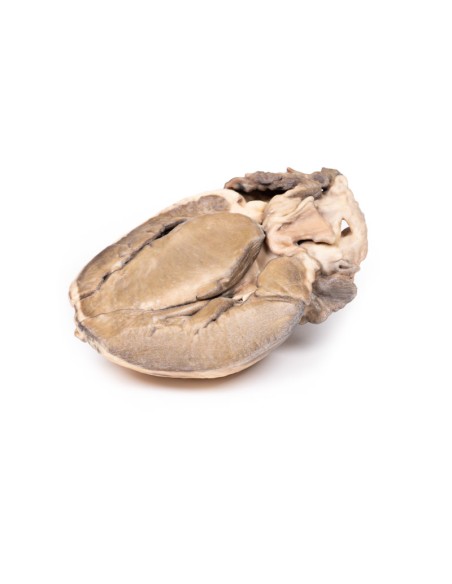
Hypertrophic Subaortic Stenosis - Erler Zimmer 3D anatomy Series MP2036
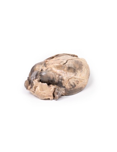
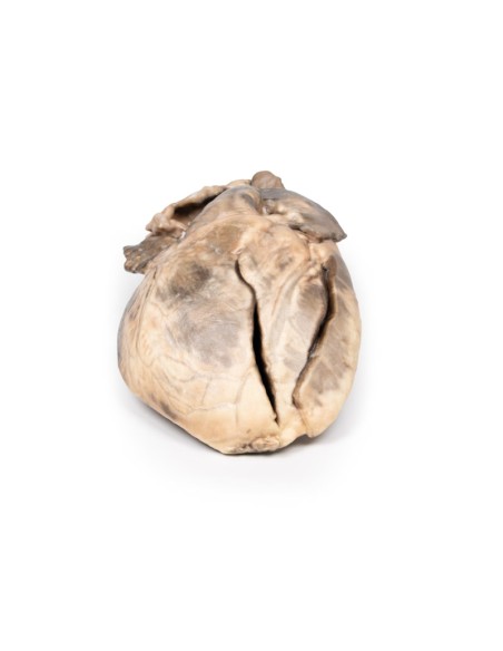
This dissection model highlighting hypertrophic subaortic Stenosis is part of the exclusive Monash 3D anatomy series, a comprehensive series of human dissections reproduced with ultra-high resolution color 3D printing.
Clinical History.
A thin 42-year-old American tourist was found dead in his hotel room. An autopsy by a medical examiner was performed.
Pathology
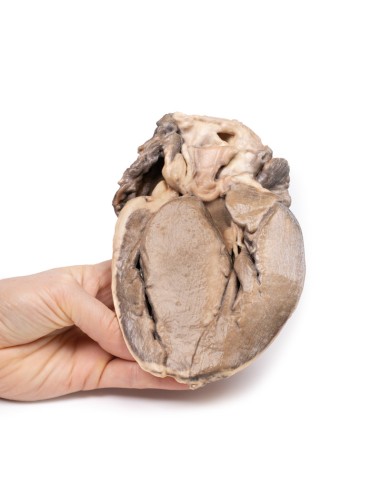
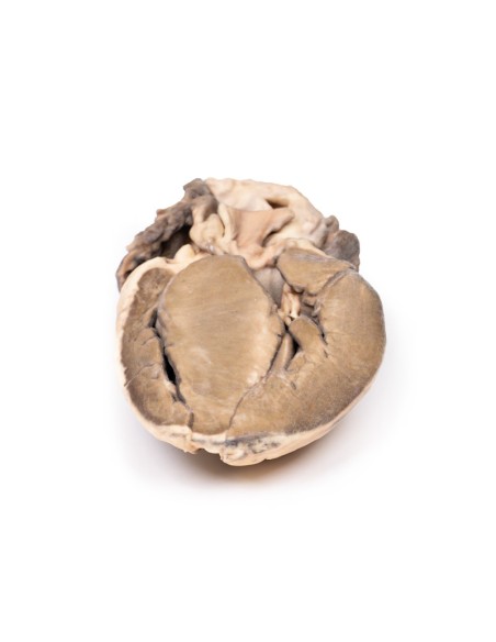
This is a longitudinal section of the heart showing the left and right ventricles and the interventricular septum. The notable abnormality is a grossly thickened interventricular septum and left ventricular hypertrophy. Visible aortic cusps appear insignificant, as does the mitral valve. The ventricular septum is so large that it invades the lumen of the left ventricle.
Diagnosis
Idiopathic hypertrophic subaortic stenosis, also known as hypertrophic cardiomyopathy.
Additional Information.
Subaortic stenosis is considered acquired rather than congenital and is suggested to result from an underlying defect in the architecture of the left ventricular outflow tract (LVOT). The defect may be such that the resulting turbulent blood flow leads to progressive thickening and fibrosis of the LVOT and aortic valve. Progression of the disease often leads to hypertrophic cardiomyopathy secondary to the increased aortic pressure required to be overcome by the left ventricle. Mild or moderate stenosis is often asymptomatic. As the disease progresses and the stenosis becomes severe, symptoms such as exertional dyspnea and syncope may occur. Investigation and subsequent diagnosis are often induced by the presence of a systolic ejection murmur on objective examination. Echocardiography is used to confirm the diagnosis.
Nowadays, the definitive treatment of subaortic stenosis consists of surgical correction of the obstruction.
.
What advantages does the Monash University anatomical dissection collection offer over plastic models or plastinated human specimens?
- Each body replica has been carefully created from selected patient X-ray data or human cadaver specimens selected by a highly trained team of anatomists at the Monash University Center for Human Anatomy Education to illustrate a range of clinically important areas of anatomy with a quality and fidelity that cannot be achieved with conventional anatomical models-this is real anatomy, not stylized anatomy.
- Each body replica has been rigorously checked by a team of highly trained anatomists at the Center for Human Anatomy Education, Monash University, to ensure the anatomical accuracy of the final product.
- The body replicas are not real human tissue and therefore not subject to any barriers of transportation, import, or use in educational facilities that do not hold an anatomy license. The Monash 3D Anatomy dissection series avoids these and other ethical issues that are raised when dealing with plastinated human remains.
No reviews
Tap to zoom
