
Your cart
There are no more items in your cart


Tetralogy of Fallot - Erler Zimmer 3D anatomy Series MP2037
erler zimmer
EZ-MP2037
Last items in stock
€405.28
Tax included
Made in ultra-high resolution 3D printing in full color.
Tetralogy of Fallot - Erler Zimmer 3D anatomy Series MP2037
This dissection model highlighting a Tetralogy of Fallot is part of the exclusive Monash 3D anatomy series, a comprehensive series of human dissections reproduced with ultra-high resolution color 3D printing.
Clinical History.
A 21-month-old child was admitted with a history of exhaustion and exertional dyspnea in the previous 2-3 months. During this period there had been several attacks of acute dyspnea each lasting two minutes. Examination revealed central cyanosis, mild finger clubbing, and a maximal systolic murmur at the left sternal margin. Cardiac catheterization led to a diagnosis of tetralogy of Fallot and severe pulmonary edema. Surgical correction (Willis-Potts anastomosis between the aorta and the origin of the left pulmonary artery) was performed. The child developed acute dyspnea and left lobar consolidation 12 hours after surgery and died despite treatment.
Pathology
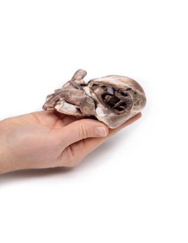
The child's heart is seen from the anterior aspect. The anterior wall of the right ventricle was excised to reveal prominent right ventricular hypertrophy and a narrowed pulmonary outflow tract. The pulmonary valve ring is also small, with a bicuspid stenosed valve. There is a patch of endocardial fibrosis in the outflow tract below the pulmonary valve. The origin of the aorta overlies a high ventricular septal defect. A probe could be passed into the aorta from the hypertrophied right ventricle. The additional probe was able to be passed from the narrowed pulmonary trunk into a dilated, thin-walled left pulmonary artery and through surgical anastomosis into the descending aorta. Examination of the posterior aspect of the specimen reveals an open right atrium and left ventricle. When viewed from the right side of the heart, There is a large interatrial defect (ASD), 8 mm in diameter at the site of the foramen ovale (large arrow). Another small ASD (small arrow), 3 mm in diameter is present posterior to the superior border of the large ASD. Note that the wall of the left ventricle is slightly thinner than the wall of the right ventricle.
Further information
The four features of tetralogy of Fallot are: 1. Ventricular septal defect (VSD); 2. An aorta that straddles the VSD with the latter communicating with both ventricles (overlying aorta) rather than just the left ventricle; 3. Pulmonary stenosis or obstruction of the right ventricular overflow tract; 4. Right ventricular hypertrophy. This condition usually causes cyanosis early in life. Its severity depends on the degree of obstruction to pulmonary outflow, which determines whether a left-right or right-left shunt is present. In some patients, pulmonary blood flow is increased due to the presence of a pervious ductus arteriosus. Patients with this condition may survive untreated into adult life, and some may reach middle age. However, surgical correction is now possible and is desirable, as the disorder is eventually fatal. Sometimes additional cardiac abnormalities may be present. (E.g., interatrial septal defect, as was found in this case).
In most cases of tetralogy of Fallot, the cause is unknown although genetic factors play a role in some patients. For example, the condition is more common in patients with Down syndrome (trisomy 21; in association with AV canal defects) or DiGeorge syndrome (22q11 deletion).
.
What advantages does the Monash University anatomical dissection collection offer over plastic models or plastinated human specimens?
- Each body replica has been carefully created from selected patient X-ray data or human cadaver specimens selected by a highly trained team of anatomists at the Monash University Center for Human Anatomy Education to illustrate a range of clinically important areas of anatomy with a quality and fidelity that cannot be achieved with conventional anatomical models-this is real anatomy, not stylized anatomy.
- Each body replica has been rigorously checked by a team of highly trained anatomists at the Center for Human Anatomy Education, Monash University, to ensure the anatomical accuracy of the final product.
- The body replicas are not real human tissue and therefore not subject to any barriers of transportation, import, or use in educational facilities that do not hold an anatomy license. The Monash 3D Anatomy dissection series avoids these and other ethical issues that are raised when dealing with plastinated human remains.
No reviews
Tap to zoom
