
Your cart
There are no more items in your cart


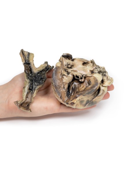
Hydatid disease affecting the heart and aorta - Erler Zimmer 3D anatomy Series MP2035
erler zimmer
EZ-MP2035
€701.87
Tax included
Made in ultra-high resolution 3D printing in full color.
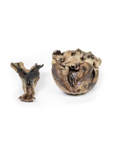
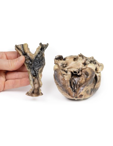
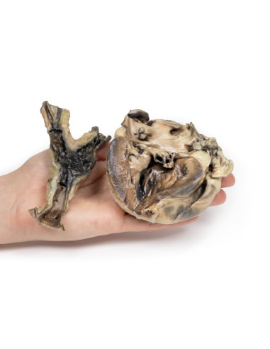
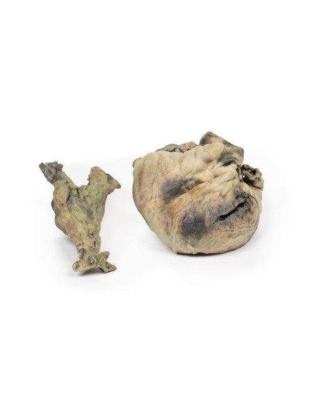
Hydatid Disease Affecting the Heart and Aorta - Erler Zimmer 3D anatomy Series MP2035
This dissection model highlighting a Hydatid Disease affecting the heart and aorta is part of the exclusive Monash 3D anatomy series, a comprehensive series of human dissections reproduced with ultra-high resolution color 3D printing.
Medical history
This 11-year-old girl had an 18-month history of hydatid disease (see below). A total of 17 cysts were removed from the child's brain during craniotomy on three occasions, and subsequently cysts were found in the kidneys, mesentery, and abdominal aorta at its bifurcation. X-ray of the heart showed a calcified cyst, and the patient was sent to a tertiary hospital for its removal. The patient deteriorated and died following open-heart surgery during which a dead hydatid cyst was found in the left ventricle.
Pathology
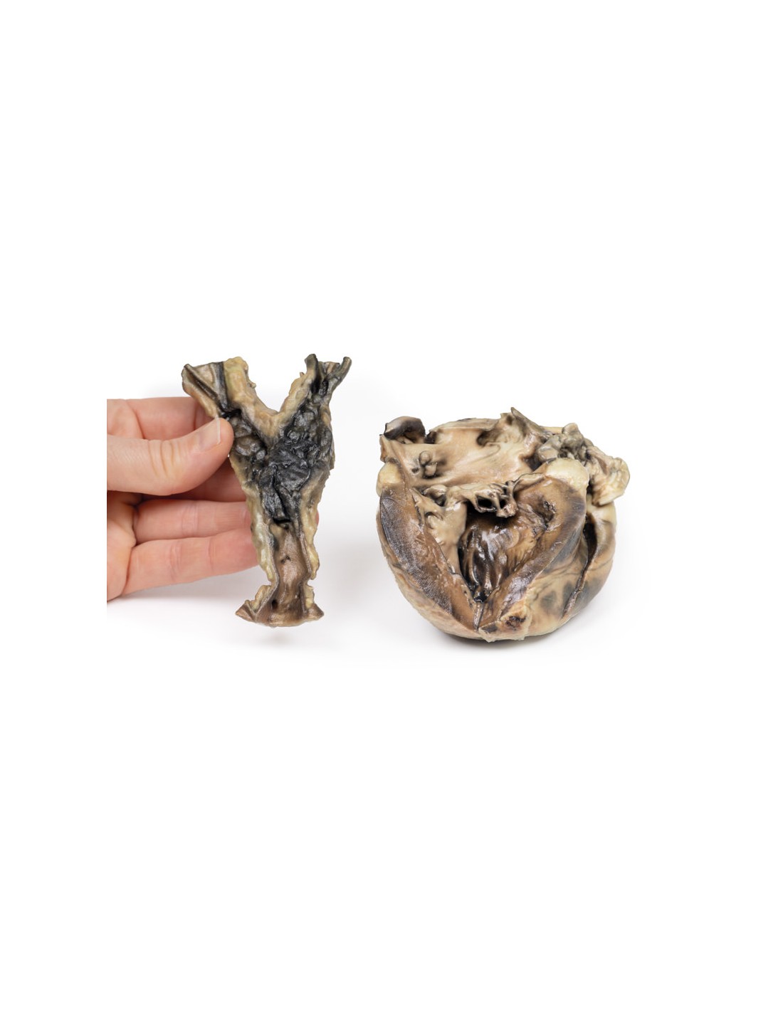
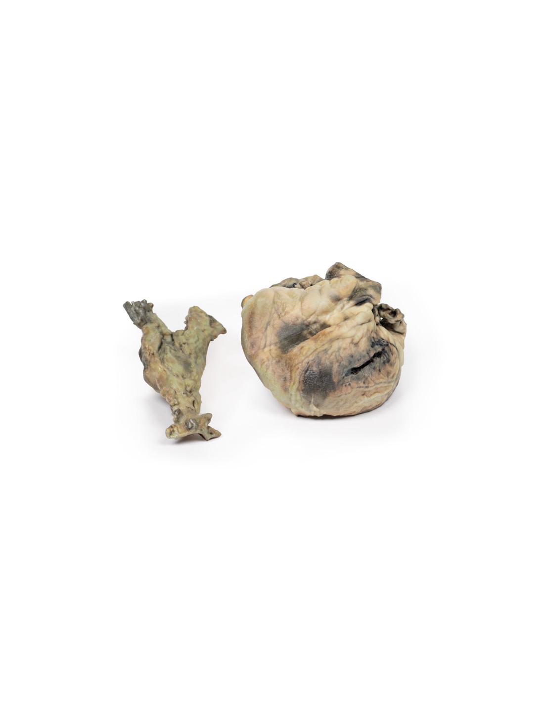
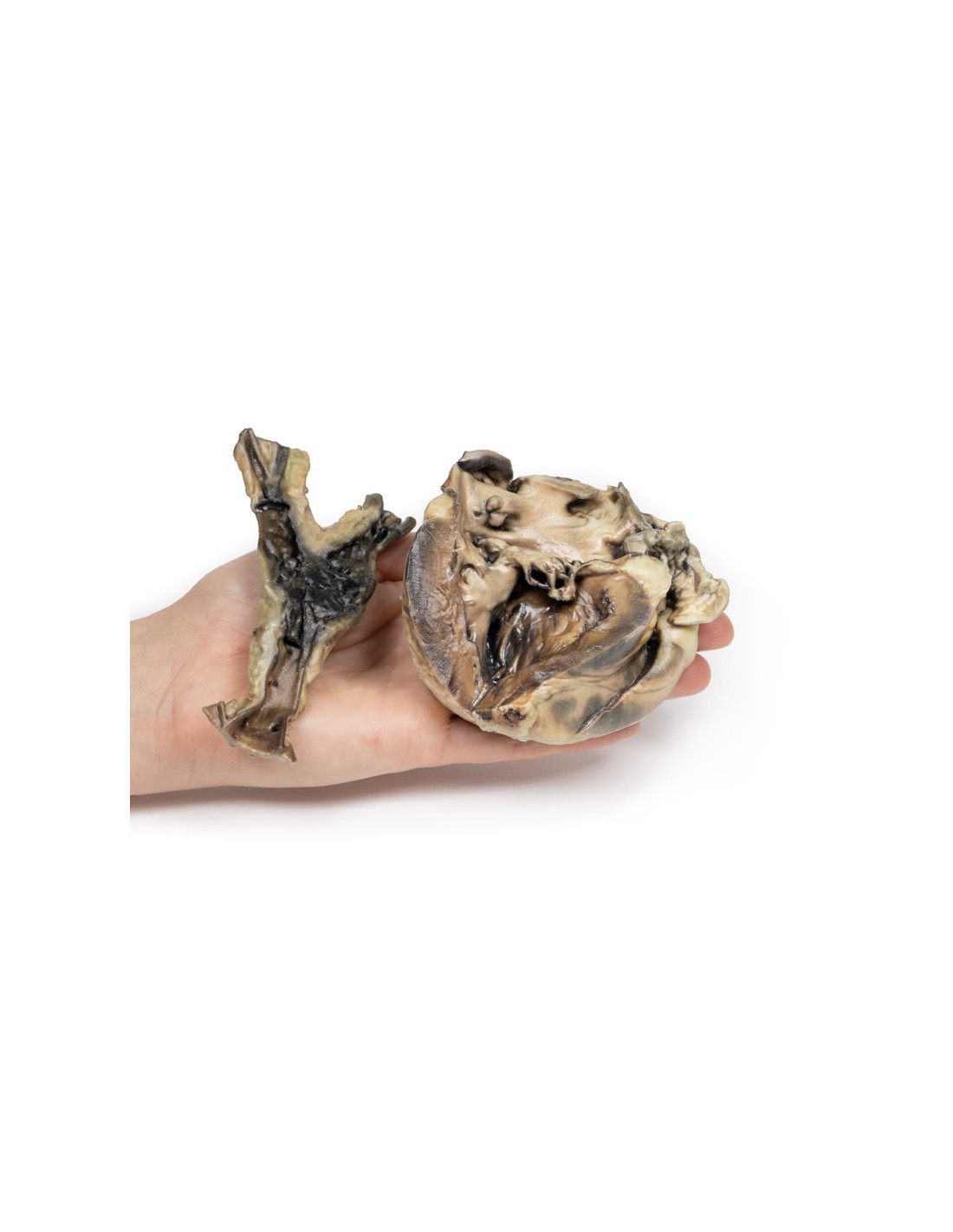
The specimens are of the heart, with the left ventricle open, and the aorta at its common iliac bifurcation. The aorta shows some atheromatous deposition in the upper part. There is a large mass of ante mortem clot at the site of the iliac bifurcation with extension along both common iliac arteries.
The heart shows left ventricular wall hypertrophy and abnormal communication between the left ventricle and atrium flowing through the posterior cusp of the mitral valve through the papillary muscle into the left ventricular cavity. This channel is surrounded by thickened fibrous tissue. The posterior cusp of the valve has been divided. Sutured surgical incisions are visible on the posterior face of the specimen and on the ventricular wall and in the left atrial appendage. Hydatid cysts occupy the abdominal aorta at its bifurcation and the canal joining the left ventricle and left atrium.
Histology demonstrated cysts within the wall of the aorta composed of 3 layers: an outermost pericystic fibrous layer; a middle layer of ectocysts that was laminated, hyaline, and acellular; and the inner endocyst in the germinal layer, consisting of daughter cysts and brood capsules with scolex. A focal granulomatous palisade reaction was also present within the wall of the aorta.
Further Information.
Hydatid cyst is a human parasitic disease caused by the larval stage of the cestode tapeworm Echinococcus granulosus, which infests the intestines of dogs, its definitive hosts. Humans can serve as incidental hosts by ingesting ova in vegetables or water contaminated with dog feces. Humans are infected by ingestion of eggs passed in dog feces. Oncospheres released from the eggs penetrate the intestinal mucosa and, through the portal system, are deposited in the liver, lungs, muscles or other organs, where hydatid cysts form.
Hydatid disease is endemic in livestock breeding areas of the world, particularly in Mediterranean countries, the Middle East, South America, Australia and New Zealand. Although no part of the body can be spared from hydatid cysts, they mainly affect the liver and lungs. Cardiac involvement is a much rarer but potentially fatal condition and comprises 0.5-2% of all hydatid cases. Cardiac complications and presentation vary depending on the location, size, and integrity of the cysts. Left ventricular myocardium most frequently involved. Pericardial involvement occurs mainly in multifocal cardiac echinococcosis. Cyst growth causes them to be pushed to a weaker side of the cardiac wall, the epicardium or endocardium. HCs in the left ventricle are usually found at the subepicardial level, so they rarely rupture into the pericardial space.
Although E. granulosus is still found in sheep and rural dogs in Australia, the prevalence of transmission is less common than it was. The marked reduction in prevalence in rural domestic dogs, and also in sheep, is the result of the inclusion of the highly effective cestocidal drug, praziquantel, in all inexpensive, generic and readily available dog dewormers and the development of inexpensive commercial dry dog food [2].
References
1. Oraha et al. Ann Med Surg (Lond). 2018 18-21
2. Jenkins et al. Int J Parasitol Parasites Wildl. 2019: 256-259.
.
What advantages does the Monash University anatomical dissection collection offer over plastic models or plastinated human specimens?
- Each body replica has been carefully created from selected patient X-ray data or human cadaver specimens selected by a highly trained team of anatomists at the Monash University Center for Human Anatomy Education to illustrate a range of clinically important areas of anatomy with a quality and fidelity that cannot be achieved with conventional anatomical models-this is real anatomy, not stylized anatomy.
- Each body replica has been rigorously checked by a team of highly trained anatomists at the Center for Human Anatomy Education, Monash University, to ensure the anatomical accuracy of the final product.
- The body replicas are not real human tissue and therefore not subject to any barriers of transportation, import, or use in educational facilities that do not hold an anatomy license. The Monash 3D Anatomy dissection series avoids these and other ethical issues that are raised when dealing with plastinated human remains.
No reviews
Tap to zoom
