
Your cart
There are no more items in your cart


Abdominal aortic aneurysm - Erler Zimmer 3D anatomy Series MP2030
erler zimmer
EZ-MP2030
€858.88
Tax included
Made in ultra-high resolution 3D printing in full color.
Abdominal Aortic Aneurysm - Erler Zimmer 3D anatomy Series MP2030
This dissection model highlighting an Abdominal Aortic Aneurysm is part of the exclusive Monash 3D anatomy series, a comprehensive series of human dissections reproduced with ultra-high resolution color 3D printing.
Clinical history
This 70-year-old man with a past history of mild gastroesophageal reflux presented to Alfred Hospital with a sudden onset of severe upper abdominal pain that radiated to the tip of his left shoulder. On examination, he was distressed and hyperventilating, heart rate 87/min and blood pressure 140/90 mm Hg. Abdominal examination revealed board-like rigidity and reduced bowel sounds. At emergency laparotomy, no evidence of bowel rupture was found; the pancreas appeared normal and an unruptured abdominal aortic aneurysm was noted. Endoscopy the next day showed a ruptured esophageal ulcer, and a Celestin tube was inserted. The patient developed localized infectious complications, pulmonary edema, and congestion and died 19 days after admission.
Pathology
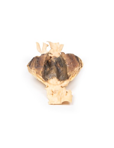
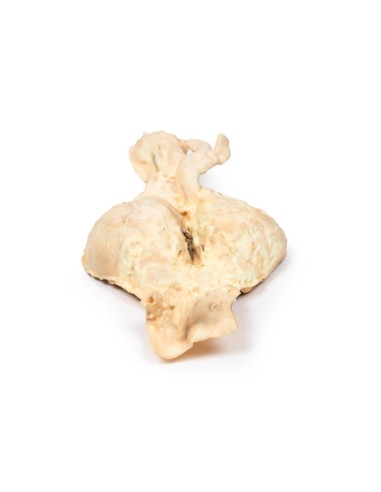
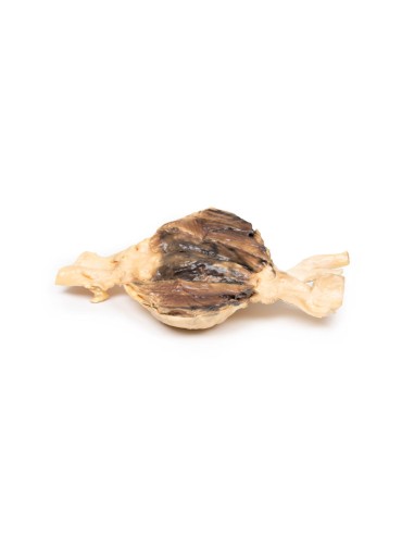
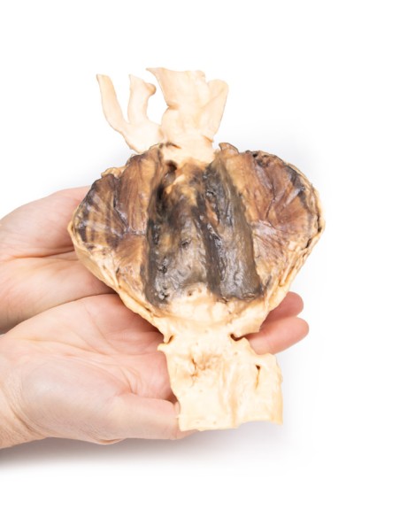
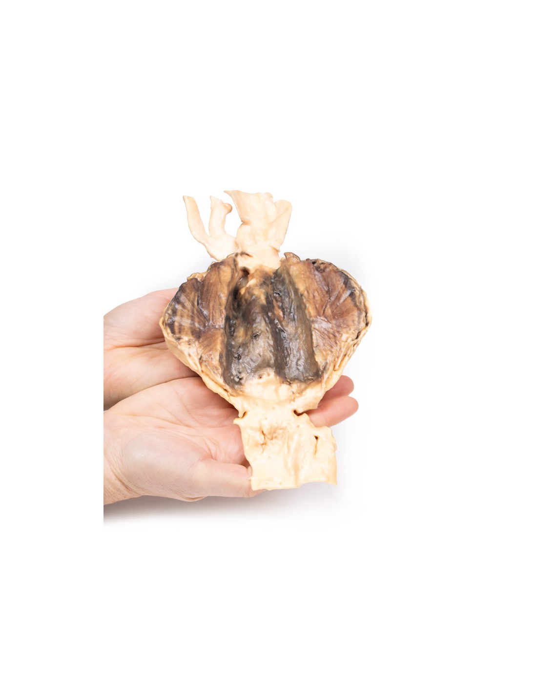
The specimen consisted of the lower abdominal segment of the aorta along with the common iliac vessels and the proximal portions of the internal and external iliac arteries. A large aneurysm measuring 10 x 7 cm is located below the origin of the renal arteries extending to the aortic bifurcation. The aneurysm with its severe thinning of the wall of the abdominal aorta is partly lined with a laminated thrombus, indicating chronicity of the process. There is evidence of a recent thrombus on the luminal surface. There also appears to be some aneurysmal dilatation of the common and (open) proximal left external iliac artery. The abdominal aorta at the upper end of the specimen shows focally multiple ulcerated atheromatous plaques. There is no evidence of rupture.
Additional Information.
Abdominal aortic aneurysm (AAA or triple A) represents a localized enlargement of the abdominal aorta (diameter >3 cm or more than 50% larger than normal)[1]. They are usually asymptomatic except during rupture[1]. Large aneurysms may be palpable on abdominal examination. Occasionally, abdominal, back, or leg pain may occur depending on location and size. Rupture may cause pain in the abdomen or back, sudden drop in blood pressure with loss of consciousness, and often leads to death[1]. AAAs occur most commonly in people older than 50 years, in men, and among those with a family history of this disease. Additional risk factors include smoking, hypertension, and other diseases of the heart or blood vessels. They are also found in genetic abnormalities, including Marfan syndrome and Ehlers-Danlos syndrome.
What advantages does the Monash University anatomical dissection collection offer over plastic models or plastinated human specimens?
- Each body replica has been carefully created from selected patient X-ray data or human cadaver specimens selected by a highly trained team of anatomists at the Monash University Center for Human Anatomy Education to illustrate a range of clinically important areas of anatomy with a quality and fidelity that cannot be achieved with conventional anatomical models-this is real anatomy, not stylized anatomy.
- Each body replica has been rigorously checked by a team of highly trained anatomists at the Center for Human Anatomy Education, Monash University, to ensure the anatomical accuracy of the final product.
- The body replicas are not real human tissue and therefore not subject to any barriers of transportation, import, or use in educational facilities that do not hold an anatomy license. The Monash 3D Anatomy dissection series avoids these and other ethical issues that are raised when dealing with plastinated human remains.
No reviews
Tap to zoom
