Your cart
There are no more items in your cart


Retrosternal goiter - Erler Zimmer 3D anatomy Series MP2102
erler zimmer
EZ-MP2102
€491.17
Tax included
Made in ultra-high resolution 3D printing in full color.
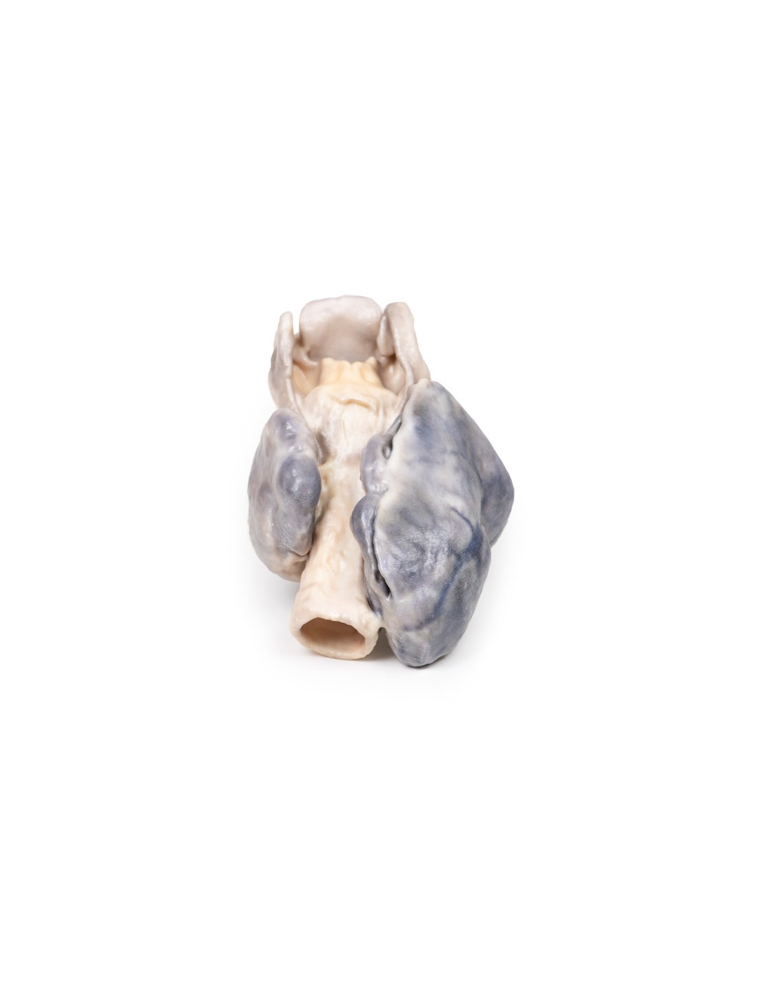
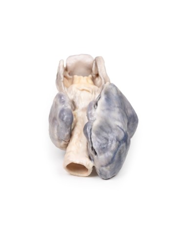
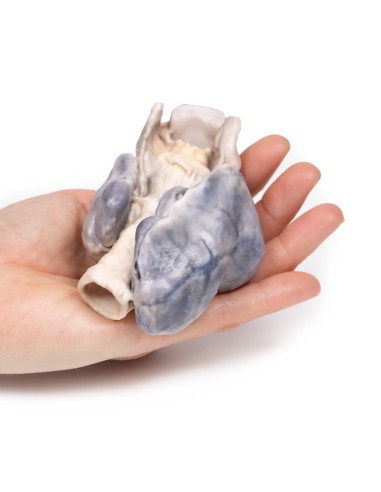
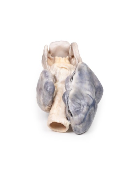
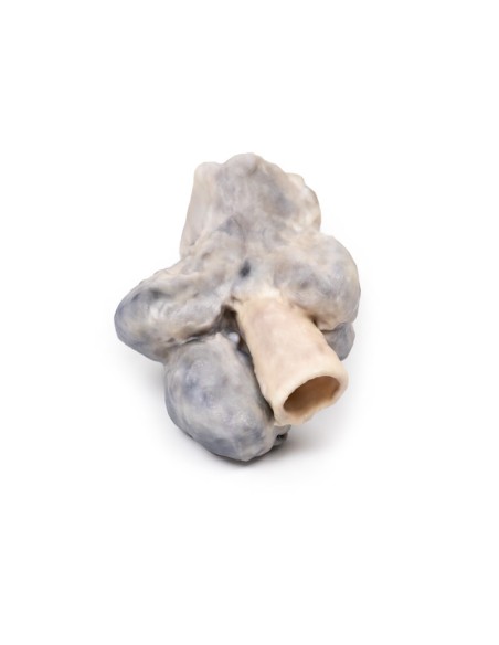
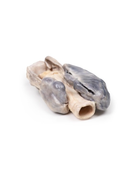
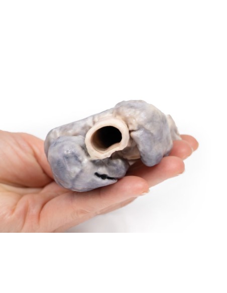
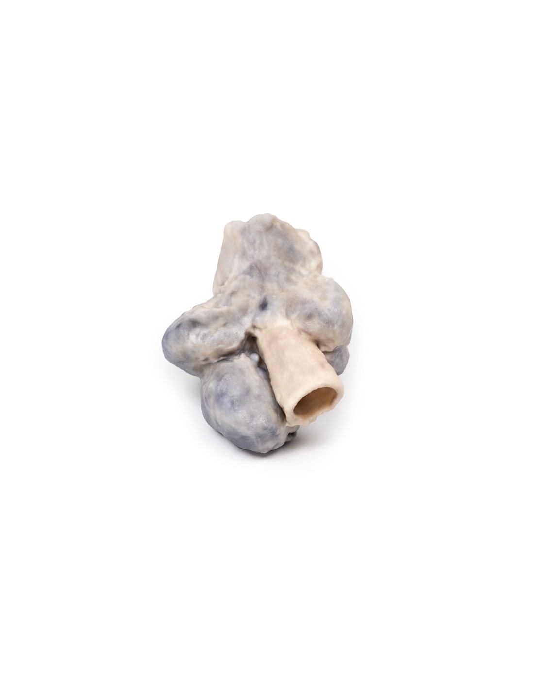
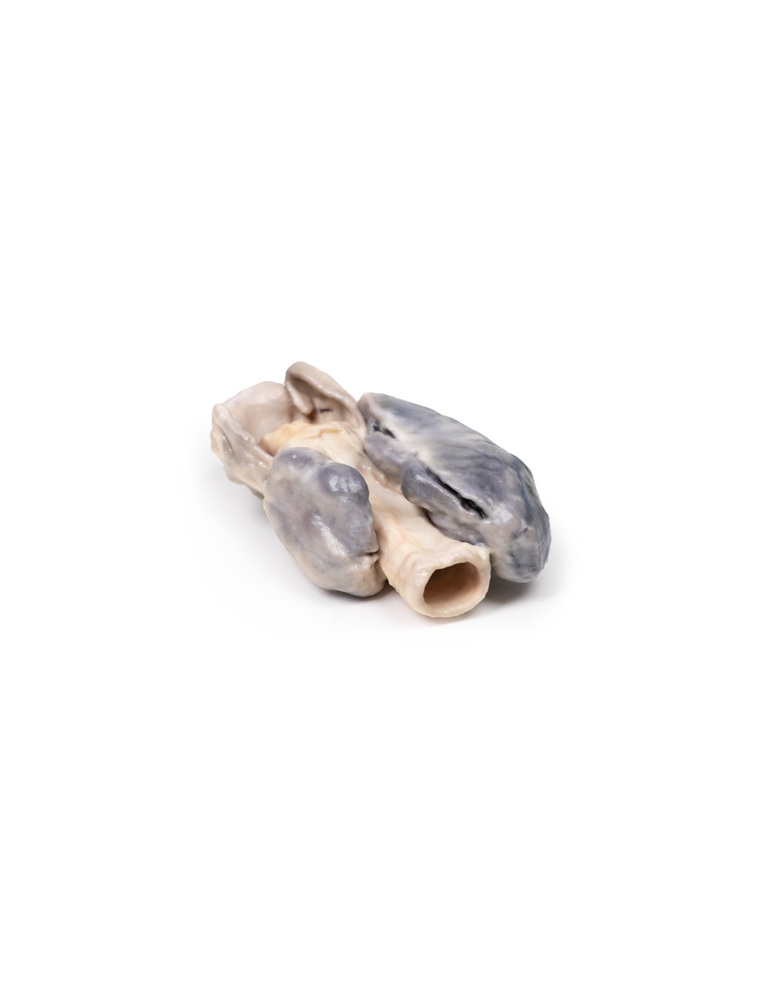
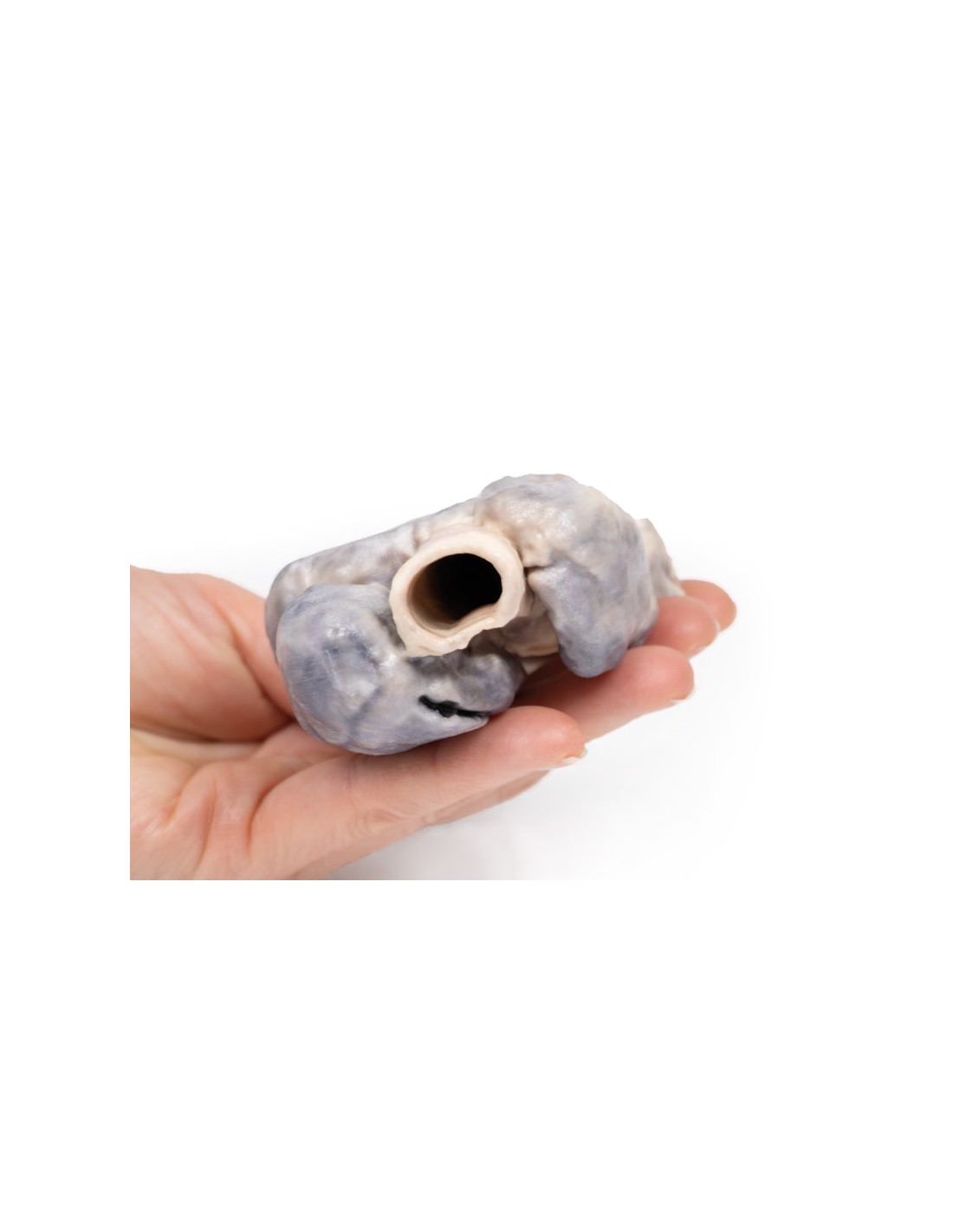
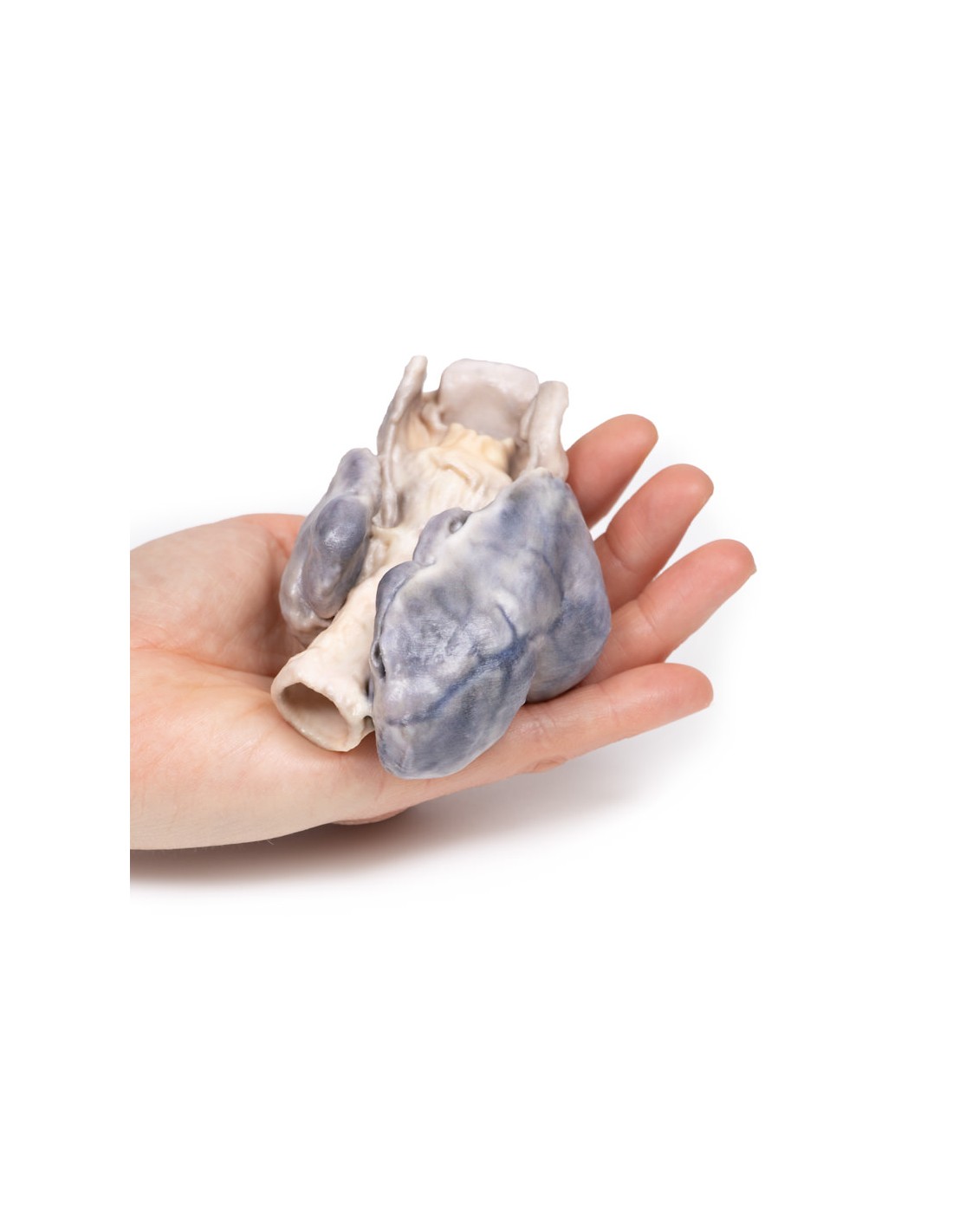
Retrosternal Goiter - Erler Zimmer 3D anatomy Series MP2102
This dissection model highlighting a Retrosternal Goiter is part of the exclusive Monash 3D anatomy series, a comprehensive series of human dissections reproduced with ultra-high resolution color 3D printing.
Clinical presentation
A 60-year-old woman presented with abnormal neck swelling, persistent cough and difficulty swallowing. She had gained weight in recent years. She died of unrelated cardiovascular disease, and the specimen was obtained postmortem.
Pathology
The specimen, taken postmortem, includes the larynx, trachea, and large multilobular thyroid gland. The thyroid gland is grossly enlarged particularly the right lobe, which has two large lobes extending superiorly and inferiorly for an interval of 7-8 mm, well beyond its normal margins when viewed on the anterior aspect. Posteriorly, the esophagus has been opened to expose the posterior wall of the trachea. The right lobe appears larger than the anterior perspective, and the abnormal growth appears to be mainly the lower pole of the right lobe. The surfaces show no major pigmentary changes. Prominent veins are visible on the surface of the right lobe.
Additional Information.
Goiter is often detected simply as a mass or swelling in the neck, but depending on the size and location of the growth it may produce symptoms of pressure on the trachea and esophagus. There may be difficulty breathing, dysphagia, coughing, and hoarseness. Recurrent laryngeal nerve palsy may occur due to an expanding goiter, but this is rare. Symptoms suggesting obstruction of the trachea may occur, including coughing, stridor, and shortness of breath. Occasionally, tenderness and a sudden increase in goiter size arise due to cystic expansion and hemorrhage in a nodule [1].
Causes of goiter include autoimmune diseases (Hashimoto's thyroiditis, Grave's disease), formation of one or more thyroid nodules, and iodine deficiency. Goiter occurs when there is reduced thyroid hormone synthesis secondary to biosynthetic defects and/or iodine deficiency, leading to an increase in thyroid-stimulating hormone (TSH). This stimulates thyroid growth as a compensatory mechanism to overcome the reduced hormone synthesis. Elevated TSH is also believed to contribute to thyroid enlargement in the goiter form of Hashimoto's thyroiditis in combination with fibrosis secondary to the autoimmune process in this condition. In Grave's disease, goiter results mainly from TSH receptor antibody stimulation[2].
Scientific references.
1. Hughes et al. (2012) Goitre: Causes, investigation and management.
2. Aust Family Physician, 41, 572-576.
.
What advantages does the Monash University anatomical dissection collection offer over plastic models or plastinated human specimens?
- Each body replica has been carefully created from selected patient X-ray data or human cadaver specimens selected by a highly trained team of anatomists at the Monash University Center for Human Anatomy Education to illustrate a range of clinically important areas of anatomy with a quality and fidelity that cannot be achieved with conventional anatomical models-this is real anatomy, not stylized anatomy.
- Each body replica has been rigorously checked by a team of highly trained anatomists at the Center for Human Anatomy Education, Monash University, to ensure the anatomical accuracy of the final product.
- The body replicas are not real human tissue and therefore not subject to any barriers of transportation, import, or use in educational facilities that do not hold an anatomy license. The Monash 3D Anatomy dissection series avoids these and other ethical issues that are raised when dealing with plastinated human remains.
No reviews
Tap to zoom