
Your cart
There are no more items in your cart


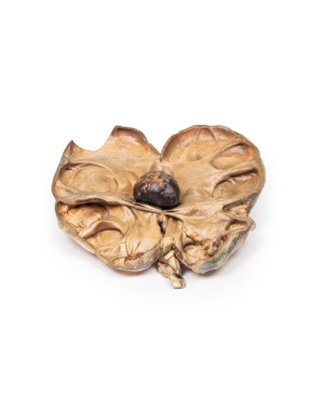
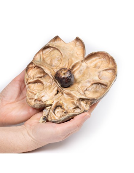
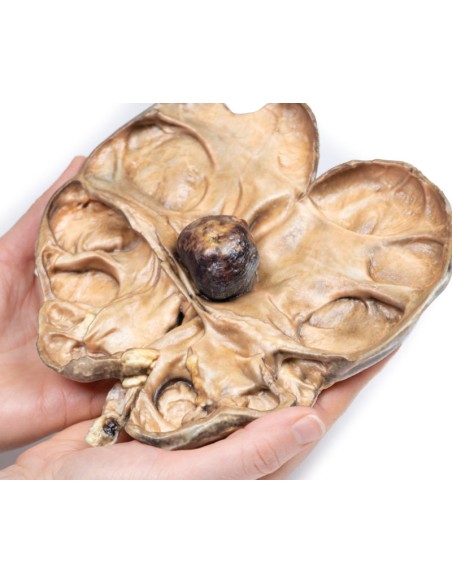
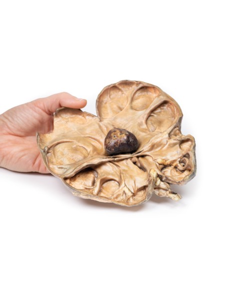
Hydronephrosis and hydroureter caused by obstruction by a kidney stone - Erler Zimmer 3D anatomy Series MP2094
erler zimmer
EZ-MP2094
€771.65
Tax included
Made in ultra-high resolution 3D printing in full color.
Hydronephrosis and hydroureter caused by obstruction from a kidney stone - Erler Zimmer 3D anatomy Series MP2094
This dissection model highlighting hydronephrosis and hydroureter caused by obstruction from a kidney stone is part of the exclusive Monash 3D anatomy series, a comprehensive series of human dissections reproduced with ultra-high resolution color 3D printing.
Clinical History.
A 72-year-old woman presented with flank colic and increasing malaise. Intermittent hematuria was noted. Biochemical investigations revealed significantly reduced renal function. CT abdomen showed congenital renal agenesis of the left kidney and hydronephrosis and hydroureter on the right side, due to obstruction by a smaller stone. A percutaneous lithotomy was attempted to relieve the obstruction, but the patient died from a cardiac event during the procedure.
Pathology
The specimen is the patient's right kidney, which is grossly and partially divided in two. There is a large dilatation of the pelvic-caliceal system visible and significant atrophy of the renal tissue, particularly in the cortex. There is a large brown tartar visible in the renal pelvis at the ureteropelvic junction.
Further information
Urolithiasis (kidney stones) is a very common disease that affects up to 1 in 10 people during their lifetime. Stone formation can occur anywhere along the urinary tract, but most commonly occurs within the kidneys. Risk factors for stone formation include male sex; any condition that affects urine composition, such as hypercalciuria or elevated urinary oxalate; systemic metabolic disorders, such as cystinuria and taste; dietary factors, such as high intake of oxalate and animal protein ??low fluid intake; and environmental factors, such as high dry temperatures. Eighty percent of kidney stones are unilateral.
Symptoms of urolithiasis include stabbing pain, hematuria, nausea, vomiting, fainting, dysuria, and urgency. Symptoms depend on the size and location of the stone. Urolithiasis can be asymptomatic especially if stones form and remain within the renal pelvis or bladder. Symptoms occur when the stones move into the ureter. Stone pain is usually colic and typically severe in nature; occurring in paroxysms. The flank is the most common site for pain, but pain can occur anywhere along the urinary tract and in the genitals. The pain resolves as the stone passes. Hematuria may be large or microscopic.
Diagnosis can be made on the basis of history and examination. Radiological tools frequently used to aid diagnosis include CT scan without contrast medium or ultrasound of the kidneys and bladder. Less commonly used imaging methods include abdominal radiography, intravenous pyelogram, and MRI.
If left untreated, renal damage and eventually renal failure from progressive obstruction and hydronephrosis will occur. If the obstructing tartar is not relieved, pressure buildup proximal to the obstruction will occur. This pressure is transmitted back through the collecting ducts to the cortex causing progressive atrophy of the renal parenchyma with dilation of the renal calyces and pelvis. The pressure also compresses the vasculature in the medulla leading to ischemic medullary damage. Glomerular filtration persists in the affected kidney until the end of the disease process, when it will gradually decrease. Obstruction triggers an interstitial inflammatory process leading to fibrosis. Kidney stones also predispose patients to infection secondary to the obstruction and the trauma they cause to the urothelium.
Treatment in acute patients includes supportive treatment to allow passage of the stone. Medical treatment used includes analgesia, commonly NSAIDs and opioids, and agents to promote stone passage, such as alpha-blockers, calcium channel blockers, and antispastics. Surgery may be necessary if there are serious complications from stones or if the stone is large and cannot be expelled with conservative treatment. Surgical interventions include lithotripsy (using laser or electricity), laparoscopic stone removal, or percutaneous stone removal. Rarely, open surgery is required.
.
What advantages does the Monash University anatomical dissection collection offer over plastic models or plastinated human specimens?
- Each body replica has been carefully created from selected patient X-ray data or human cadaver specimens selected by a highly trained team of anatomists at the Monash University Center for Human Anatomy Education to illustrate a range of clinically important areas of anatomy with a quality and fidelity that cannot be achieved with conventional anatomical models-this is real anatomy, not stylized anatomy.
- Each body replica has been rigorously checked by a team of highly trained anatomists at the Center for Human Anatomy Education, Monash University, to ensure the anatomical accuracy of the final product.
- The body replicas are not real human tissue and therefore not subject to any barriers of transportation, import, or use in educational facilities that do not hold an anatomy license. The Monash 3D Anatomy dissection series avoids these and other ethical issues that are raised when dealing with plastinated human remains.
No reviews
Tap to zoom
