Your cart
There are no more items in your cart


hydronephrosis and hydroureters - Erler Zimmer 3D anatomy Series MP2093
erler zimmer
EZ-MP2093
€786.41
Tax included
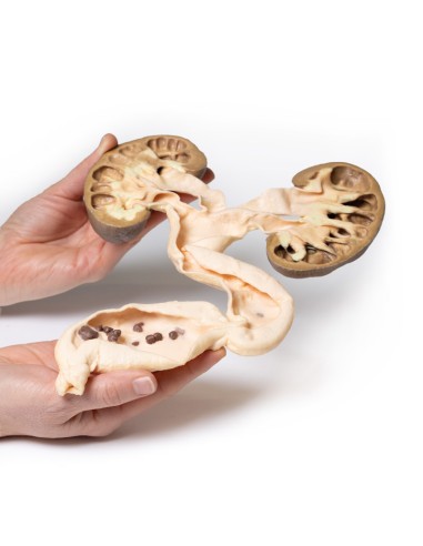
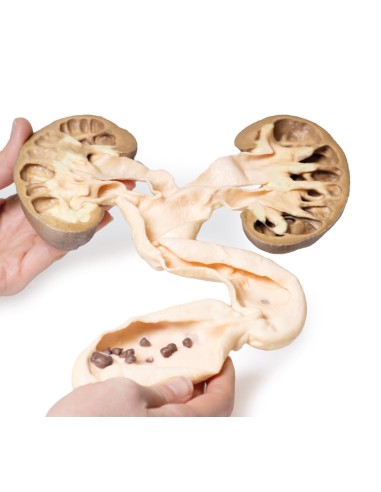
Made in ultra-high resolution 3D printing in full color.
hydronephrosis and hydroureters - Erler Zimmer 3D anatomy Series MP2093
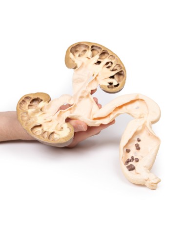
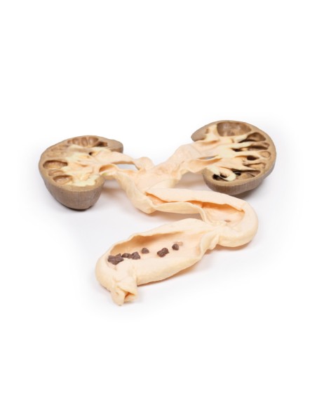
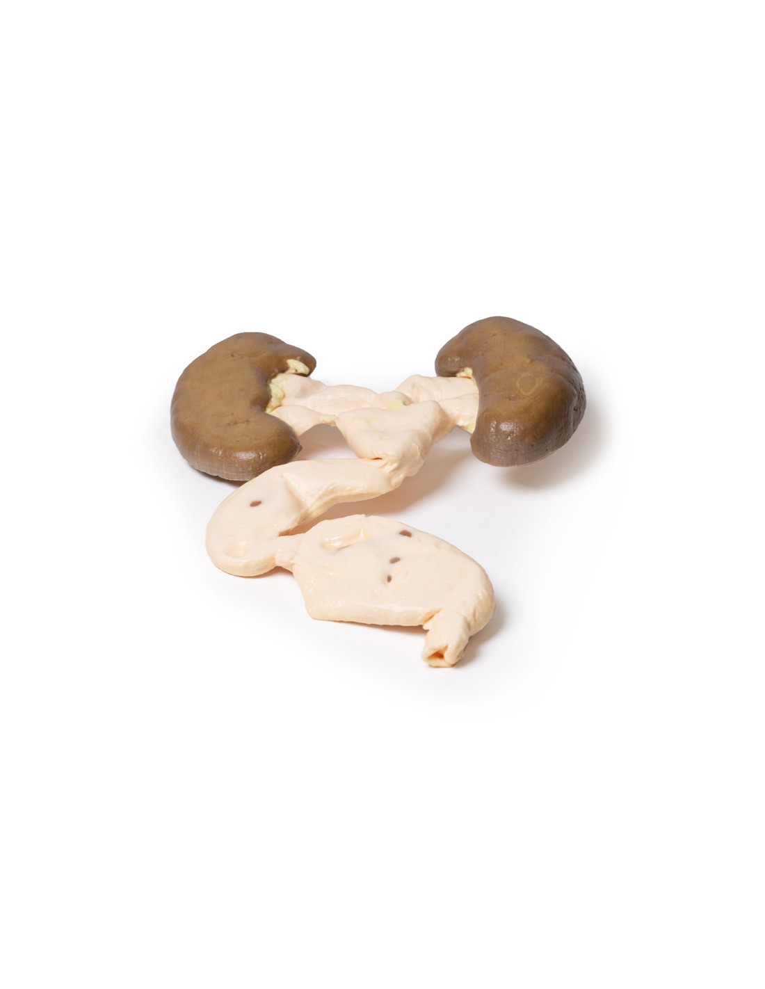
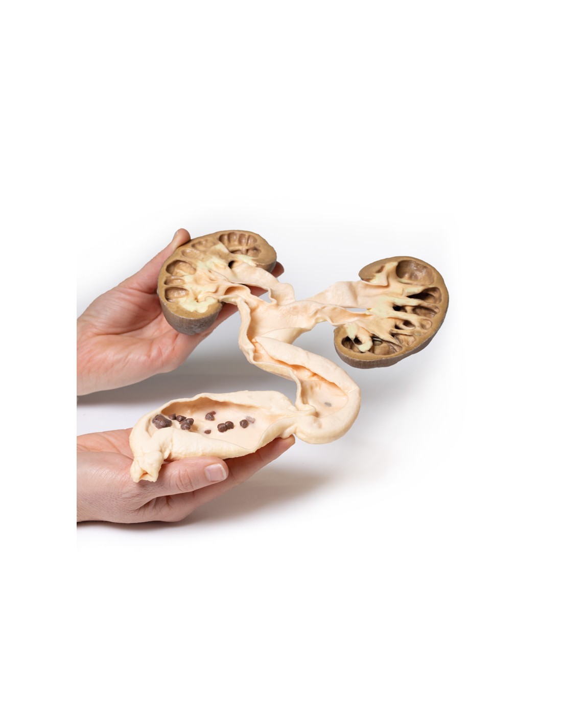
This dissection model highlighting hydronephrosis and hydroureters is part of the exclusive Monash 3D anatomy series, a comprehensive series of human dissections reproduced with ultra-high resolution color 3D printing.
Clinical History.
A 49-year-old man presented with a 6-week history of malaise, urinary frequency and hematuria. Further questioning revealed intermittent pain in the left flank. Abdominal ultrasonography showed severe hydronephrosis and hydroureter, secondary to multiple ureteric stones obstructing the uretero-vesical junction. He underwent a left nephrectomy and ureterectomy and recovered successfully.
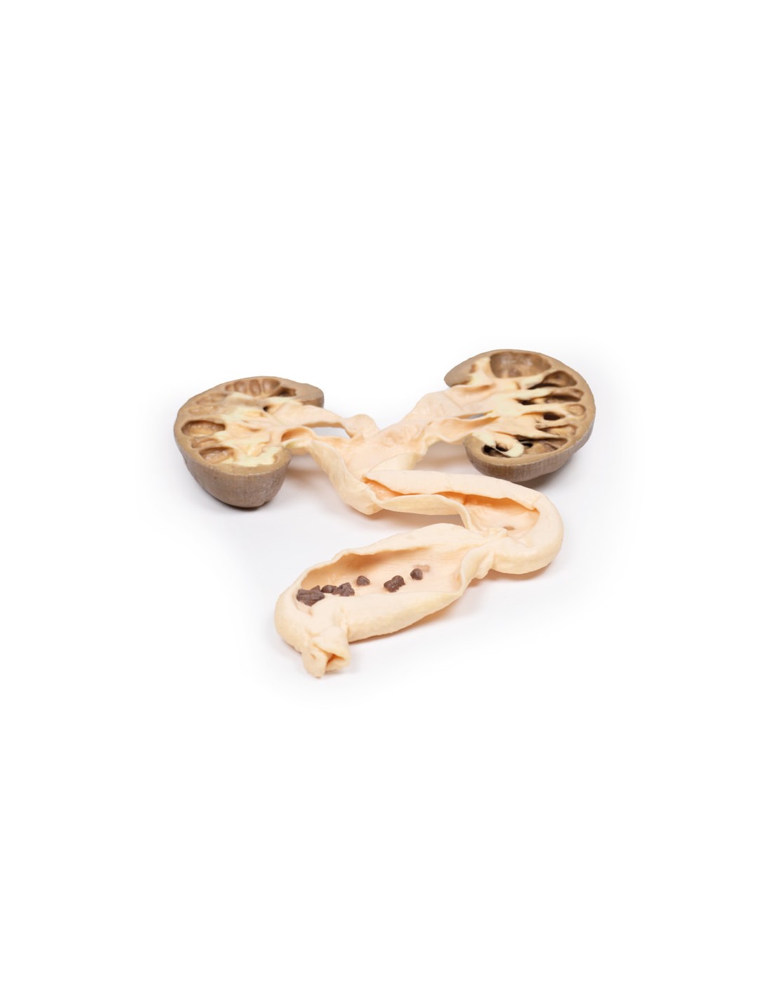
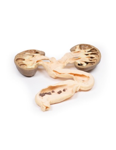
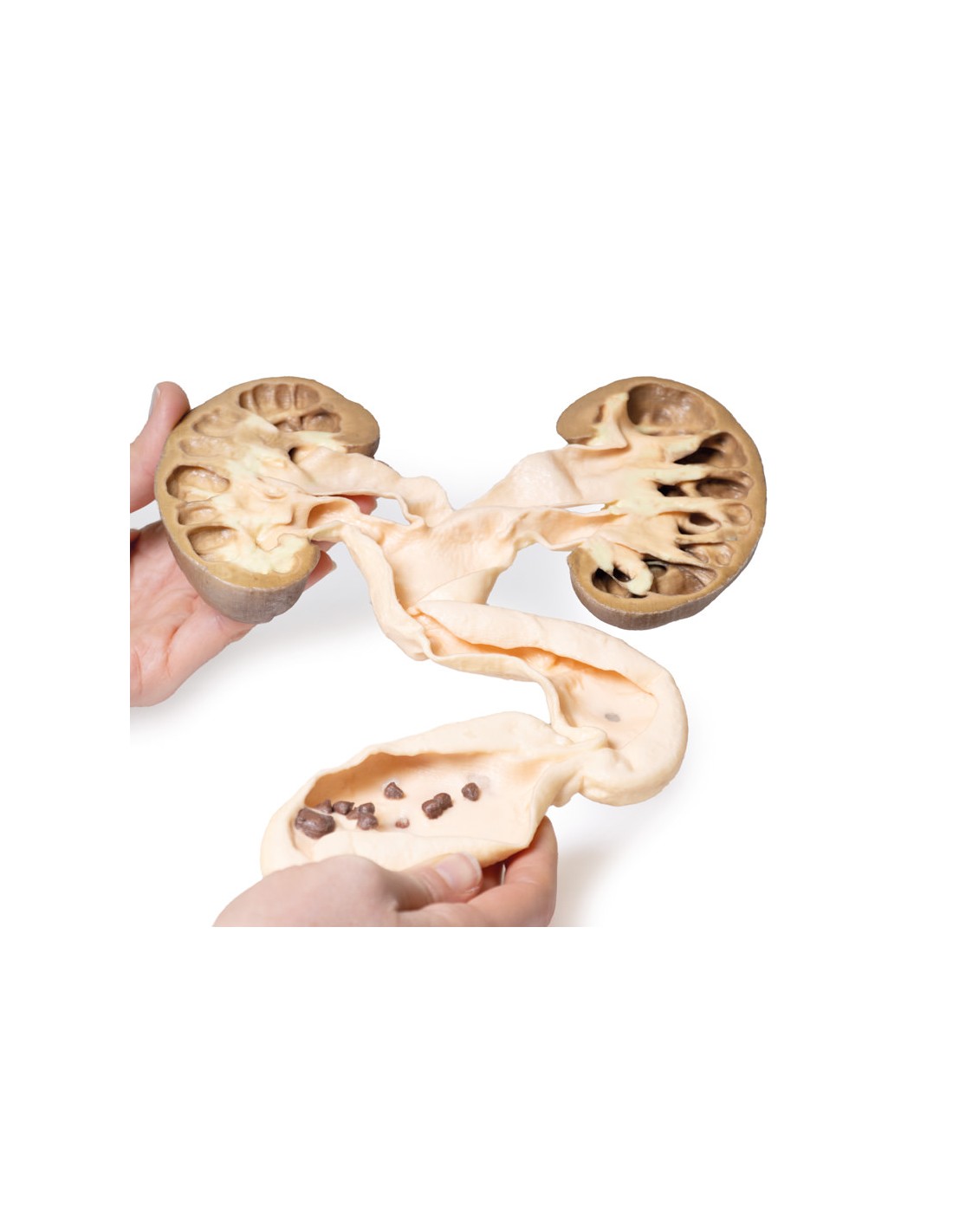
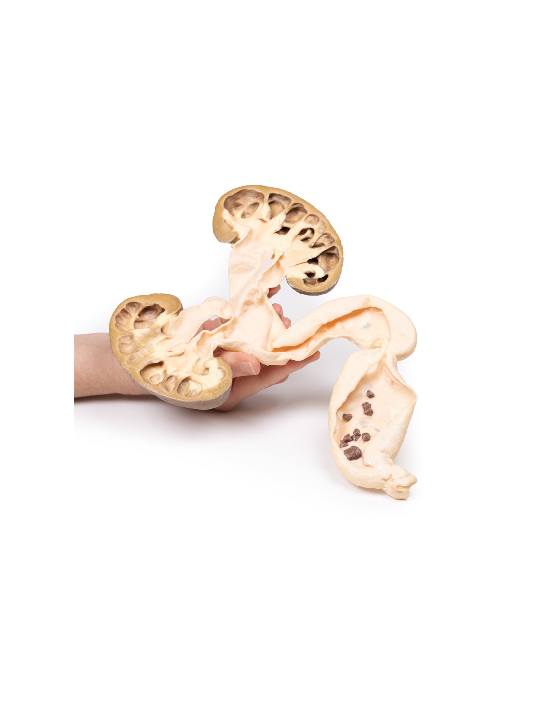
Pathology
This is the patient's left nephrectomy and ureterectomy specimen. The kidney has been divided in two and the cut surface of both halves is shown, mounted in continuity with the ureter, which has been opened. The kidney is grossly hydronephrotic and there is considerable atrophic thinning and loss of renal parenchymal tissue. The ureter is extremely dilated and distally contains numerous small brownish-black stones with irregular, sharp surface protrusions. These are calcium oxalate stones.
This is an example of hydronephrosis and hydroureter due to stones obstructing the lower end of the ureter.
More information
Hydronephrosis, or obstructive uropathy, is dilatation of the renal pelvis and calyces caused by an obstruction in the outflow of urine. Obstruction can occur anywhere in the urinary tract. Any lesion, intrinsic (within the outflow system) or extrinsic (outside the ureter), that impedes urine flow can lead to hydronephrosis. Common causes include: congenital abnormalities, urinary stones, urinary tract tumors, urinary tract inflammation, prostatic hypertrophy, and prostate tumors. The symptoms of hydronephrosis relate to the pathology causing the obstruction (e.g., renal colic pain with stones), the time period of the obstruction (acute or chronic), the site (unilateral or bilateral), and whether it is complete or partial.
If the obstruction is not cleared, it will eventually cause pressure buildup proximal to the obstruction. This pressure is transmitted retrogradely through the collecting ducts to the cortex causing progressive atrophy of the kidney with dilation of the renal calyces and pelvis. The pressure also compresses the vascular system in the medulla leading to ischemic medullary damage. Glomerular filtration persists in the affected kidney until the end of the disease process, when filtration gradually decreases or ceases. Obstruction triggers an interstitial inflammatory process that leads to fibrosis. Ultrasound is the key diagnostic tool followed by CT scan or urogram. Most obstructive lesions require surgical intervention to relieve the blockage. Surgical interventions depend on each individual cause,
.
What advantages does the Monash University anatomical dissection collection offer over plastic models or plastinated human specimens?
- Each body replica has been carefully created from selected patient X-ray data or human cadaver specimens selected by a highly trained team of anatomists at the Monash University Center for Human Anatomy Education to illustrate a range of clinically important areas of anatomy with a quality and fidelity that cannot be achieved with conventional anatomical models-this is real anatomy, not stylized anatomy.
- Each body replica has been rigorously checked by a team of highly trained anatomists at the Center for Human Anatomy Education, Monash University, to ensure the anatomical accuracy of the final product.
- The body replicas are not real human tissue and therefore not subject to any barriers of transportation, import, or use in educational facilities that do not hold an anatomy license. The Monash 3D Anatomy dissection series avoids these and other ethical issues that are raised when dealing with plastinated human remains.
No reviews
Tap to zoom