Your cart
There are no more items in your cart


Renal cell carcinoma - Erler Zimmer 3D anatomy Series MP2097
erler zimmer
EZ-MP2097
€575.72
Tax included
Made in ultra-high resolution 3D printing in full color.
Renal Cell Carcinoma - Erler Zimmer 3D anatomy Series MP2097
This dissection model highlighting Renal Cell Carcinoma is part of the exclusive Monash 3D anatomy series, a comprehensive series of human dissections reproduced with ultra-high resolution color 3D printing.
Clinical History.
A 64-year-old man presents with a 5-month history of generalized malaise, weight loss, and dull pain in his right side. On examination, there is a palpable abdominal mass on the right side. He is known to be hypertensive. Urinalysis reveals microscopic hematuria. The patient underwent right nephrectomy.
Pathology
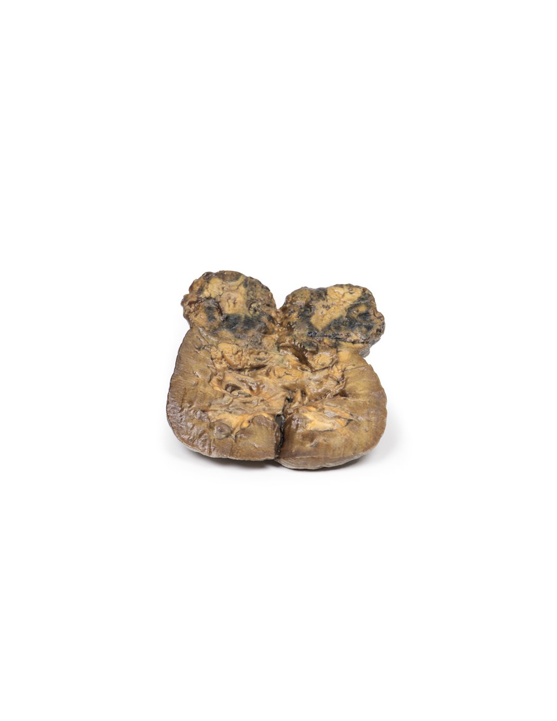
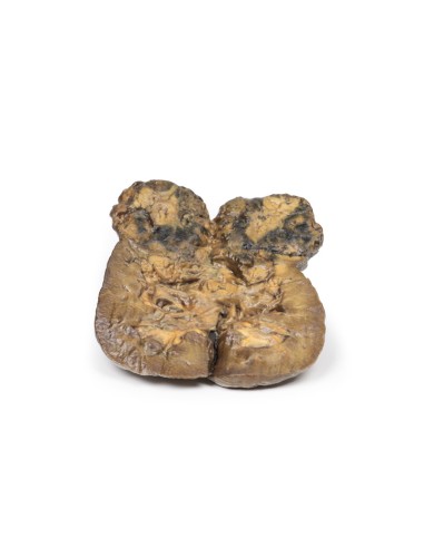
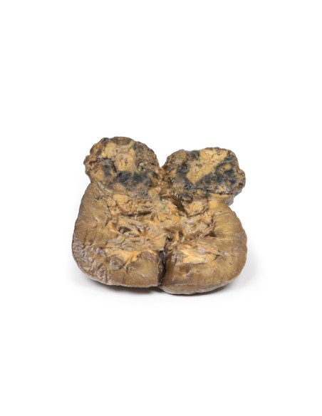
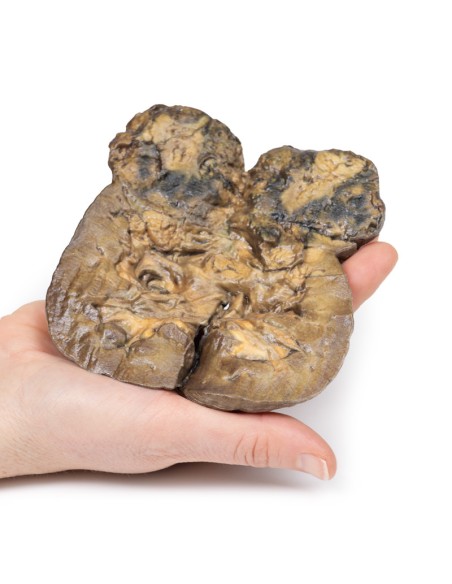
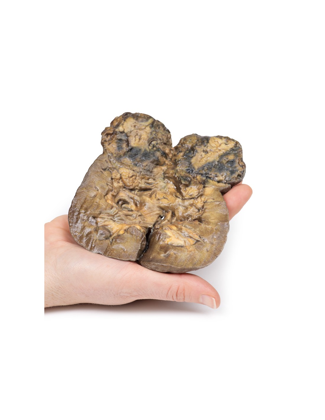
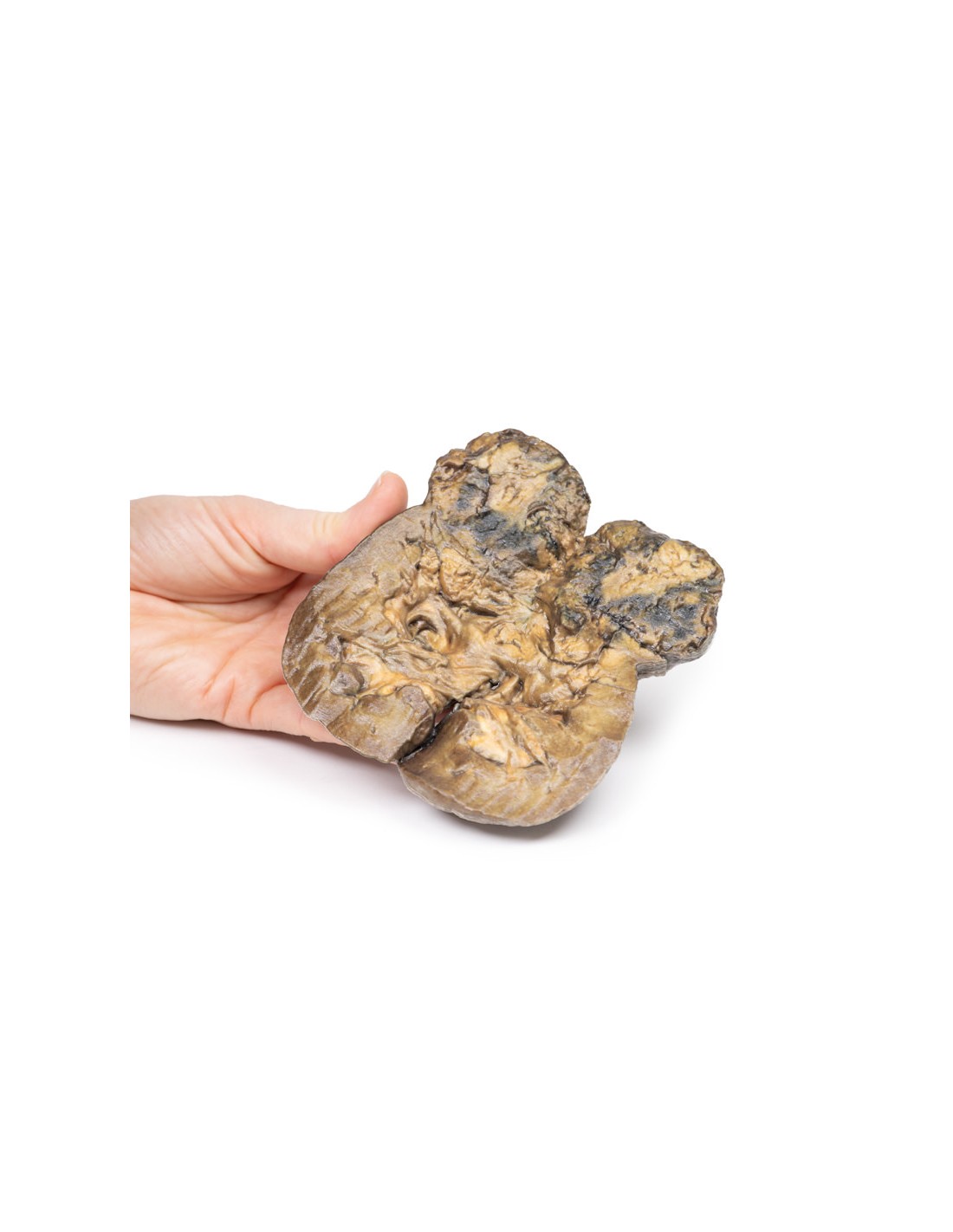
The specimen is a kidney, which was incompletely sectioned in the coronal plane and mounted to visualize the cut surface. The lower pole of the kidney was replaced by a rounded, ill-defined irregular mass 5 cm in diameter, which compressed and distorted the overlying renal parenchyma. The cut surface of the tumor has a variegated appearance caused by areas of hemorrhage and necrosis. Several small pale yellow tumor nodules are present in the cortex and medulla above and separated from the lower pole tumor. These are intrarenal metastases. The renal pelvis appears slightly dilated with some blunting of the renal papillae, suggesting a degree of hydronephrosis. The capsular surface is finely nodular with some gross scarring and contains several small simple cysts (see back of specimen). Histologically,
Further Information.
Renal cell carcinoma (RCC) comprises 85% of primary renal neoplasms. They originate within the renal cortex. The risk of developing RCC is doubled in males. It occurs most commonly in the sixth decade of life. Other risk factors for RCC include smoking, obesity, hypertension, unchallenged estrogen therapy, as well as exposure to asbestos, petroleum, and heavy metals. Most RCCs are sporadic, but about 5 percent are due to autosomal dominant familial tumors, such as Von Hippel Lindau syndrome, hereditary leiomyomatosis, and Birt-Hogg-Dubé syndrome.
There are several main types of primary renal tumors according to the genetic and histological features of the tumor: clear cell carcinoma (70-80%), papillary carcinoma (10-15%), chromophobe carcinoma (5-10%), oncitic carcinoma (3-7%), and collector duct (Bellini) carcinoma (<1%).
Clear cell carcinoma typically has a deletion of chromosome 3p and arises from the proximal tubule. They can be solid or less commonly cystic. They occur in association with Von Hippel Lindau and sporadically. Papillary carcinomas arise from the proximal tubule. They are associated with trisomies 7 and 17; loss of Y in male patients; and MET kinase domain mutations. They are often multifocal in origin. Chromophobe carcinoma originates from intercalated cells of the collecting ducts. They are associated with multiple chromosomal losses and hypodiploidy. They have a low risk of disease progression.
Renal oncocytic carcinomas typically consist of well-differentiated cells with prominent eosinophilic granular cytoplasm; they are associated with a good prognosis. In contrast, collector duct (Bellini) carcinoma of the kidney is a highly aggressive tumor with an extremely poor prognosis because it does not respond well to chemotherapeutic drugs used for renal cell carcinoma and progresses and spreads more rapidly. It is a variety of renal cell carcinoma (RCC) that originates from the distal segment of Bellini's collecting ducts in the renal medulla.
The typical clinical features of RCC are costovertebral pain, palpable mass, and hematuria. RCC is the great mimic in medicine producing many manifestations including: polycythemia, hypercalcemia, hypertension, pyrexia, Cushing's syndrome, eosinophilia, and amyloidosis. RCC tends to metastasize before producing local symptoms. The most common sites of distal spread are the lungs (50%) and bones (33%) followed by lymph nodes, adrenal glands, and brain. RCC has a tendency to invade
the renal vein and extend over it like a tumor thrombus, growing as a solid column extending to the inferior vena cava.
Ultrasound and CT scan are the most common investigations used to evaluate renal lesions and diagnose renal carcinoma. In some patients, a tissue biopsy may be necessary. An increasing number of patients are diagnosed with RCC because of incidental renal lesions detected on abdominal CT scan required for other medical reasons.
The average 5-year survival rate for RCC is 70%. Treatment depends on the stage of the tumor. Radical nephrectomy is the usual surgical option. Medical treatment includes chemotherapeutic drugs as well as vascular endothelial growth factor (VEGF) inhibitors and tyrosine kinase inhibitors in patients with metastatic disease.
.
What advantages does the Monash University anatomical dissection collection offer over plastic models or plastinated human specimens?
- Each body replica has been carefully created from selected patient X-ray data or human cadaver specimens selected by a highly trained team of anatomists at the Monash University Center for Human Anatomy Education to illustrate a range of clinically important areas of anatomy with a quality and fidelity that cannot be achieved with conventional anatomical models-this is real anatomy, not stylized anatomy.
- Each body replica has been rigorously checked by a team of highly trained anatomists at the Center for Human Anatomy Education, Monash University, to ensure the anatomical accuracy of the final product.
- The body replicas are not real human tissue and therefore not subject to any barriers of transportation, import, or use in educational facilities that do not hold an anatomy license. The Monash 3D Anatomy dissection series avoids these and other ethical issues that are raised when dealing with plastinated human remains.
No reviews
Tap to zoom