Your cart
There are no more items in your cart


Renal Infarction - Erler Zimmer 3D anatomy Series MP2098
erler zimmer
EZ-MP2098
€232.17
Tax included
Made in ultra-high resolution 3D printing in full color.
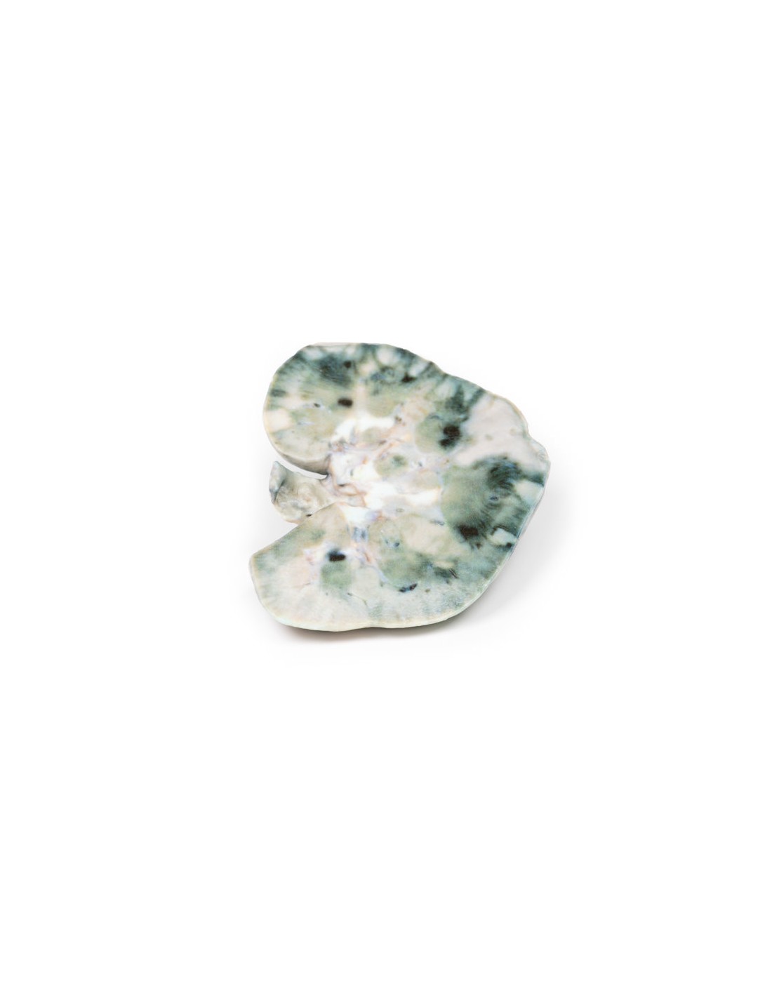
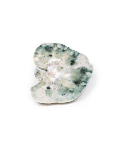
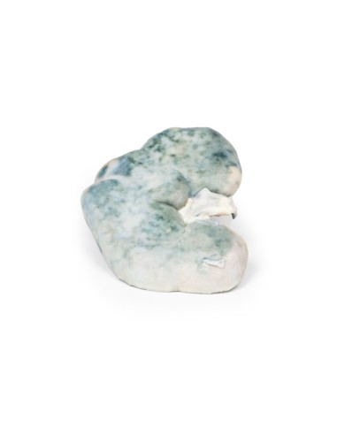
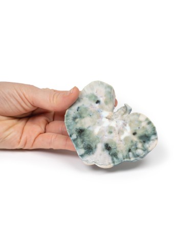
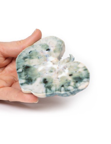
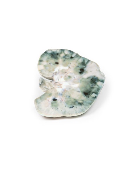
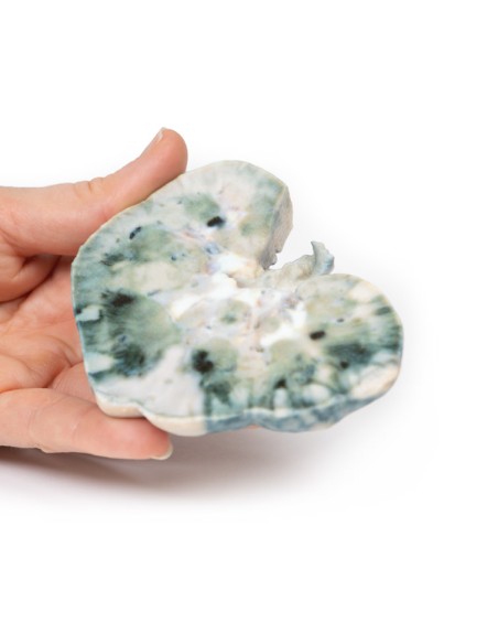
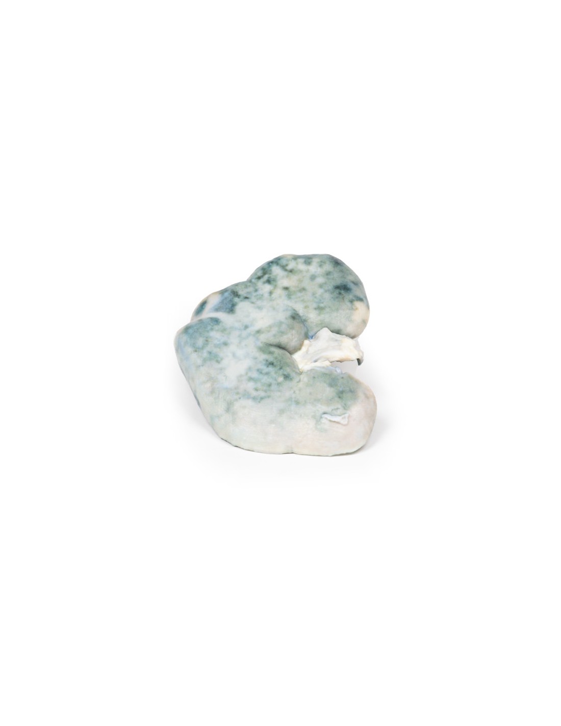
Renal Infarction - Erler Zimmer 3D anatomy Series MP2098
This dissection model highlighting a renal infarct is part of the exclusive Monash 3D anatomy series, a comprehensive series of human dissections reproduced with ultra-high resolution color 3D printing.
Clinical history
A 54-year-old male patient presents with flank pain. He is an active intravenous drug user. Further questioning reveals a history of intermittent hematuria, fevers, malaise, and vomiting. On examination he is hypertensive and pyrexic. Inspection of the limbs reveals Janeway lesions in the extremities and evidence of recent intravenous drug use. Auscultation of the chest reveals a systolic murmur. Blood tests reveal elevated inflammatory markers, decreased renal function, elevated LDH and multiple bacterial blood cultures. Echocardiogram shows a large mobile tricuspid vegetation. Treatment for infective endocarditis was started but he later died of sudden cardiac arrest.
Pathology
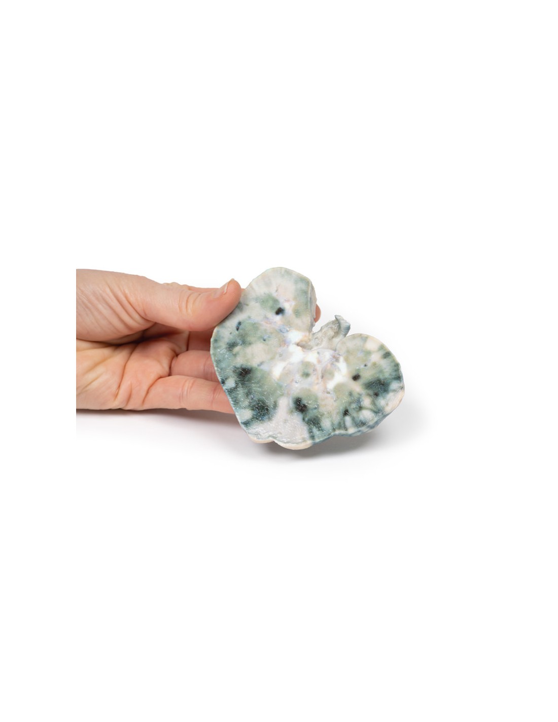
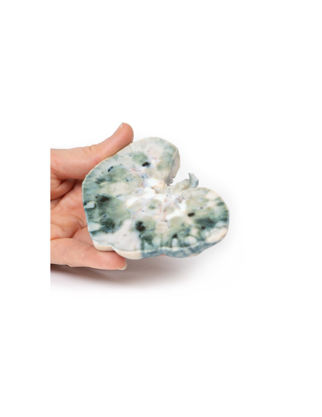
The specimen is the patient's kidney from the postmortem examination. The kidney has been divided in two with a cut half surface showing. Within the cortex are multiple well-demarcated yellow-white areas in the shape of a wedge. The base of these pyramids lies against the cortical surface and extends along the renal columns with the apex facing the medulla. The largest is evident the lateral upper pole of the kidney. These pale areas are infarcted renal tissue. There are dark areas of irregular shape representing areas of hemorrhage.
Further information
Renal infarction results from a disruption of blood flow to the kidney. The kidneys receive nearly a quarter of the cardiac output but have limited collateral circulation. The cortex is the area most susceptible to infarction since blood flow goes from proximal to distal. The main causes of disruption of this circulation are cardioembolic diseases, renal artery damage, hypercoagulant states, or idiopathic.
Cardioembolic causes are the most common. These include mural thrombi post-myocardial infarction, septic emboli from infective endocarditis, and emboli from mechanical valves. Idiopathic renal infarction is the second most common cause. Renal artery damage is the third most common cause and includes renal artery dissection, acute vasculitis from polyarteritis nodosa, trauma or post endovascular surgery. Hypercoagulable states are the rarest cause of renal infarcts such as hereditary thrombophilia and antiphospholipid antibody syndrome. The infarction is bilateral in about 15% of cases.
The presentation of renal infarction depends on the underlying etiology. It can be clinically silent. Common manifestations include costovertebral angle pain, hematuria, hypertension due to increased renin release, nausea, vomiting, and sometimes fever.
Laboratory tests used to aid diagnosis include urinalysis for hematuria and serum creatinine levels, which may be elevated, especially in bilateral disease. CT scan of the abdomen with contrast medium is the radiological investigation of first choice. A wedge-shaped perfusion defect is the classic finding. Treatment varies depending on the cause of the infarction, but generally involves supportive therapy and treatment of the underlying pathology.
.
What advantages does the Monash University anatomical dissection collection offer over plastic models or plastinated human specimens?
- Each body replica has been carefully created from selected patient X-ray data or human cadaver specimens selected by a highly trained team of anatomists at the Monash University Center for Human Anatomy Education to illustrate a range of clinically important areas of anatomy with a quality and fidelity that cannot be achieved with conventional anatomical models-this is real anatomy, not stylized anatomy.
- Each body replica has been rigorously checked by a team of highly trained anatomists at the Center for Human Anatomy Education, Monash University, to ensure the anatomical accuracy of the final product.
- The body replicas are not real human tissue and therefore not subject to any barriers of transportation, import, or use in educational facilities that do not hold an anatomy license. The Monash 3D Anatomy dissection series avoids these and other ethical issues that are raised when dealing with plastinated human remains.
No reviews
Tap to zoom