Your cart
There are no more items in your cart


Transitional cell papillary carcinoma of the renal pelvis - Erler Zimmer 3D anatomy Series MP2099
erler zimmer
EZ-MP2099
€645.50
Tax included
Made in ultra-high resolution 3D printing in full color.
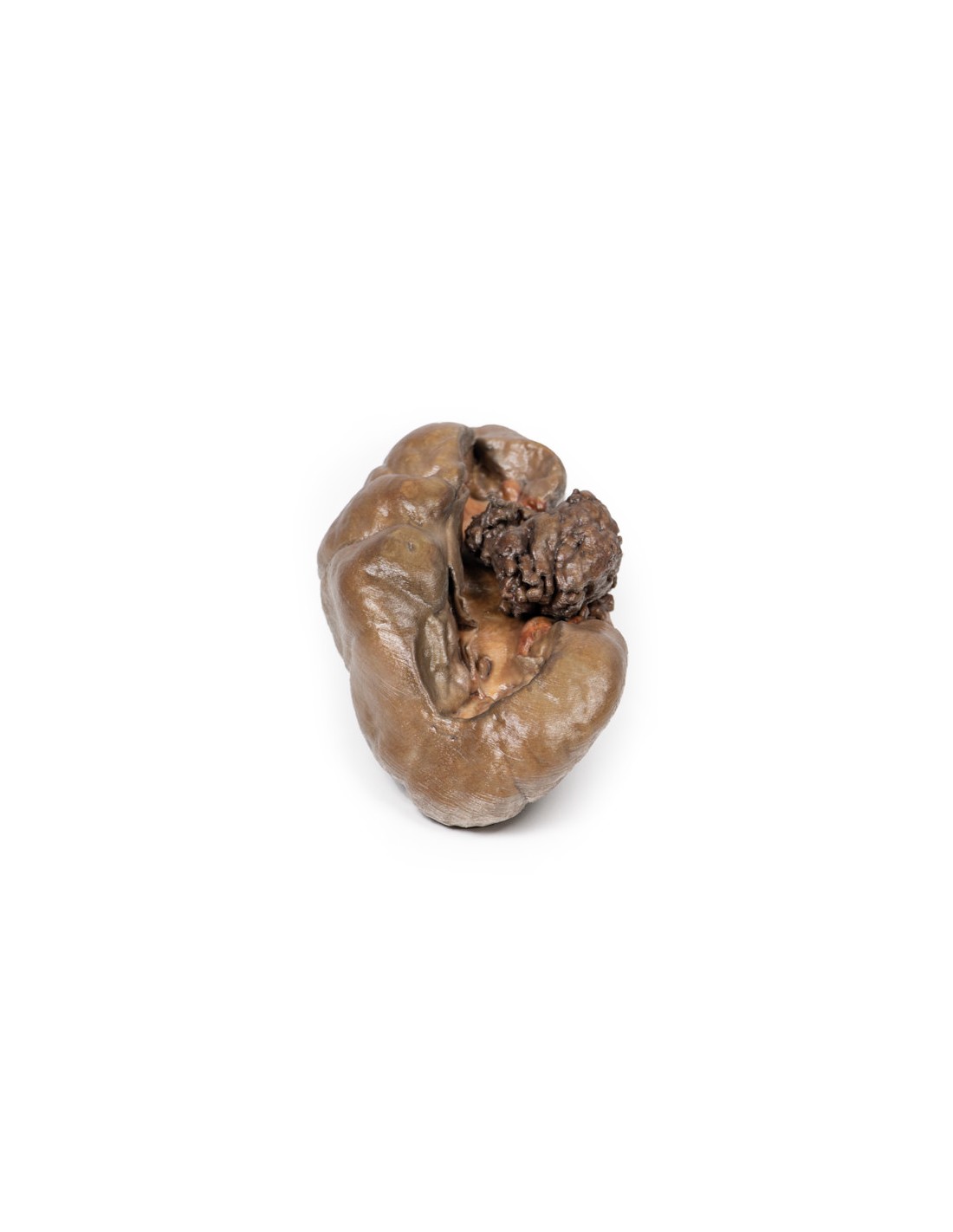
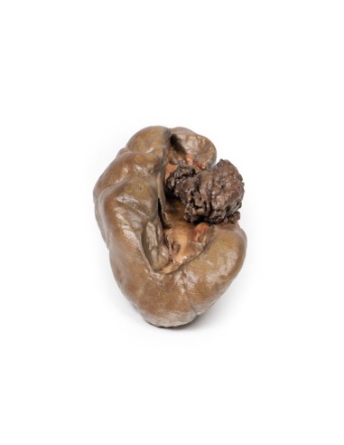
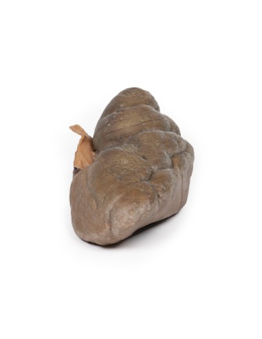
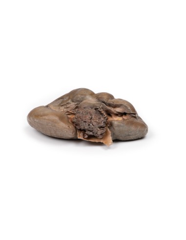
Papillary Transitional Cell Carcinoma of the Renal Pelvis - Erler Zimmer 3D anatomy Series MP2099
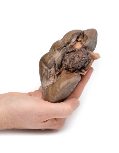
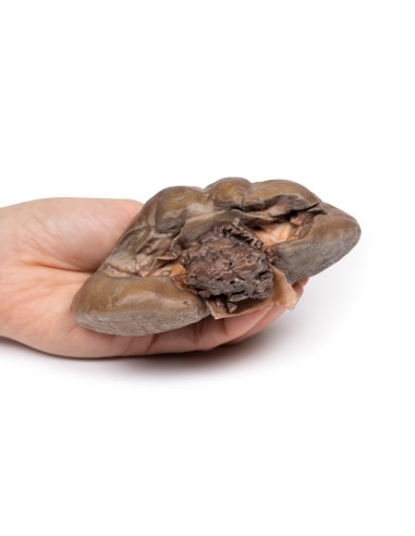
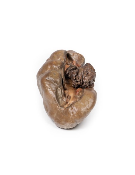
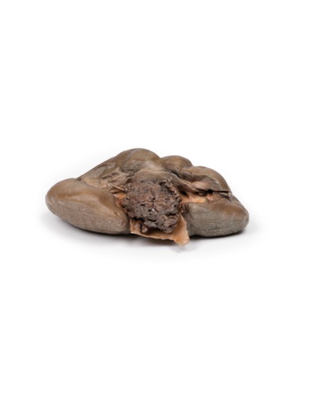
This dissection model highlighting a Papillary Transitional Cell Carcinoma of the Renal Pelvis is part of the exclusive Monash 3D anatomy series, a comprehensive series of human dissections reproduced with ultra-high resolution color 3D printing.
Clinical Story.
A 60-year-old man who had worked in a paint factory for 40 years developed painless hematuria for one month. CT scan showed a suspected tumor in the left renal pelvis. He underwent nephrectomy.
Pathology
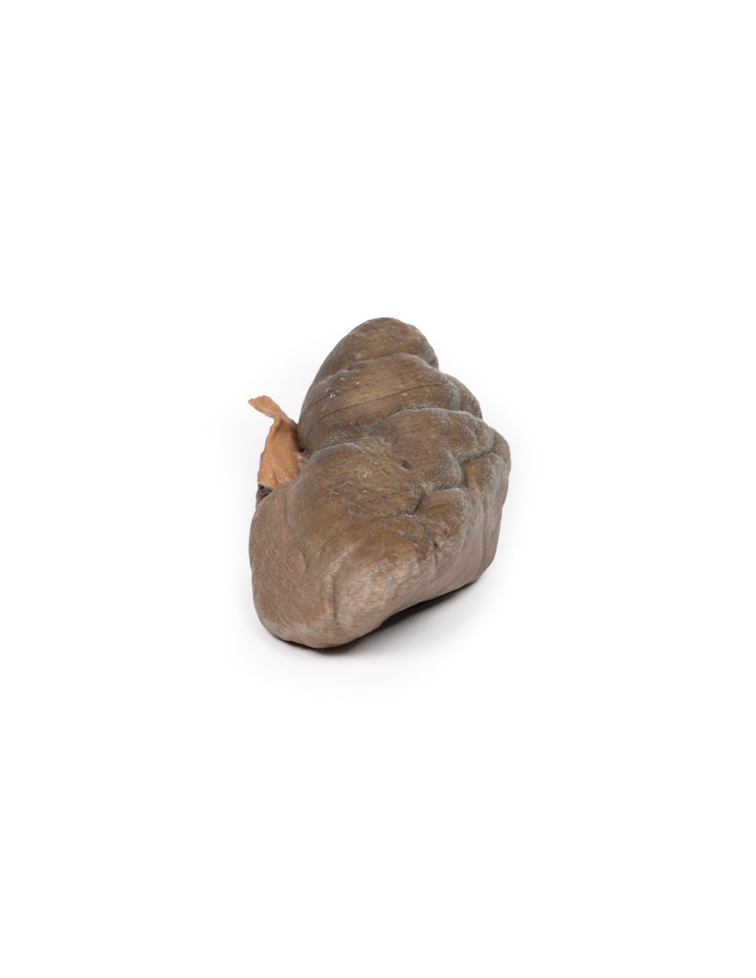
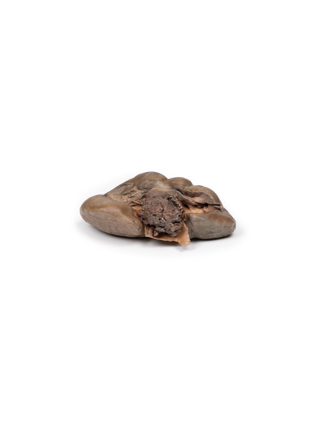
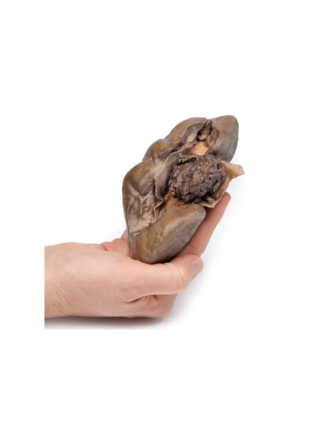
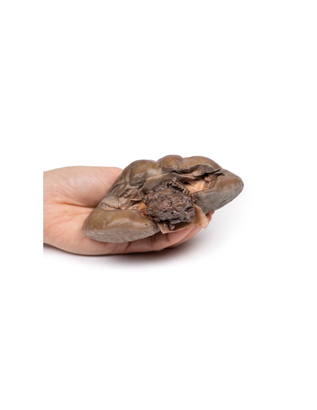
This is the post-nephrectomy kidney. Note that the kidney retains its fetal lobulation. There is a 35 mm diameter friable papillary tumor protruding into the renal pelvis. The renal pelvis is visibly dilated due to this obstructing tumor. Histological examination revealed that it is papillary transitional cell carcinoma arising in the renal pelvis.
Further Information.
Between 5-10% of primary renal tumors arise in the urothelium lining the renal pelvis and calyces. These are similar to tumors that can arise in the ureter and urinary bladder. These tumors range from benign papillomas (rare) to well-differentiated papillary carcinomas, which are common, and poorly differentiated tumors that may be papillary or flat and infiltrating.
Symptoms of these tumors of the renal pelvis tend to occur early. Because of the friable nature of the tumors, hematuria is common. As the tumors grow, obstructive symptoms such as palpable hydronephrosis and flank pain may be noted. Tumors can sometimes be multiple; involving the pelvis, ureter, and bladder.
There is an increased risk of developing urothelial tumors in individuals with Lynch syndrome and analgesic nephropathy. Smoking significantly increases the risk of developing urothelial tumors. Industrial chemicals called aromatic amines, such as benzidine and beta-naphthylamine, which are sometimes used in the dye industry, can lead to urothelial tumors.
Infiltration of the pelvic wall and calyces is common in these tumors. The prognosis with infiltration is not good. Five-year survival rates range from 50-100% for low-grade, noninvasive lesions to 10% for high-grade infiltrating tumors.
.
What advantages does the Monash University anatomical dissection collection offer over plastic models or plastinated human specimens?
- Each body replica has been carefully created from selected patient X-ray data or human cadaver specimens selected by a highly trained team of anatomists at the Monash University Center for Human Anatomy Education to illustrate a range of clinically important areas of anatomy with a quality and fidelity that cannot be achieved with conventional anatomical models-this is real anatomy, not stylized anatomy.
- Each body replica has been rigorously checked by a team of highly trained anatomists at the Center for Human Anatomy Education, Monash University, to ensure the anatomical accuracy of the final product.
- The body replicas are not real human tissue and therefore not subject to any barriers of transportation, import, or use in educational facilities that do not hold an anatomy license. The Monash 3D Anatomy dissection series avoids these and other ethical issues that are raised when dealing with plastinated human remains.
No reviews
Tap to zoom