
Your cart
There are no more items in your cart


Polycystic kidney disease of the adult - Erler Zimmer 3D anatomy Series MP2091
erler zimmer
EZ-MP2091
€603.90
Tax included
Made in ultra-high resolution 3D printing in full color.
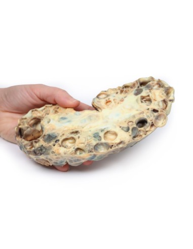
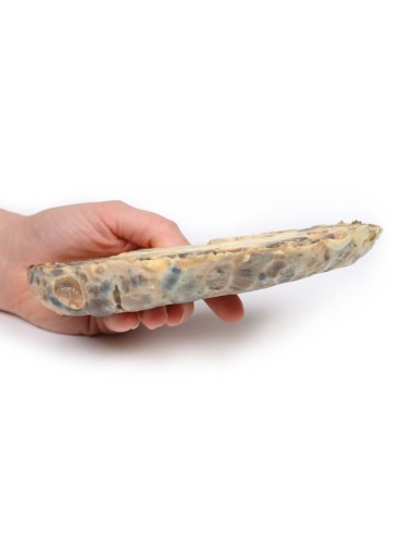
Adult Polycystic Kidney Disease - Erler Zimmer 3D anatomy Series MP2091
This dissection model highlighting Polycystic Kidney Disease of the Adult is part of the exclusive Monash 3D anatomy series, a comprehensive series of human dissections reproduced with ultra-high resolution color 3D printing.
Clinical history
A 40-year-old man visits his family physician complaining of 2-week-old hematuria and new onset of headache with blurred vision. His family physician detects a blood pressure of 260/110 and refers the patient to the hospital. The patient collapses on arrival at the hospital. A brain CT scan shows a large subarachnoid hemorrhage from a ruptured "berry" aneurysm. The patient dies shortly after admission.
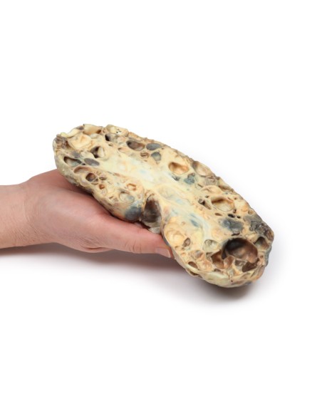
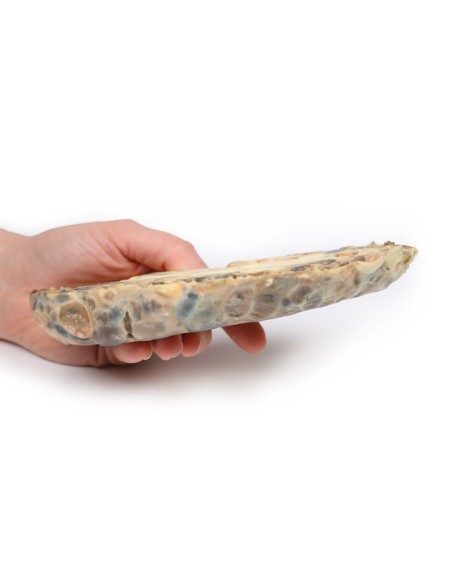
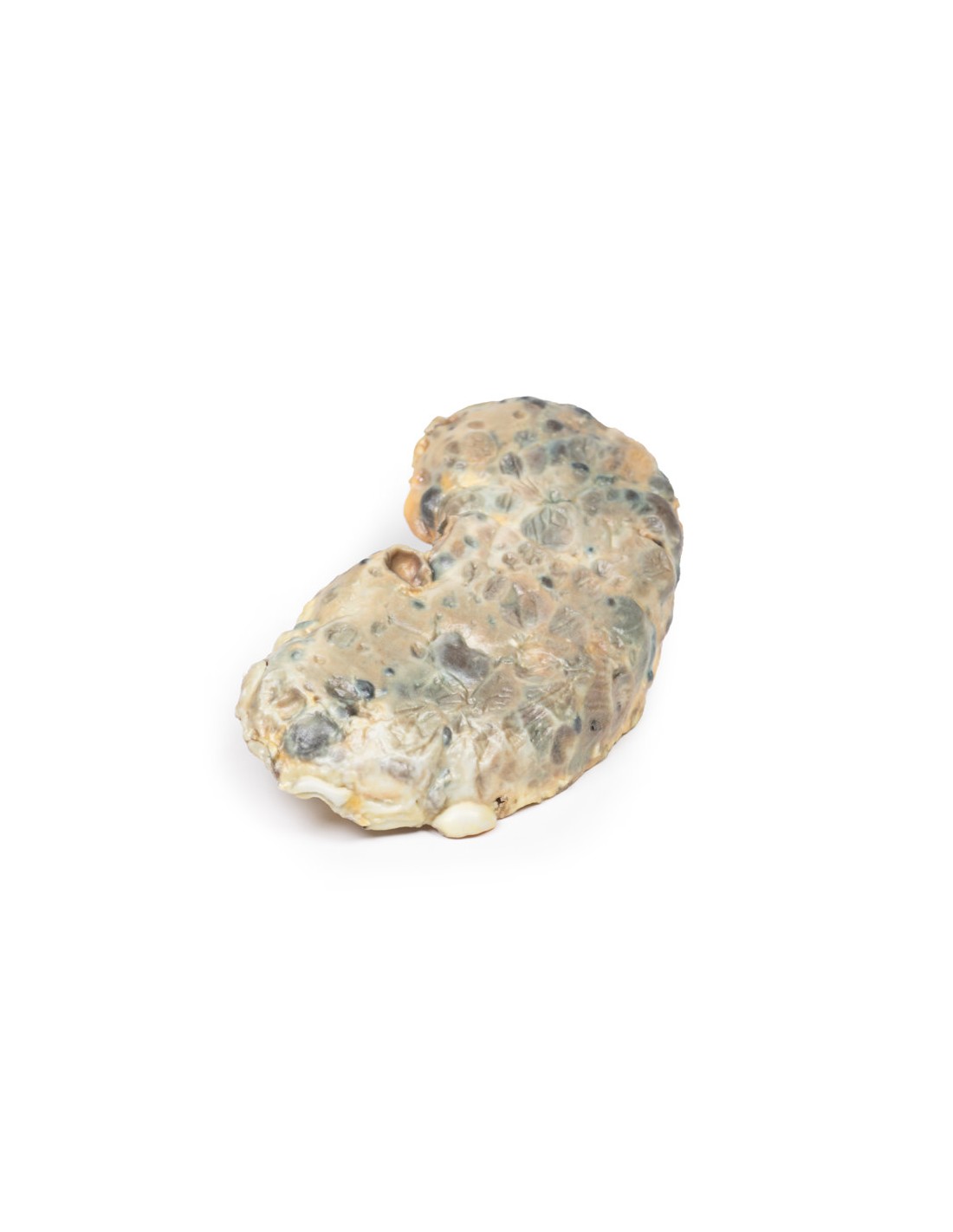
Pathology
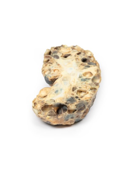
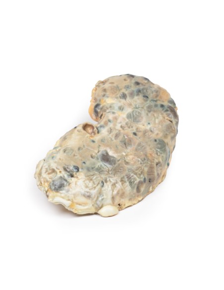
The specimen is an enlarged kidney. The renal parenchyma has been almost completely replaced by numerous dilated cysts of variable size, up to 3 cm in diameter. The cysts have thin, translucent walls, and some cysts contain material of varying colors, giving a "marble-like" appearance to the cut surface of the kidney. The varying colors are caused by secretions within the cysts, which may be mixed with hemorrhage. The outer surface appears lobulated due to multiple protruding cysts. Any residual renal parenchyma is severely atrophic caused by pressure from the numerous cysts. This is an example of adult polycystic kidney disease.
Further information
Adult polycystic kidney disease (APKD) is an autosomal dominant disease characterized by the presence of multiple cysts within the renal parenchyma. The cysts develop from the altered renal tubule epithelium. The cysts expand, destroying the glomeruli, causing ischemia, pressure atrophy, and eventually leading to renal failure.
APKD occurs in 1 in 40 to 1000 live births. Mutations in the PKD1 gene on chromosome 16p13.3 and the PKD2 gene on chromosome 4q21 have been described as causative mutations. These encode for the membrane proteins ??polycystin 1 and 2, respectively. Patients with PKD1 mutation are more common and have a more severe phenotype. End-stage renal disease (ESRD) occurs at an average age of 74.0 years in PKD2 compared with 54.3 years in PKD1.
Common symptoms of APKD include hematuria from hemorrhage to cysts and pain or a dragging sensation from cyst expansion and kidney enlargement. Many patients remain asymptomatic until features of renal failure such as proteinuria, polyuria, hypertension and uremia occur. Extrarenal manifestations of the disease include intracranial "berry" aneurysms, liver and pancreatic cysts, as well as mitral valve prolapse and other types of heart valve disease. Renal ultrasound is the most common investigation used to diagnose APKD. CT and MRI scans can also be used as diagnostic tools. Patients with a positive family history of APKD may be offered renal ultrasound screening and genetic testing in some cases. Treatment includes renal replacement therapy for ESRD and renal transplantation (if a donor can be found).
Finally, more than one-third of patients die from renal failure and one-third from coronary artery disease or hypertensive disease. About 1% of patients die from subarachnoid hemorrhage, due to rupture of the berry aneurysm (as in this case). The remaining deaths are due to unrelated causes.
.
What advantages does the Monash University anatomical dissection collection offer over plastic models or plastinated human specimens?
- Each body replica has been carefully created from selected patient X-ray data or human cadaver specimens selected by a highly trained team of anatomists at the Monash University Center for Human Anatomy Education to illustrate a range of clinically important areas of anatomy with a quality and fidelity that cannot be achieved with conventional anatomical models-this is real anatomy, not stylized anatomy.
- Each body replica has been rigorously checked by a team of highly trained anatomists at the Center for Human Anatomy Education, Monash University, to ensure the anatomical accuracy of the final product.
- The body replicas are not real human tissue and therefore not subject to any barriers of transportation, import, or use in educational facilities that do not hold an anatomy license. The Monash 3D Anatomy dissection series avoids these and other ethical issues that are raised when dealing with plastinated human remains.
No reviews
Tap to zoom
