Your cart
There are no more items in your cart


Osteosarcoma of the femur - Erler Zimmer 3D anatomy Series MP2116
erler zimmer
EZ-MP2116
€465.67
Tax included
Made in ultra-high resolution 3D printing in full color.
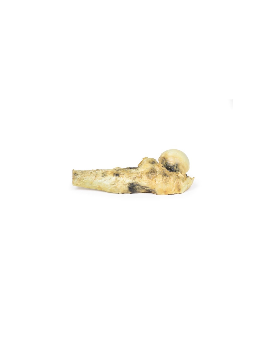
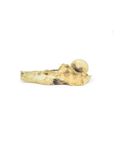
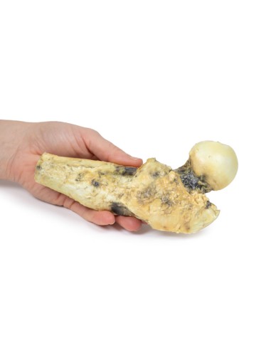
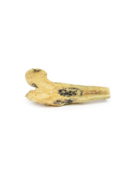
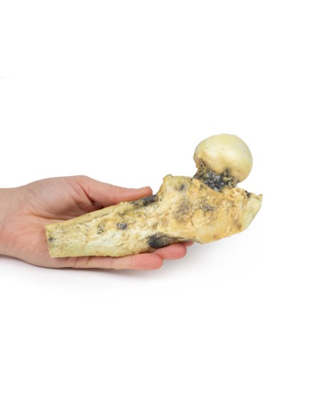
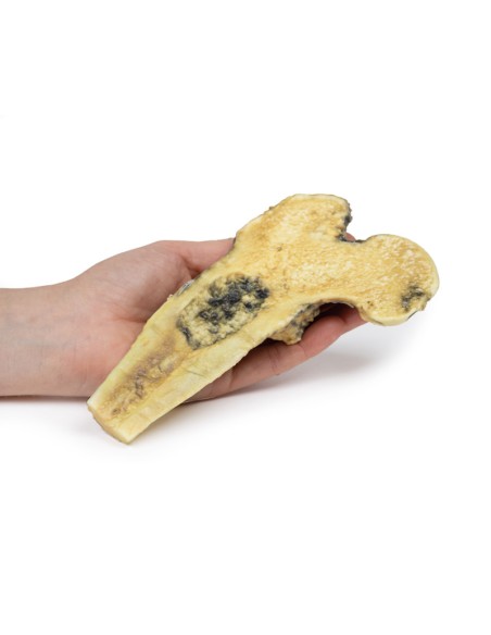
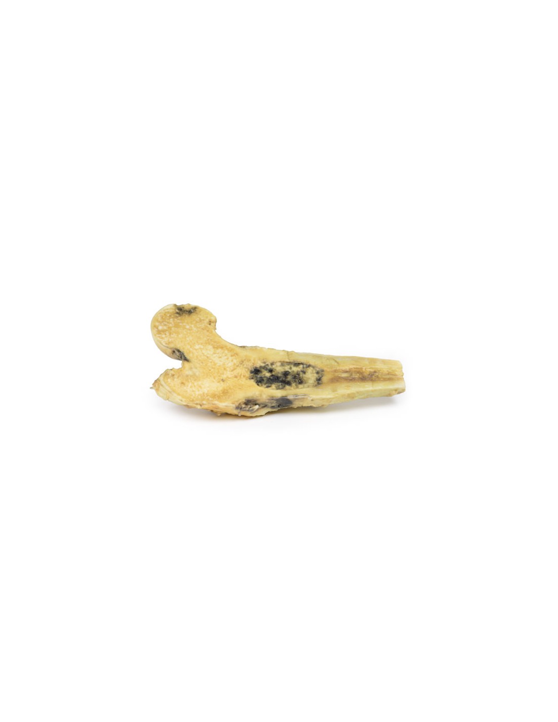
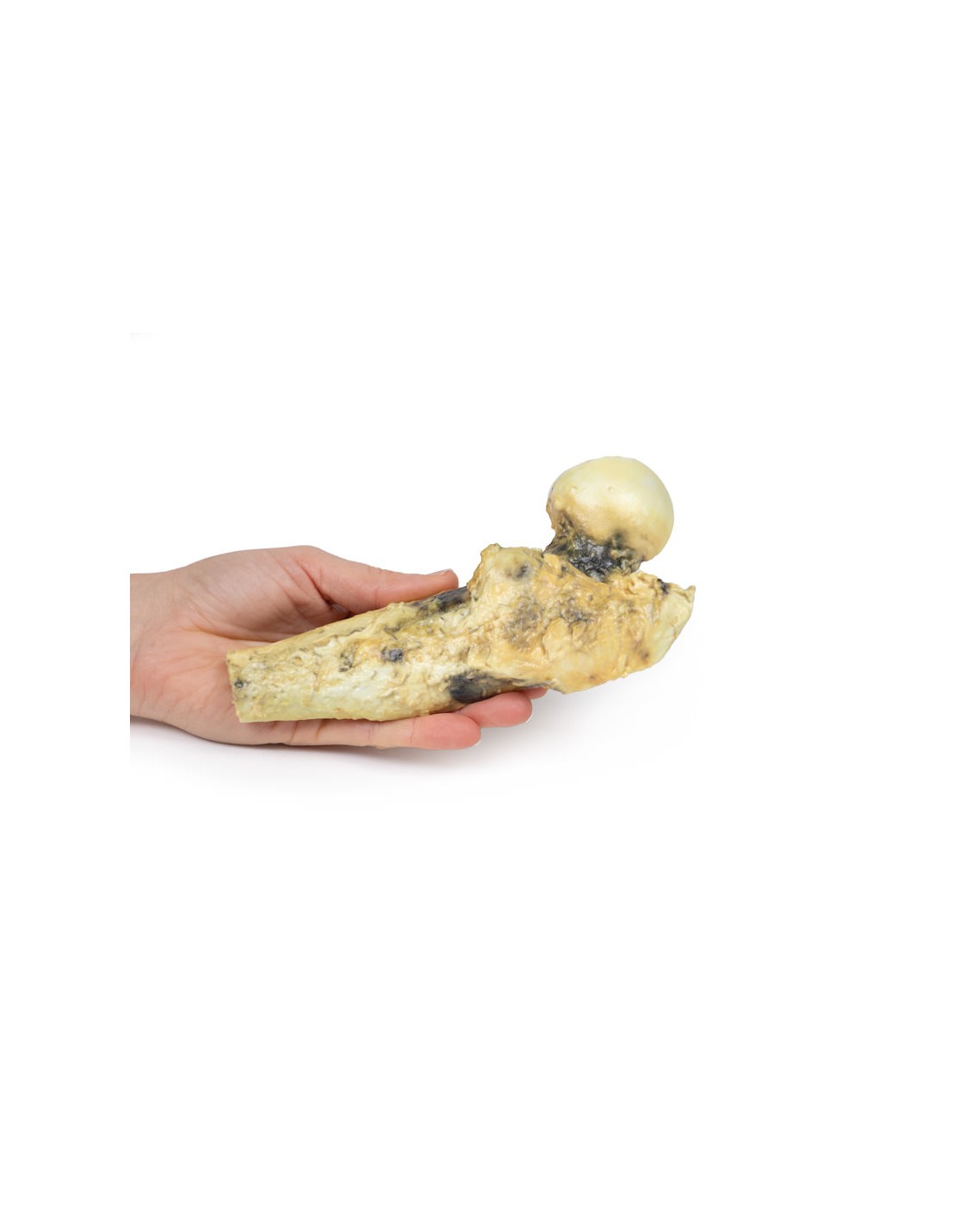
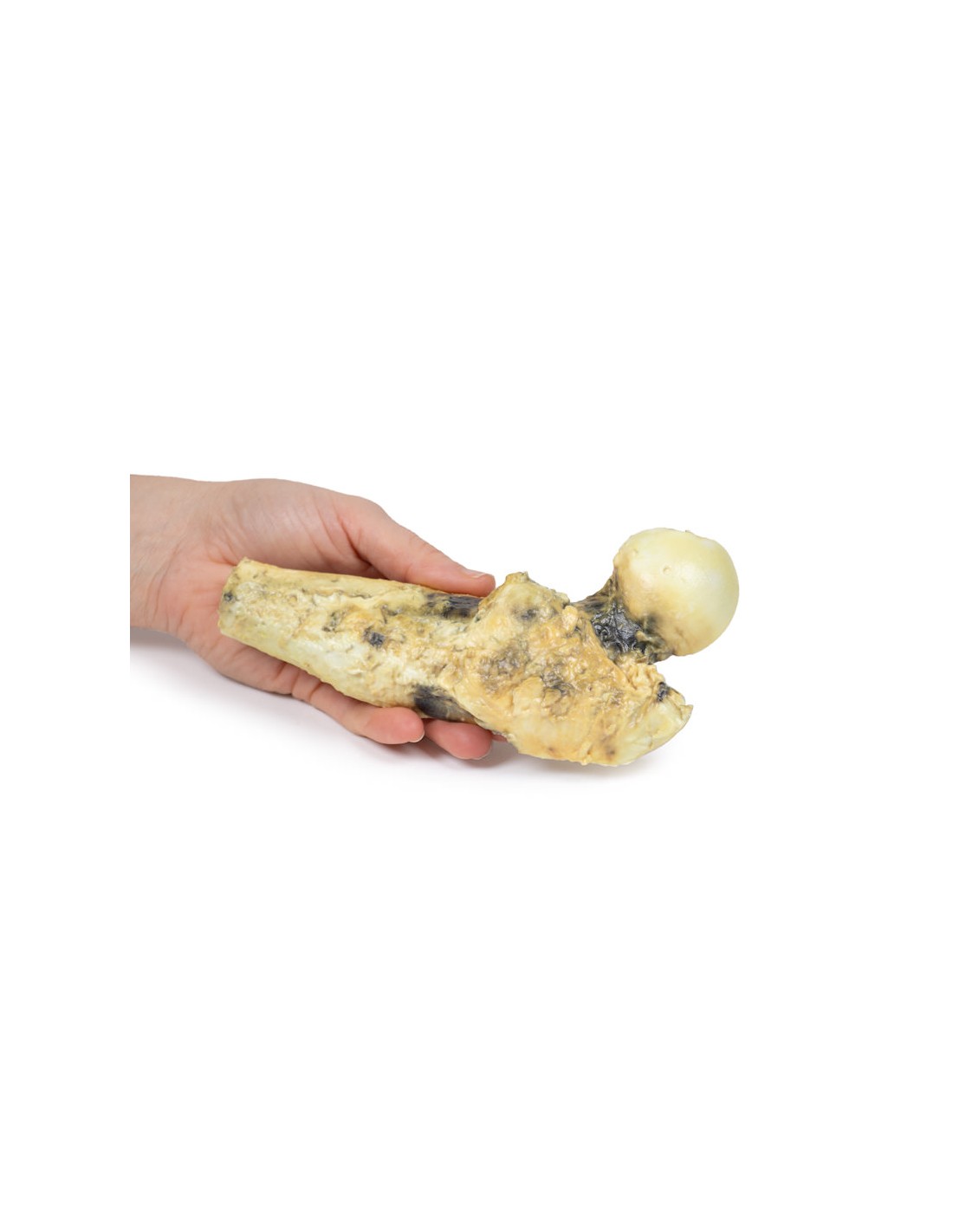
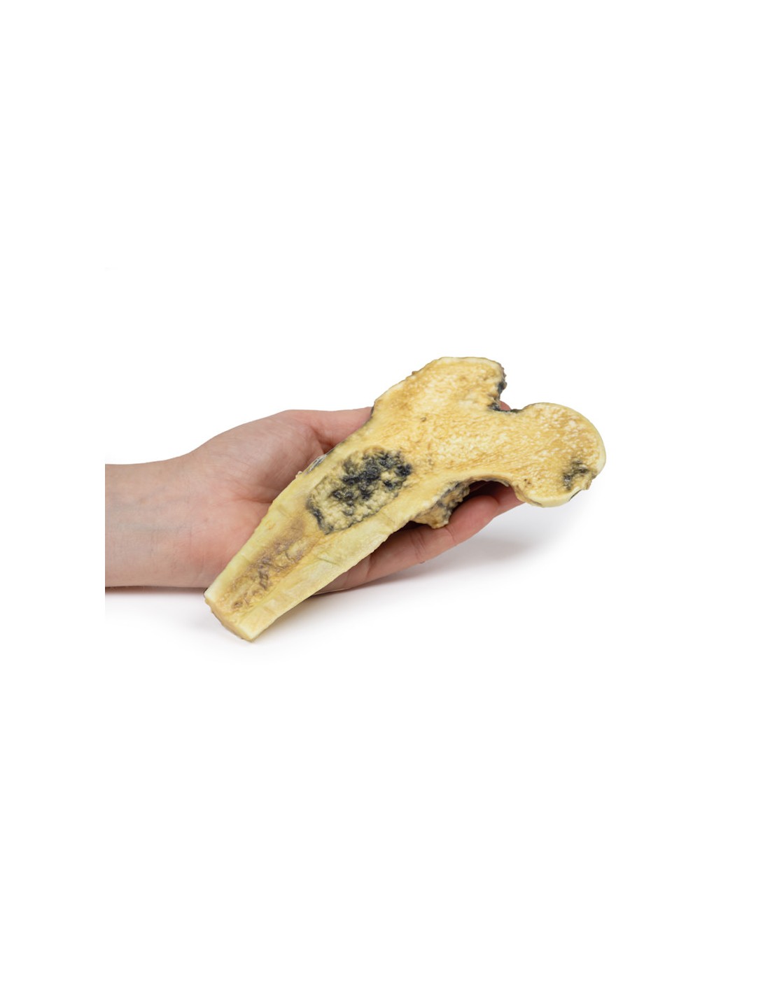
Osteosarcoma of the Femur - Erler Zimmer 3D anatomy Series MP2116
This dissection model highlighting an Osteosarcoma of the femur is part of the exclusive Monash 3D anatomy series, a comprehensive series of human dissections reproduced with ultra-high resolution color 3D printing.
Clinical History.
A 57-year-old man presents complaining of recurrent pain in the right thigh. On examination, there is no palpable abnormality in the thigh. A radiograph of the limb showed bone absorption associated with expansion and periosteal reaction to the proximal right femur. A CT scan of the limb showed a mass in the proximal right femur. A biopsy of the lesion was performed. He subsequently had excision of the right upper femur followed by insertion of a prosthesis.
Pathology
The specimen includes the head, neck and upper third of the shaft of the right femur, sawn longitudinally to show the cut surface. In the medullary cavity of the upper portion of the stem is an ovoid tumor with a maximum diameter of 6.5 cm. The tumor is not encapsulated and has a hemorrhagic cut surface with pale hyaline and cystic areas. Histologically, this is a low-grade chondrosarcoma.
Additional Information.
Chondrosarcomas are malignant bone tumors that produce cartilage. These are the third most common primary bone neoplasm after myeloma and osteosarcoma. Conventional tumors are the most common subtype of chondrosarcoma, constituting 90% of cases. Less frequently diagnosed subtypes include clear cell, dedifferentiated, and mesenchymal chondrosarcomas.
Some chondrosarcomas arise from preexisting benign lesions, such as enchondroma or osteochondroma. Common mutations in chondrosarcomas are point mutations in the IDH1 and IDH2 genes and silencing of the oncosuppressor gene CDKN2A. Chondrosarcomas occurring in multiple osteochondroma syndrome have mutations in the EXT oncosuppressor genes.
Men are twice as likely to develop chondrosarcoma as women. The axial skeleton is more frequently affected than the appendicular skeleton. About 20% affect the femur. These are largely slow-growing tumors. They usually present as painful and gradually expanding masses. At diagnosis, most are low-grade tumors that rarely metastasize. The lungs are the most common site of distant spread. Grade 1 tumors have a 5-year survival rate of nearly 90%, while with grade 3 chondrosarcoma the 5-year survival rate drops to 43%.
CT is the optimal radiological investigation for diagnosis even with MRI frequently used. Biopsies may be taken to aid in diagnosis. Treatment depends on the grade and location of the tumor. Complete surgical resection is the standard treatment. Generally, chondrosarcomas do not respond to chemotherapy or radiation therapy because they are very slow-growing tumors.
What advantages does the Monash University anatomical dissection collection offer over plastic models or plastinated human specimens?
- Each body replica has been carefully created from selected patient X-ray data or human cadaver specimens selected by a highly trained team of anatomists at the Monash University Center for Human Anatomy Education to illustrate a range of clinically important areas of anatomy with a quality and fidelity that cannot be achieved with conventional anatomical models-this is real anatomy, not stylized anatomy.
- Each body replica has been rigorously checked by a team of highly trained anatomists at the Center for Human Anatomy Education, Monash University, to ensure the anatomical accuracy of the final product.
- The body replicas are not real human tissue and therefore not subject to any barriers of transportation, import, or use in educational facilities that do not hold an anatomy license. The Monash 3D Anatomy dissection series avoids these and other ethical issues that are raised when dealing with plastinated human remains.
No reviews
Tap to zoom