
Your cart
There are no more items in your cart


Made in ultra-high resolution 3D printing in full color.
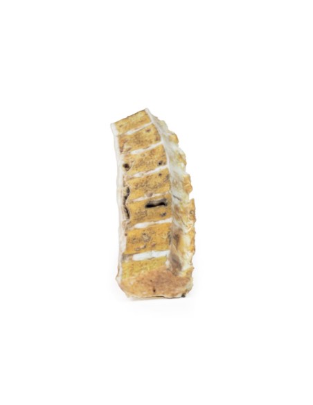
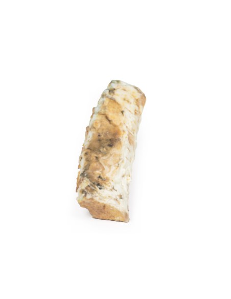
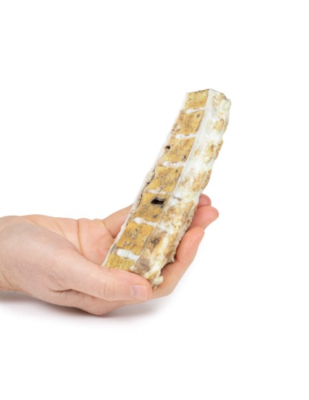
Tuberculosis - Erler Zimmer 3D anatomy Series MP2111
This dissection model highlighting a spinal column with tuberculosis degeneration is part of the exclusive Monash 3D anatomy series, a comprehensive series of human dissections reproduced with ultra-high-resolution color 3D printing.
Clinical history.
A 37-year-old woman presents with increasing thoracic back pain. She has a history of untreated human immunodeficiency virus (HIV) infection and pulmonary tuberculosis. History revealed ongoing low-grade fevers, chills, and weight loss. Examination revealed a cachexic patient with multi-level tender thoracic vertebrae. Blood analysis showed elevated serum calcium and erythrocyte sedimentation rate. Radiography of the spine showed lytic areas in the thoracic vertebrae. During her hospitalization, she developed urosepsis and died.
Pathology
The specimen is a portion of the patient's thoracic spine that was sawed longitudinally and mounted to visualize the cut surface of 7 thoracic vertebrae. Osteolytic areas, ranging in diameter from 1 to 12 mm, are present in all vertebrae, containing degenerative caseous material* (mostly now lost) and surrounded by a thin zone of dense bone. The tubercular inflammatory process has extended into one of the intervertebral discs and has also spread outside the vertebral bodies to form collections of caseous material under the anterior longitudinal ligament. This is an example of mycobacterial tuberculous osteomyelitis of the spine with paravertebral extension, also known as Pott's disease.
Additional Information.
Tuberculosis (TB) is a chronic pulmonary and systemic infectious disease caused by Mycobacteria tuberculosis. Transmission most commonly occurs through inhalation of aerosolized droplets of M. tuberculosis. Risk factors for contracting tuberculosis include being a resident of a "developing" country where the disease may be endemic, immunosuppression (e.g., HIV, steroid use, anti-TNF use, and diabetes), chronic lung disease (e.g., silicosis), alcoholism, and malnutrition.
After initial lung infection with M. tuberculosis, the clinical manifestation varies. In 90% of individuals with an intact immune system, they enter a phase of asymptomatic latent infection. This latent tuberculosis can reactivate at any time in the patient's life. In the other 10% of patients, especially in the immunocompromised population, they develop primary disease, which is an immediate active TB infection. Manifestations of primary TB include symptoms of pulmonary infection (e.g., consolidation, effusion, and hilar adenopathy) and extrapulmonary symptoms-lymphadenopathy, meningitis, and disseminated miliary tuberculosis. Secondary tuberculosis occurs when there is reactivation of a previous latent tuberculous infection. About 10 percent of latent tuberculosis is usually reactivated during periods of weakened host immunity. Typical symptoms of reactivation are cough, hemoptysis,
Bone infection occurs in 1-3% of patients with tuberculosis infection. There is a higher incidence of developing bone disease in patients from developing countries and immunocompromised patients. Tuberculosis usually spreads hematogenously from the site of active disease. Pott's disease accounts for 40% of tuberculosis bone infections. The infection is destructive, eroding vertebral discs and vertebrae leading to compression fractures, which can cause symptoms of cord or nerve root compression. Symptoms include pain at the site of the disease, fevers, chills, weight loss, compression symptoms, and spinal deformities, such as kyphosis and scoliosis.
The diagnosis of tuberculosis is usually made with a clinical history, chest X-ray and multiple sputum cultures. Mantoux skin tuberculin test and serum interferon gamma release test can also be used to help screen for infection. Biopsies can be taken from the site of suspected infection for culture to aid in diagnosis.
Treatment involves prolonged courses of multiple antibiotics, depending on the antibiotic resistance of the infecting mycobacterial species.
* Caseous degeneration or necrosis is a unique form of cell death in which the tissue retains a cheese-like appearance.
What advantages does the Monash University anatomical dissection collection offer over plastic models or plastinated human specimens?
- Each body replica has been carefully created from selected patient X-ray data or human cadaver specimens selected by a highly trained team of anatomists at the Monash University Center for Human Anatomy Education to illustrate a range of clinically important areas of anatomy with a quality and fidelity that cannot be achieved with conventional anatomical models-this is real anatomy, not stylized anatomy.
- Each body replica has been rigorously checked by a team of highly trained anatomists at the Center for Human Anatomy Education, Monash University, to ensure the anatomical accuracy of the final product.
- The body replicas are not real human tissue and therefore not subject to any barriers of transportation, import, or use in educational facilities that do not hold an anatomy license. The Monash 3D Anatomy dissection series avoids these and other ethical issues that are raised when dealing with plastinated human remains.
No reviews
Tap to zoom
